Sign Up
You are not currently logged in. Please log in to CEUfast to enable the course progress and auto resume features.

This Course Has Expired

Sorry, but this course is no longer active. We are keeping the course material here for research puposes. View our full course library
REQUEST THIS COURSE BE RENEWED
Author:    Jeff Thompson (Senior Developer)

Introduction

Asthma is a lung disease that is characterized by inflammation of the airways which is either completely or partially reversible (bronchoconstriction/bronchospasm), and mucus production. This disease process is often a response to a trigger or stimulus and can lead to labored breathing, dyspnea, chest tightness, cough, and wheezing. Severe attacks of asthma can become potentially threatening to the survival of the patient. Stimuli that can trigger an attack can include things like allergens, smoke, extreme air temperatures, dander (from pets), exercise, illness, or even stress. The inflammation that is a response to such triggers can often be controlled with pharmaceutical measures and prevention. It can also be simply defined as reversible airway obstruction (Merck, 2005; Wikipedia, 2008; Friedman, 2007).

Epidemiology

Here are some interesting epidemiological facts about asthma:

  • The prevalence of asthma is growing, especially in developed countries and in children where it affects 1 in 4 children who live in urban environments (Merck, 2005; Wikipedia, 2008).
  • It affects more than 17 million Americans (Friedman, 2007) and 4% to 7% of the world's population (Merck, 2005).
  • The prevalence is higher for those under the age of 18 than it is for those from the ages of 18 to 64 (Merck, 2005).
  • There is a higher chance for males before puberty to develop asthma and for girls to develop it after puberty (Merck, 2005).
  • It is a leading cause for hospitalization and the number 1 condition accounting for elementary school absenteeism (Merck, 2005).
  • In the United States, Puerto Ricans, African Americans, Filipinos, and Native Hawaiians are the most likely to develop asthma and Mexicans and Koreans are the least likely (Wikipedia, 2008).
  • Being born in the United States increases one’s chances of getting asthma than those who were born in other countries and migrated here (Wikipedia, 2008).
  • Asthma is highest among low-income populations which are also most likely to live near industrial areas (Wikipedia, 2008).
  • Asthma has been strongly correlated with the presence of cockroaches in living quarters (Wikipedia, 2008).
  • Severe persistent asthma is greater in low income communities compared with communities with more access to treatment (Wikipedia, 2008).
  • Asthma is more prevalent in athletes, although this may be due to those with even mild disease may be more likely to pursue diagnosis based on a decrease in ability to perform (Wikipedia, 2008).

Risk factors for Asthma

Common risk factors associated with the development (not necessarily exacerbation) of asthma include:

1. Poor air quality.
   
2. Tobacco smoke inhalation.
   
3. A lack of exposure to viral respiratory infections as a young child.
   
4. High antibiotic use early in life.
   
5. C-sections (it is thought that delivery via C-section changes the normal exposure of the child to bacteria which alters the immune system than when delivered vaginally).
   
6. High stress levels in a child’s caregiver (it is thought that high stress can lead to behaviors that can affect asthma such as smoking, but it is also hypothesized that stress itself can affect the immune system directly).
   
7. A family history of asthma.
   
8. Premature birth or low birth weight.
   
9. Maternal smoking.
   
10. Being male for asthma in prepubertal children or female past puberty and into adulthood.
   
11. Genetic disposition. There are 25 genes that have been routinely associated with the development of asthma, most recently noted is the ADAM33 gene. These genes are also linked to inflammatory/immune responses. However, it is not clear if they are directly related to the disease because their associations vary in that not all genes are present in all cases.
   
12. Gene-environmental association. Some research suggests that some genetic predispositions may only be effective in their association with asthma when combined with environmental factors.

(Wikipedia, 2008)

Etiology of Asthma

There are many genetic and environmental factors that have been associated with the development/prevalence of asthma, but it has not been singled out which ones are directly causative, or if they are only effective in combination (Wikipedia, 2008). The ADAM33 gene was most recently identified which is thought to stimulate airway smooth muscle and fibroblast proliferation or may play a role in cytokine production. Susceptibility genes for the T-helper cells and their respective cytokines (IL4, IL5, IL9, and IL 13) are thought to possibly play a role as well. Although infection and early endotoxin exposure can help in younger children inducing tolerance to asthma, household and environmental allergens are implicated in asthma development in older children and in adults. Even though air pollution can trigger asthmatic reactions, it is not linked in the development of the disease. Obesity, diets low in vitamins C and E, omega-3 fatty acids, young maternal age, poor maternal nutrition, prematurity, low birthweight, lack of breastfeeding, and exposure to volatile organic compounds are also implicated in asthma development (Merck, 2005). The hygiene hypothesis is one in which it is supposed that cleaner environments for young children can lead to an increased risk for asthma (Wikipedia, 2008).

Pathophysiology of Asthma

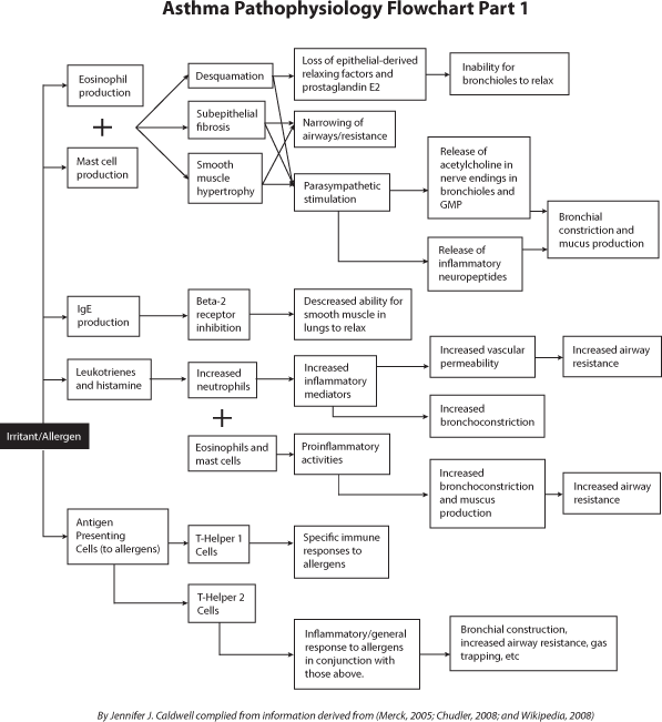
Both genetics and environment may play a role in the development of T-helper cells. It is thought that infants are born with a predisposition towards producing more T-helper 2 immune responses, known as the humoral immune system. This type of immunity predisposes the patient towards more of an allergic and inflammatory response (Merck, 2005). It is more of a general response than the adaptive immune system (which arises from T-helper 1 cells) and is associated with IgE production and activation of eosinophils. The T-helper 1 immune response is very specific against specific antigens and does not induce the inflammation and general response that the humoral system does (Wikipedia, 2008—Immune System). It is thought that early childhood exposure to infection, both bacterial and viral, can shift the body to perform more T-helper 1 responses instead of T-helper 2 responses. As the T-helper 2 responses are diminished, the patient is less predisposed to having allergic/inflammatory responses to pathogens. Therefore, as more affluent countries begin having fewer children, quicker medical treatment (more readily providing antibiotic treatment), and early vaccination, this T-helper-2-suppressing mechanism may be reduced (Merck, 2005).

T-helper 2 cells, eosinophils, and mast cells, among others, form infiltrates in the epithelial and smooth muscle layers in the airways. This leads to desquamation (shedding of the epidermal layer), subepithelial fibrosis, and smooth muscle hypertrophy. This narrows the airways and increases the reactivity of these tissues to allergens and irritants. It also increases reactivity to parasympathetic stimulation (and the release of inflammatory neuropeptides), and other substances that trigger bronchoconstriction (Merck, 2005).

In parasympathetic stimulation, bronchial constriction and mucus production increases (Chudler, 2008). During stimulation, impulses travel to the vagal center of the brainstem, down the vagal afferent pathway back to the bronchus where acetylcholine is released from the nerve endings. This release of acetylcholine results in the formation of cyclic GMP (guanine monophosphate) which leads to bronchoconstriction (Wikipedia, 2008). Epithelial-derived relaxing factors and prostaglandin E2 inhibit bronchoconstriction and are lost in asthmatics as are some substances that deteriorate endogenous bronchoconstrictors (endopeptidases). This is due to the desquamated epithelium and mucosal edema (Merck, 2005). In simplest terms, asthmatics become hypersensitive to certain stimuli and have an allergic reaction in their airways (Wikipedia, 2008).

Leukotrienes are naturally produced and may be responsible for the effects of an inflammatory response. In asthma, they can cause airway obstruction, increased secretion of mucus, mucosal accumulation, bronchoconstriction, and infiltration of inflammatory cells into the airway wall. Their production by the body is part of a complex reaction that includes the production of histamine. Some leukotrienes have a chemotactic effect on neutrophils (which release inflammatory mediators) and can increase vascular permeability and bronchoconstriction. Cysteinyl leukotriene receptors are present on mast cells, eosinophils, and endothelial cells. When these are reacted with, proinflammatory activities are stimulated (Wikipedia, 2008—Leukotrienes in Asthma).

When allergens are inhaled into the respiratory system, they are taken up by antigen presenting cells which actually present pieces of the allergen to T-helper cells. These antigens are usually ignored by the immune system, but in asthmatics, these cells transform into T-helper 2 cells which form the humoral immune response system. They produce antibodies against that specific antigen and when the allergen is reintroduced into the body, these antibodies react with the allergen, causing inflammation, chemical production to cause airway constriction, and mucus production (Wikipedia, 2008).

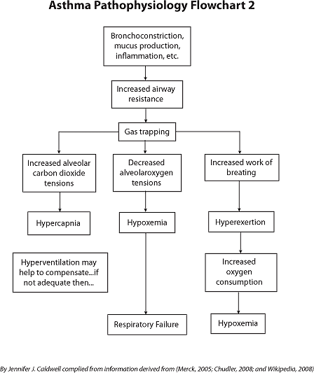
When a stimulus is present to cause an asthmatic attack, the airways narrow and produce excess mucus (Wikipedia, 2008). This reaction to the airways is totally or partially reversible. The lungs are not evenly ventilated, even though they remain adequately perfused. Therefore, alveolar oxygen tensions decrease (as the blood carries available oxygen away and adequate amounts of oxygen are not able to be inhaled), and carbon dioxide tensions increase (as the waste carbon dioxide is released and not able to be completely expelled). Hyperventilation occurs and patients are often able to expel the majority of excess carbon dioxide, but in severe cases, gas trapping through significant bronchoconstriction can lead to hypoxemia and hypercapnia. Gas trapping also causes the respiratory muscles to be mechanically disadvantaged which increases the work of breathing for the patient in order to generate adequate inspiratory force. Hyperexertion of the patient increases oxygen consumption, making hypoxemia worse. In severe enough cases, the hypoxemia can lead to respiratory and cardiac failure (Merck, 2005).

Beta-2 receptors, when stimulated, cause smooth muscle relaxation in the bronchioles. Andor Szentivanvi demonstrated that IgE blocks these receptors. IgE overproduction is commonplace in atopic (allergic) diseases. This led to his Beta Adrenergic Theory of Asthma. It was also demonstrated by researchers at Harvard that asthma may be secondary to the overproduction of a special type of “killer” T-cell (Wikipedia, 2008).

-See Asthma Pathophysiology Flowcharts 1 and 2-

Asthma Pathophysiology

Asthma Pathophysiology


Classification of Asthma

Asthma is classified according to the patient’s symptoms. Despite how severe the patient’s asthma may be categorized, the patient can have mild to severe exacerbations at any time. Those with mild asthma may have very severe exacerbations with long periods in between. Status asthmaticus is a term that describes very severe asthmatic attacks of bronchospasm that is resistant to treatment (Merck, 2005).

According to the National Heart, Lung, and Blood Institute, asthma is categorized into 4 categories, depending on the patient’s symptoms. These are described in the table below:

Type of Asthma

Symptom Frequency

Nighttime Symptoms

Other


Mild Intermittent

Twice a week or less.

Twice a month or less

Between episodes there are no symptoms and lung function is normal.

Mild Persistent

More than twice a week, but no more than once a day.

More than twice a month.

Asthma attacks may affect your activity.

Moderate Persistent

Every day.

More than once a week.

Asthma attacks may affect your activity.

Severe Persistent

Throughout the day.

Often.

Physical activity is limited.

(National Heart, Lung, and Blood Institute, 2008)


Another classification for asthma comes from the American Academy of Allergy, Asthma, and Immunology and classifies asthma into four groups:

1. Allergic asthma which is caused by airway inflammation when the patient is exposed to allergens.
   
2. Exercise-induced asthma which is when exercise induces the airways to narrow during vigorous activity.
   
3. Cough-variant asthma which is a chronic cough that is not accompanied by shortness of breath, due to asthmatic reaction.
   
4. Occupational asthma which is related to working in an occupational environment. When exposed to the particular occupational/work environment, the patient’s symptoms are worse.
 

(Friedman, 2007)

Stimuli for Asthmatics

There are many common stimulants/triggers for asthma attacks, both environmental and physiological. Here is a list of the most common:

1. Allergens from nature (dust mites, pollens, mold spores, pet dander) (Wikipedia, 2008; Merck, 2005; Friedman, 2007).
   
2. Volatile organic compounds which are organic chemicals that, under normal conditions, can vaporize and enter the atmosphere (Wikipedia, 2008—Volatile Organic Compounds). (These can be found in perfumes, soaps, detergents, paper tissue, paper towels, toilet paper, creams, deodorants, etc.) (Wikipedia, 2008).
   
3. Medications (Aspirin is a common trigger, beta blockers, some antibiotics) (Wikipedia, 2008; Merck, 2005; Friedman, 2007).
   
4. Food allergies (milk, peanuts, eggs, etc.) (Wikipedia, 2008; Friedman, 2007).
   
5. Fossil fuels (ozone, smog, nitrogen dioxide, sulfur dioxide, etc.) (Wikipedia, 2008).
   
6. Various chemicals (sulfites, chloramines—chlorinated substances) (Wikipedia, 2008).
   
7. Early childhood infections, especially viral. Even though it can be protective against asthma, those already suffering with the dysfunction find this a trigger for an attack (Wikipedia, 2008; Merck, 2005; Friedman, 2007).
   
8. Exercise, although the effects are more limited and it is found that exercising actually helps to prevent asthma (Wikipedia, 2008; Merck, 2005; Friedman, 2007).
   
9. Hormonal changes (especially in adolescent girls and with older women around their menstrual cycles or pregnancy) (Wikipedia, 2008).
   
10. Emotional stress (Wikipedia, 2008; Merck, 2005; Friedman, 2007).
   
11. The weather (cold, high humidity) can make it harder for asthmatics to breathe (Wikipedia, 2008; Merck, 2005; Friedman, 2007).
   
12. Gastroesophageal reflux disease can cause an asthmatic attack when stomach acid is aspirated into the lungs, causing bronchoconstriction (Merck, 2005).

Signs and Symptoms of Asthma

Patients with mild asthma may be mostly asymptomatic between episodes/attacks. Those having an attack experience dyspnea, chest tightness, wheezing, and coughing. Some patients only have a cough with their asthma and this is called cough-variant asthma. Sometimes symptoms are only related to triggers/stimuli, other times they can simply follow a rhythmic pattern and may be worse during sleep. Those with more severe asthma experience nocturnal awakening (Merck, 2005; Wikipedia, 2008; Friedman, 2007).

Pulsus paradoxus (decrease of systolic blood pressure during inspiration), tachypnea, tachycardia, use of accessory muscles to breathe, and wheezing (even though wheezing may not be present in severe bronchospasm due to markedly limited airflow), are all signs of an asthma attack. Often the patient cannot easily speak due to the feeling of dyspnea. Due to air trapping, the expiratory phase of respiration is prolonged, often at a ratio of 1:3. If the patient is not able to compensate readily and/or does not receive treatment adequately, they may progress towards respiratory failure and may begin to have an altered state of consciousness, cyanosis, a greater degree of pulsus paradoxus, decrease in blood O2 level, increase in CO2, and hyperinflation. In patients who have longstanding asthma that is not well controlled, they begin to exhibit a barrel-shaped chest due to prolonged hyperinflation (Merck, 2005, Wikipedia, 2008). Most of these signs and symptoms are reversible with early treatment, but some types of asthma, such as in status asthmaticus, where asthma is perhaps only partially reversible, respiratory arrest and death can result (Wikipedia, 2008).

Diagnosis of Asthma

The diagnosis of asthma is based on many factors including history and physical, pulmonary function testing, diagnosis of causes of asthma, and excluding other diseases that may have similar symptoms of asthma (Merck, 2005). Alternative diagnoses may include: use of bronchoconstrictors (medicine), chronic obstructive pulmonary disease (COPD), or pulmonary aspiration. Asthma may be triggered by gastroesophageal reflux disease in some patients and can be treated with antacids. Asthma is strongly suggested in patients who suffer from certain allergic conditions that suggest atopic allergy or in one who has a family history of asthma. Actual airway function measurement is easier for adults—many children are unable to perform such tests and are diagnosed based on analysis of the patient’s medical history and improvement with inhaled bronchodilators (Wikipedia, 2008).

Testing for Asthma

Peak flow monitoring: Testing one’s peak flow at rest and after exercise can help the patient to determine the severity of reaction, especially in those with exercise-induced asthma (Wikipedia, 2008). Patients can monitor their peak expiratory flows at home with a hand-held spirometer (Merck, 2005).

A 20% or greater increase on a peak flow meter following treatment from 10 minutes of inhaled beta agonist, 6 weeks of inhaled corticosteroids, or 14 days of 30 mg of prednisone may suggest asthma. Also a 20% or greater decrease in peak flow following exposure to a certain trigger can be an indicator for asthma (Wikipedia, 2008).

Pulmonary function testing: Pulmonary function testing confirms and quantifies the severity and reversibility of airway obstruction. Bronchodilators should be stopped before the test, if possible, in order to get an accurate reading of the patient’s baseline. Spirometry should be obtained before and after inhaling a short-acting bronchodilator. Signs of airway obstruction before bronchodilator use include reduced forced expiratory volume in the first second (FEV1), and reduced ratio of FEV1 to forced vital capacity (FVC) which is read as FEV1/FVC. The FVC itself may also be decreased. Due to lung trapping, asthmatics may have reduced lung volume measurements. If the patient has an improvement of their FEV1 of greater than 12% or 0.2 L after inhaling a bronchodilator, this confirms reversible airway obstruction (Merck, 2005).

Flow volume loops can be used to diagnose or eliminate the possibility of vocal cord dysfunction which may cause upper airway obstruction that can mimic asthma with reduced ability to draw deep breaths, initiation of a cough, or wheezing (Merck, 2005).

A methacholine challenge test is often used to provoke bronchoconstriction in those who are still suspected of having asthma, but have normal spirometry, normal flow-volume loops, and who have no contraindications for the procedure. Contraindications include those patients who have an FEV1 loess than 1 L or less than 50%, a recent MI or stroke, and severe hypertension (systolic blood pressure greater than 200, diastolic greater than 100). A decrease in the patient’s FEV1 of greater than 20% indicates the patient has asthma. COPD needs to be ruled out as it will also cause similar response to this test as asthma (Merck, 2005).

Other testing: Other testing for asthma may include DLCO testing (diffusing capacity for carbon monoxide) which can help to distinguish it from COPD. In asthma, these levels are elevated and in COPD they are usually decreased, especially if the patient has emphysema (Merck, 2005).

A chest x-ray may rule out heart failure or pneumonia as alternative diagnoses. A chest x-ray in an asthmatic patient is usually normal or shows evidence of hyperinflation or segmental atelectasis (secondary to mucus plugging). If the patient has infiltrates on their x-ray, this may indicate allergic bronchopulmonary aspergillosis (Merck, 2005).

Allergy testing is indicated for those who have a suggestion of allergic stimuli/triggers. Children are potentially eligible for immunotherapy, and adults and children can be more aware of their triggering factors. Anti-IgE antibody therapy may be useful in patients with certain allergic responses. Both skin testing and radioallergosorbent testing (RAST) can identify allergic triggers. Elevated serum eosinophils (greater than 400 cells/µL and nonspecific IgE (greater than 150 international units) may suggest allergic asthma (Merck, 2005).

Large eosinophil counts in sputum is suggestive of asthma, but is no longer practiced because it is not sensitive or specific to the disease (Merck, 2005).

Treatment

Overall treatment of asthma centers primarily on preventing attacks by controlling trigger factors, treating with medications suited to the patient’s disease progression, and monitoring the response/progression of disease. The goal is to prevent exacerbations as much as possible, minimize the need for emergency intervention/hospitalizations, and maintain adequate pulmonary function/activity (Merck, 2005; Wikipedia, 2008).

Controlling Triggering Factors: Managing trigger factors simply depends on what the source of stimulus is for the patient. Sometimes use of devices that prevent dust buildup like mattress covers, frequent sheet washing, and use of synthetic fiber pillows can help eliminate dust triggers. Sometimes removal of carpeting, pets, plush toys, etc. is necessary to cut down on the accumulation of dust particles. Since humidity can be a trigger, a dehumidifier can be beneficial for some, and it can also be useful in the prevention of mold buildup in the living environment. Extermination is important for removal of cockroaches and other vermin that can introduce triggering factors. Those who are sensitive to sulfites will have to keep their diets free from food containing sulfites, as is in many prepared foods and red wine. Other irritants to the airway such as cigarette or cigar smoke (along with fireplace smoke), perfumes, cold temperatures, etc. can be controlled as by avoiding environments with these triggers. Those who are sensitive to certain medications such as aspirin or beta blockers can avoid their use by using alternative medicines such as acetaminophen or more cardioselective medications such as metoprolol which may not have as significant of adverse effect (Merck, 2005).

Medication Therapy: Medications given for asthma are usually either taken orally or inhaled and include:

1.Bronchodilators (beta agonists). These relax bronchial smooth muscle, decrease mast cell degranulation and histamine release which inhibits microvascular leakage, and increase mucociliary clearance (Merck, 2005). Mucociliary clearance refers to the cilia (small hairs) which line the airways which are covered with mucus. They beat quickly in a rhythmic fashion in order to propel trapped particles and bacteria out of the airways and into the pharynx. When this mechanism is impaired, the respiratory tract is predisposed to infection (Cann, 2004). Bronchodilators come in both short and long acting forms. Oral beta agonists have more systemic effects than the inhaled forms and are less commonly used. Tachycardia and tremor are the most common adverse effects and are dose related. Increased use of beta agonists indicates that the patient’s asthma is not well controlled and other measures need to be taken to prevent attacks (Merck, 2005; Wikipedia, 2008). Bronchodilators are often available in metered-dose inhalers (MDIs) and are sometimes difficult to use properly. The young or elderly may have difficulty using them due to inability to hold one’s breath for more than 10 seconds at a time or be able to coordinate dispensing and inhaling the medication properly. Spacers are often used to facilitate proper use of these inhalers which allow the drug to be dispersed in order to be inhaled in smaller breaths. Nebulizers provide larger and more continuous doses by processing the mediation into a vapor which the patient inhales until the medication dose is used. Since patients having an acute attack may not be able to inhale deeply, a nebulizer may be more helpful than an inhaler as the patient can inhale the vapor continuously, whereas the medication may not be adequately delivered to the periphery of the lung without deep inhalation. As the patient receives smaller doses of the medication with a nebulizer, consequent inhalations may allow more of the medication to be delivered more distally in the lung (Wikipedia, 2008).
  
a.   Short acting bronchodilators. The most common short-acting bronchodilator is albuterol and it is inhaled 2 to 8 puffs as needed for relieving acute bronchoconstriction. It is also used as a prophylactic measure for preventing exercise-induced bronchoconstriction. It takes effect within minutes and lasts for up to 8 hours, depending on the preparation (Merck, 2005). These are recommended in all types of asthma (Wikipedia, 2008).
  
b.   Long-acting bronchodilators are usually inhaled at bedtime to prevent nighttime awakenings and/or are taken every 12 hours to prevent exacerbations. The use of long-acting bronchodilators can help the patient by reducing the amount of corticosteroids the patient needs to take (Merck, 2005; Wikipedia, 2008).
  
2.Anticholinergics. These relax smooth muscle by blocking the activation of muscarinic cholinergic receptors (which allow muscle to contract). Ipratropium has a minimal effect alone, but in conjunction with short acting beta agonists has an additive effect. These are rarely used in prevention of an attack, rather are used after one has occurred (Merck, 2005; Wikipedia, 2008).
  
3.Corticosteroids inhibit inflammation, block the synthesis of leukotrienes, inhibit cytokine production, reverse the down-regulation of beta receptors, and inhibit adhesion protein activation which limits the adverse effects of inflammation as it resolves (Merck, 2005; Busse, et al., 2000). Down-regulation is the process by which a cell decreases sensitivity to a molecule or hormone in order to decrease stimulation by that stimulus (Wikipedia, 2008, Downregulation and Upregulation). By reversing down regulation, the cells in the airways are more likely to respond to administered medications rather than block their responses. Corticosteroids work well on the late response, but not the early responses to allergens. Early administration of systemic corticosteroids can often abort an exacerbation and prevent possible hospitalization. These can also be administered intravenously, but oral administration is equally effective. Inhaled corticosteroids are not helpful in acute exacerbations; however, are effective for longterm suppression and control of symptoms. Oral thrush and alterations in voice are side effects of inhaled corticosteroids which can be minimized with the use of a spacer and washing the mouth with water after its use. Adverse systemic effects usually occur with doses greater than 800 mcg a day and include osteoporosis, cataracts, suppression of the adrenal-pituitary system, etc. Untreated asthma leads to deterioration in pulmonary function, and treatment with corticosteroids can slow or halt this deterioration (Merck, 2005; Wikipedia, 2008). Natural killer T-cells seem to be resistant to the use of corticosteroids (Wikipedia, 2008).
  
4.Mast cell stabilizers (Merck, 2005; Wikipedia, 2008). These inhibit mast cells from releasing histamine, reduce airway hyperresponsiveness, and block early and late allergen responses. They are given prophylactically only as they are not effective after symptoms are already present. They are considered the safest, but most ineffective of all asthmatic treatment medications (Merck, 2005).
  
5.Leukotriene modifiers. These are taken orally and can be used for prevention and control of asthmatic symptoms with mild to severe asthma by blocking the production of leukotrienes which contribute to airway inflammation (Merck, 2005; Wikipedia, 2008; Friedman, 2007).
  
6.Methylxanthines are used to relax smooth muscle in the bronchioles. They may improve myocardial and diaphragmatic contractility, inhibit the intracellular release of calcium, decrease microvascular leakage into the airways, and inhibit the late response to allergens. They decrease eosinophil infiltration into the bronchial mucosa and of T-lymphocytes into the epithelium. They are used for longterm control along with beta agonists. There are many medications and conditions that can alter the metabolism and elimination of methylxanthines and since theophylline has so many adverse side effects, it is not readily used any longer. Patients on theophylline must be monitored to keep levels between 5 and 15 mcg/mL (28 to 83 µmol/L) (Merck, 2005).
  
7.Other medications are used in certain circumstances and may include antihistamines (Wikipedia, 2008), immunotherapy, corticosteroid-sparing medications, methotrexate, gold, cyclosporine, omalizumab, nebulized lidocaine, nebulized heparin, colchicine, and IV immunoglobulin (Merck, 2005). Hyposensitization is sometimes also performed when a suspected certain allergy is thought to be the cause of the attacks (Wikipedia, 2008).

Monitoring Response to Treatment: Pulmonary function tests (PFTs) help to establish a baseline and assessment of improvement or decline in a patient’s respiratory function. Peak expiratory flows measure airflow and obstruction and help to determine the severity of an asthma exacerbation. One can keep a log of expiratory flows in order to monitor response to therapy and trends in certain environmental settings to determine triggering factors (Merck, 2005).

Treatment of an Acute Asthma Exacerbation

When an acute attack occurs, treatment focuses on relieving the symptoms and maintaining baseline PFTs. The first order of treatment is to use short-acting beta agonists for acute exacerbations. Patients who feel better after 2 to 4 puffs administered up to 3 times up to 20 minutes apart and who have an expiratory flow of greater than 80% of baseline can usually manage the attack from home. If a patient does not readily respond or has severe symptoms, he/she may need to go to the emergency department for further intervention. Patients who do not return to baseline within 4 hours, have a significant decrease in pO2, or increase in pCO2 may need to be hospitalized (Merck, 2005). Pulse oximetry and either peak expiratory flows or FEV1 measurements help to establish the severity of the exacerbation and help to document treatment response. A 15% to 20% reduction from baseline indicates a significant attack. When the patient’s baseline is not known, predicted values are used (Merck, 2005).

Inhaled beta agonists and anticholinergics are the drugs of choice in the emergency room (Merck, 2005; Wikipedia, 2008). Although the effect is not as great in adults, nebulized treatment (especially heliox) is preferred in children because of the difficulties coordinating proper inhalation of inhalers (Merck, 2005). Methylxanthines are also occasionally effective when usual medications do not work (Wikipedia, 2008).

Subcutaneous administration of epinephrine, terbutaline (a beta-2-agonist) is an alternative treatment for children. Although beta-agonist administration is avoided in adults subcutaneously due to potential cardiac effects, it may benefit patients who do not respond to maximal inhaled therapy or for those who have difficulty with nebulized treatment. Nebulized ipratropium can be administered with albuterol for patients who do not respond to albuterol alone (Merck, 2005).

Systemic corticosteroids are given for all cases except the mildest ones and are not necessary if 1 or 2 doses of a bronchodilator reverse the patient’s symptoms. Tapering usually begins after 1 week to 10 days and lasts from 2 to 3 weeks (Merck, 2005).

Antibiotics are given only when infection is suspected (Merck, 2005). Oxygen is administered when the patient’s blood oxygen saturation is less than 90% and is given by nasal cannula or face mask (Merck, 2005; Wikipedia, 2008). Anxiety is controlled via reassurance and anxiolytics are avoided if possible as they may increase the need for mechanical ventilation (Merck, 2005).

Continued deterioration despite aggressive therapy may indicate the need for positive pressure ventilation or intubation and mechanical ventilation (Merck, 2005; Wikipedia, 2008). Volume-cycled ventilation in assist control mode is used because it provides constant alveolar ventilation with changing airway resistances. Example: A rate of 8 to 14, greater than 60 to 80 L/min volume to prolong exhalation and to minimize auto-positive end-expiratory pressure (PEEP), tidal volumes of 10 to 12 mL/kg. If peak pressures (plateau) exceed 30 to 35 cm H20, then the tidal volumes can be reduced to 5 to 7 mL/kg to limit injury to the patient (Merck, 2005).

Other measures may include the use of heliox to decrease the work of breathing, magnesium sulfate to relax smooth muscle, and general anesthesia which can cause bronchodilation for unknown reasons (thought due to a direct relaxing effect on airway smooth muscle or attenuation of cholinergic tone) (Merck, 2005; Wikipedia, 2008).

Arterial blood gases are taken in patients with marked respiratory distress or suspected potential impending respiratory failure (Merck, 2005). Capnography can be used which measures the amount of exhaled carbon dioxide to determine the severity of an asthma attack as well as the response to treatment (Wikipedia, 2008).

Treatment of Chronic Asthma

For those with chronic asthma, medication administration in appropriate doses helps to keep them out of the hospital for acute exacerbations. A step-down therapy method of reducing medication dosages to the minimal dose needed for the patient is the treatment modality for all asthma types (Merck, 2005).

Patients with mild asthma may not need daily medication and may simply keep a beta agonist (albuterol) on hand for acute symptoms. Increased use of these medications (for any severity of asthma) indicates the need for more longterm therapy (Merck, 2005; Wikipedia, 2008).

Patients who have mild persistent asthma (more than 2 attacks per week) should receive antiinflammatory therapy, usually in the form in inhaled corticosteroids at low doses; however, some patients can be controlled with the use of mast cell stabilizers, leukotriene modifiers, or extended-release theophylline. Again, a beta agonist is kept on hand in case of acute attack for breakthrough symptoms. When patients begin requiring beta agonist “rescue” therapy daily, they are considered for inhaled corticosteroid or combination therapy (Merck, 2005; Wikipedia, 2008).

Patients with moderate persistent asthma (daily attacks) are treated with inhaled corticosteroids along with a long-acting inhaled beta agonist. The use of the long-acting beta agonist allows the patient to require smaller doses of corticosteroids. Some patients can be on corticosteroids alone or use leukotriene receptor antagonists or extended-release theophylline instead of the long-acting beta agonist. Antireflux measures are initiated to help prevent the need for medication in patients with reflux-induced asthma. Nasal corticosteroids can be used for those with problems with rhinitis (Merck, 2005; Wikipedia, 2008).

Patients with severe asthma require several medications in higher doses. They may include combinations of high-dose inhaled corticosteroids with long-acting beta agonists or a combination of inhaled corticosteroids, a long-acting beta agonist, and a leukotriene modifier. Again, beta agonists are used in acute attacks (Merck, 2005).

For patients with exercise-induced asthma, short-acting beta agonists or mast cell stabilizers are taken before initiation of exercise. If this does not control their symptoms enough, more longterm therapy may be indicated. Aspirin-sensitive patients avoid NSAIDs and use COX-2 inhibitors. Leukotriene modifiers may block the response to NSAIDs (Merck, 2005).

Non-medical Treatment of Asthma

Many asthmatics utilize alternative therapies to attempt to improve their asthma including chiropractic and physiotherapeutic methods, osteopathy, yoga, meditation, air ionizers, etc. These have not been found to be harmful to the patient, and there is yet to be significant evidence proving their effectiveness (Wikipedia, 2008).

Prevention of Asthma

Prevention of asthma mainly focuses on avoidance of trigger factors, although augmentation with pharmaceutics helps to prevent attacks. It is important to educate patients as they do better if they know about their disease and how to manage it at home. It is important for them to know what their triggers are, what medications to use and when, proper inhaler use, how to use a spacer if warranted, and to initiate corticosteroid use early in exacerbations. They need to be knowledgeable about washing out their spacers after use to prevent bacterial colonization and thus another possible trigger source for an attack (Merck, 2005). They need to know how to monitor their air flows with hand held spirometry units as well. Inhaled corticosteroids help to suppress inflammation in those who have frequent symptoms and can prevent an acute attack (Wikipedia, 2008). As mentioned above, in asthmatics who have exercise-induced attacks, they can take beta agonists and mast cell stabilizers prophylactically to prevent an attack. Often, when an asthmatic patient begins to feel better and have fewer attacks after being initiated on a regimen, they begin reducing the amount of preventative medication that they take and often become prone to increased attacks and decrease of their longterm management. Proper education and insistence that they consult with their physician before making changes is important so that they do not develop complications (Wikipedia, 2008).

Nursing Interventions

Nurses in the hospital setting can be useful in treating patients with asthma by identifying the signs and symptoms of an attack, and by initiating care for attacks quickly. Patients who come to the hospital for treatment of an asthmatic exacerbation for the first time or who are newly diagnosed with asthma require teaching about their disease process, how to use their medications for long and short-term use, inhaler technique for use, potential triggering factors and how to avoid them, how to monitor peak flow monitoring at home, and how often to follow up with a physician when at home. Those patients who have dealt with asthma chronically may also require re-teaching of appropriate use of medications and how to control symptoms, and should not be dismissed as being educated about their disease process just because of the chronicity of their disease. The patient’s educational needs can be assessed by simply interviewing the patient about their management techniques for asthma and knowledge about the disease.

Nurses who work in the community setting need to be able to quickly identify the signs and symptoms of an asthmatic attack, and when to seek more definitive care either from a physician’s office or from the emergency system. They can be instrumental in patient teaching regarding the use of medications appropriately, including proper inhaler technique, and using preventative measures for those who have been previously diagnosed with asthma such as use of inhalers before physical activity. They can also help patients identify their triggering factors and help them learn ways to avoid them. They can help to teach patients how to keep accurate track of their peak flows. Patients need to know when their symptoms are under control and when they need additional help.

Conclusion

In summary, although no definite marker or trigger has been located to identify a specific underlying cause for all asthmatics, it is clear that its prevalence is increasing, especially in developed countries like the United States. Until a cure can be found and the incidence can be lowered through more curative measures, proper education, identification of trigger factors, adequate management and prevention of factors that can contribute to an attack may help those already suffering with the disease and will hopefully help to decrease their rate of hospitalization, morbidity, and mortality.

References

Busse, W. W., Holgate, S. T., and Reed, C. E., (2000), Asthma and Rhinitis, 2nd Edition, Published by Blackwell Publishing, [Online], (View Source).

Cann, A. J., (2004), Microbiology Bytes, ‘Primary Ciliary Dyskinesia’, [Online], http://www.microbiologybytes.com/iandi/1c.html, [Accessed 7/17/2008].

Chudler, E. H., (2008), Neuroscience for Kids, ‘The Autonomic Nervous System’, [Online], http://faculty.washington.edu/chudler/auto.html, [Accessed 7/14/2008].

Friedman, S., (2007), ‘Topic of the Month: January 2007: Asthma Facts’, Copyright by American Academy of Allergy, Asthma, and Immunology, [Online], (View Source), [Accessed 6/25/2008].

National Heart, Lung, and Blood Institute, (2008), ‘Asthma’, [Online], (View Source), [Accessed 7/21/2008].

Merck Manual, The, (2005), ‘Asthma’, [Online], (View Source), [Accessed 6/25/2008].

Wikipedia, (2008), Wikipedia, The Free Encyclopedia, ‘Asthma’, [Online], http://en.wikipedia.org/wiki/Asthma, [Accessed 6/25/2008].

Wikipedia, (2008), Wikipedia, The Free Encyclopedia, ‘Immune System’, [Online], (View Source), [Accessed 7/14/2008].

Wikipedia, (2008), Wikipedia, The Free Encyclopedia, ‘Leukotrienes in Asthma’, [Online], (View Source), [Accessed 7/21/2008].

Wikipedia, (2008), Wikipedia, The Free Encyclopedia, ‘Volatile Organic Compounds’, [Online], (View Source), [Accessed 7/15/2008].

Wikipedia, (2008), Wikipedia, The Free Encyclopedia, ‘Downregulation and Upregulation’, [Online], http://en.wikipedia.org/wiki/Upregulation, [Accessed 7/17/2008].