Sign Up
For the best experience, choose your profession & state.
You are not currently logged in. Please log in to CEUfast to enable the course progress and auto resume features.

Course Library

Wound Series Part 4: Lymphedema and Chronic Wounds

1.5 Contact Hours
CEUfast OwlGet one year unlimited nursing CEUs $39Sign up now
This peer reviewed course is applicable for the following professions:
Advanced Practice Registered Nurse (APRN), Certified Nurse Practitioner, Clinical Nurse Specialist (CNS), Licensed Practical Nurse (LPN), Licensed Vocational Nurses (LVN), Nursing Student, Occupational Therapist (OT), Occupational Therapist Assistant (OTA), Physical Therapist (PT), Physical Therapist Assistant (PTA), Registered Nurse (RN), Registered Nurse Practitioner
This course will be updated or discontinued on or before Sunday, August 13, 2023

Nationally Accredited

CEUFast, Inc. is accredited as a provider of nursing continuing professional development by the American Nurses Credentialing Center's Commission on Accreditation. ANCC Provider number #P0274.


CEUFast, Inc. is an AOTA Provider of professional development, Course approval ID#03289. This distant learning-independent format is offered at 0.15 CEUs Intermediate, Categories: Foundational Knowledge. AOTA does not endorse specific course content, products, or clinical procedures. AOTA provider number 9575.


FPTA Approval: CE23-665497. Accreditation of this course does not necessarily imply the FPTA supports the views of the presenter or the sponsors.
Outcomes

Participants will know how to identify, how to treat, compression basics, and where and when to refer patients to lymphedema specialists. Please note, this module is not meant to be a “how-to” text on how to do lymphedema therapy. Nor is the goal of this educational program to make the reader a lymphedema specialist or therapist, but rather to provide the reader with a basic overview of the problem of lymphedema and become familiar with diagnostic, differential and treatment options.

Objectives

After completing this continuing education activity, the participants will be able to meet the following:

  1. Outline the pathophysiology of lymphedema.
  2. Describe at least one diagnostic tool to diagnose lymphedema.
  3. Differentiate between lymphedema and lipedema.
  4. Categorize common signs and symptoms of lymphedema.
  5. Describe common treatments for lymphedema.
  6. Describe national and international resources for lymphedema.
CEUFast Inc. and the course planners for this educational activity do not have any relevant financial relationship(s) to disclose with ineligible companies whose primary business is producing, marketing, selling, re-selling, or distributing healthcare products used by or on patients.

Last Updated:
To earn of certificate of completion you have one of two options:
  1. Take test and pass with a score of at least 80%
  2. Reflect on practice impact by completing self-reflection, self-assessment and course evaluation.
    (NOTE: Some approval agencies and organizations require you to take a test and self reflection is NOT an option.)
Author:    Linda J. Cowan (PHD, ARNP, FNP-BC, CWS)

Lymphedema Overview

In the past, Starling's Law, introduced by Earnest Henry Starling, described a reabsorption process in the microcirculation where it was believed that most (90%) of the fluid leaking into the interstitial spaces of the lower extremities from venous insufficiency would enter into the venous end of the capillaries (such as when compression was applied) and end up back in the blood circulation; only about 10% of this interstitial fluid would be returned to the blood circulation via the lymphatic system. However, newer evidence suggests that 0% of interstitial fluid is reabsorbed back into the venous end of the capillaries, and 100% of the interstitial fluid load is managed by the lymphatic system (Hettrick & Ehmann, 2019). Bjork & Hettrick reported, "It has been established that the endothelial glycocalyx layer (EGL) controls the movement of proteins and fluid across the blood capillary wall. Despite prevailing principles regarding Starling's Law, it is now understood that there is no reabsorption of fluid back into the venous side of blood capillaries. Rather, there is only diminishing net filtration across the capillary bed, and fluid and blood proteins are removed from tissues via reabsorption through lymphatic capillaries alone. Consequently, a new paradigm that all edemas are on a lymphedema continuum has emerged. (Bjork, 2018)."

The new evidence demonstrating the lymphatics manage 100% of interstitial fluid edema has led to the term "phlebolymphedema," which has been defined as edema "due to insufficiency of the venous or/and lymphatic system, in combination with possible systemic contributors, leading to accumulation of interstitial protein-rich fluid in the interstitial space (Ostler, 2018)."

Regardless of where it originates from when interstitial body fluids (containing foreign debris, proteins, white blood cells and microorganisms) leave the interstitial spaces and enters the lymphatic system, it is referred to as lymphatic (or lymph) fluid. This course will focus on the management of lymphedema in general. However, it is important to recognize that managing all edema (such as CVI and lymphedema) will benefit from considering evidence-based treatment methodologies aimed at improving lymphatic drainage.

The lymphatic circulatory system is similar to the cardiovascular circulatory system, having lymph vessels and channels and two types of capillaries. However, it does not have the benefit of cardiac muscle contractions to pump lymph fluid along, and there is not a continuous string of vessels that connect directly. Therefore, it is described as a relatively passive system of fluid movement. Normally, contracting and relaxing muscles in the lymphatics area help move the fluid through the system. In cases where the lymphatic system is overloaded, or the lymphatic system is damaged (or both), it needs the assistance of manual massage, mechanical pumps, compression sleeves, and stiff compression wraps and bandaging systems to help: reduce capillary filtration, shift fluid into non-compressed regions, and improve the lymphatic drainage.

Lymph vessels are typically larger than capillaries but smaller than veins. Most lymph vessels also have valves to keep the flow of lymph fluid uni-directional – toward the heart and prevent back-flow or clotting. The lymph system also has hundreds of lymph nodes that serve as filters for the lymphatic fluid (Lawenda et al., 2009). There are two separate systems of lymph drainage within the body: those found within the subcutaneous tissues, which drain lymph fluid that has diffused through capillaries from the superficial tissues and skin of the body, and those that drain lymph fluid diffused from tissues within the deeper spaces and structures of the body such as the head, neck and thorax. The fluid movement through the lymphatic drainage system occurs mainly by muscles contracting and increasing pressure against other structures, effectively "squeezing" the fluid from one place to another.

The deeper lymphatics drain the deep tissues such as muscle and follow a pathway similar to the cardiovascular blood vessels. The superficial lymphatics are in the subcutaneous fatty layer of the body and drain the subcutaneous tissues and skin. The deeper the lymphatic system connects, the more superficial the lymphatic system through perforating vessels that pass through the fascia (fibrous layer covering muscle). However, these vessels do not connect directly but have open spaces between them. The lymph fluid (containing proteins, cellular debris and fluids which have escaped from cells and tissues and semi-permeable blood vessels) passes from the intercellular spaces of tissues into the lymphatic vessels, where they are directed to lymph nodes – the filters of the body. The lymph vessels return the filtered lymph fluid to the venous system. The smallest lymph vessels that are blind at one end and collect lymph fluid in tissues and organs are also known as lymph capillaries, although they are not the same as those found in the venous-arterial circulatory system.

For more than 100 years, it was not thought that the lymphatic channels crossed the body's midline, yet research initiated back in the 1950s after the development of lymphoscintigraphy and confirmed by Fife (2014) suggests this is not the case. Fife demonstrated with fluoroscopy and radio-opaque tracers that lymphatic fluid was seen to cross the midline of the body (head, neck, chest, and trunk) during lymph massage. This information is important when identifying sentinel lymph nodes to dissect in cancer cases and for manual lymphatic massage techniques

Etiological factors of Lymphedema

A severe medical problem may occur in any part of the body when lymph fluid does not adequately drain through the lymphatic system. An excessive buildup of interstitial fluid causes lymphedema in any part of the body due to a pathological disruption in the lymphatic drainage system (Rabe et al., 2018). This may occur when lymph nodes are removed, lymphatic vessels are deformed, injured, or chronically over-taxed (such as by chronic CVI), or lymphatic vessels/nodes are blocked (such as by parasites or tumors). Lymphedema from lymph node removal, lymphatic vessel injury, or lymphatic vessels/node blockages (such as by parasites or tumors) is often unilateral in presentation.Unilateral Lymphedema

Unilateral Lymphedema

Insert bilateral lymphedema

Bilateral Lymphedema

Lymphedema may appear at any age and may be sudden (such as after surgery) or progressive and worsen over time (such as when due to parasitic filariasis) (Rabe et al., 2018). Lymphedema may be mild and hardly noticeable to the patient or clinician, or it may be severe and extremely debilitating (some lower extremities have been reported to get so large that they exceed the size of the rest of the body) and become life-threatening. Over time, if left untreated, lymphedema may result in further lymph vessel damage, chronic inflammation, fibrotic changes or irreversible skin hardening (Rabe et al., 2018). It has been suggested that the prolonged sequestering of accumulated lymph fluid leads to cross-linking of proteins and other components of the fluid leading to the fibrotic changes that, if untreated, make movement and reduction of the built-up fluid increasingly more difficult to achieve.

Lipedema

Lymphedema should not be confused with lipedema. Lipedema (lipoedema or lipodema) is a fatty tissue disorder caused by an abnormal deposition of adipose tissue with hypertrophic or hyperplastic fat cells that trap fluid in tissue. This typically occurs symmetrically in the lower extremities above the ankle but may also occur in the upper portion of the upper extremities. One of the easiest ways to clinically differentiate lipedema from lymphedema is that in lipedema, the feet are spared and will appear quite normal in shape compared to the often puffy "box foot" of someone with lymphedema.

LymphedemaLipedema
Males and females affectedExclusively females affected
Due to failure of lymphaticsDue to abnormal fat deposition>
Positive family history 10%Positive family history 20-40%
Brawny skin textureNormal skin consistency
Involves feetSparing of feet
Kaposi-Stemmer’s sign positiveKaposi-Stemmer’s sign negative

Lipedema may be hereditary and occurs almost exclusively in women over the age of puberty, which has led to theories that estrogen/progesterone may play a role in the development of this problem. One German study suggested that 11% of the general female population have lipedema and up to 17% of women with lymphedema also have lipedema.8 Persons with lipedema will typically have minimal pitting edema and a negative Stemmer sign. Checking for Stemmer’s sign is positive if you cannot pinch the skin on the anterior surface of the base of the second toe. If you can gently pinch and lift this skin, it is negative. If you cannot due to thickened skin, it is positive.

photo of positive stemmer test

Positive Steemer’s Test

In addition, persons with lipedema have pain or tenderness with pressure on the affected tissue and may experience easy bruising. They also may experience persistent enlargement despite limb elevation or weight loss. To complicate matters, persons with lipedema may experience secondary lymphedema due to damaged blood vessels and tissue (altering lymphatic flow) over time (Herbst, 2014). Unfortunately, persons with lipedema or lymphedema are often under-diagnosed, misdiagnosed or just dismissed as obese.

There are two main types of lymphedema: primary and secondary lymphedema. Primary lymphedema is typically congenital or due to a genetic disorder of the lymphatic system and is more common in women. Secondary lymphedema is much more common than primary lymphedema. It is an acquired impairment of the lymphatic system, typically due to infection, trauma or as a complication of medical treatment, especially surgery and radiation, and may also result from damage to lymphatic vessels due to chronic venous insufficiency (Vojackova et al., 2012). Cancer involving the lymphoid tissue is known as lymphoma. The lymphoma will not be discussed in depth in this module.

Multiple types of cancer and cancer treatment are responsible for many of the estimated 3 to 5 million cases of lymphedema in the United States (Lawenda et al., 2009). For instance, after breast cancer surgery and treatment, women have a 10-30% risk of developing lymphedema of the upper extremity on the side of surgery/radiation. It has been reported that the risk of developing lymphedema after cancer is 15.5%. This risk is lifelong.

Lymphedema Classification System

A universal classification system does not currently exist. Most classification systems recognize primary or secondary lymphedema as "classifications" of the disease. However, attempts have been made to refine lymphedema classification schema and staging over the past 30-40 years. As more advanced diagnostic tools evolve, so does our need for a consistent and less subjective lymphedema classification system. The most widely accepted staging system for lymphedema is a four-stage system (Stages 0 through 3) proposed by the International Society of Lymphology (ISL). This staging system is traditionally only used for staging lymphedema of the limbs (arms and legs). The following is the description of the common lymphedema staging categories.

Table 1: Common Lymphedema Staging
STAGE 0
  • Also known as the latent or preclinical stage, this is a newer stage added to the classification.
  • At this stage, the patient is at risk of developing lymphedema; however, no swelling or other visible evidence of impaired lymph transport is present.
  • Stage 0 can be present for months, or years before any more serious signs appear.
Studies indicate that through the use of bioimpedance, it is possible to identify changes in the “at-risk” limb before they become visible. When changes develop, if specialized treatment is started immediately, it may be possible to prevent the development of further stages of lymphedema. Bioimpedance is a non-invasive method of determining the composition of body tissues to evaluate the presence of body fluids such as lymphedema. Such measurements have been used to establish a baseline at the time of certain cancer treatments.
STAGE 1
  • Stage I lymphedema is an early accumulation of fluid that is relatively high in protein content.
  • There is visible swelling with protein-rich lymph, however, no palpable fibrosis.
  • This swelling can be temporarily reduced by elevation of the limb (reversible pitting edema that begins distally); however, the swelling soon returns when the limb is returned to a normal position.
  • Negative or borderline Stemmer sign.
  • The swollen tissues are soft, and pitting edema is present.
Lymphedema treatment should be begun as soon as signs are early detected. Waiting for the swelling to increase, or for an infection to develop, only makes the condition more difficult to treat. Prompt treatment of this stage can often control the condition and may prevent it from becoming more severe.
STAGE II
  • Stage II lymphedema is an increase in the swelling and a change in the tissues. (Minimally pitting or non-pitting (brawny) edema.
  • Elevation of the limb will not reduce the swelling.
  • Positive Stemmer sign.
  • Hyperkeratosis (thickening of the skin).
  • The tissues become increasing firm due to fibrosis. Fibrosis is the formation of fine scar-like structures within the tissues that cause them to harden.
  • Papillomatosis of skin (rough cobblestone appearance and texture).
  • Pressure against the limb produces only a slight indentation or an indentation at all.
  • The tissue changes at this stage increase the risks of even greater swelling, fibrosis, infections, and skin problems.
Stage II lymphedema can usually be improved with intense treatment.
STAGE III
  • Stage III lymphedema, which is also known as lymphostatic elephantiasis (massive enlargement and disfigurement of limb), is a condition in which the tissue becomes extremely swollen and thickened due to a blockage in the flow of lymph, chronic buildup of fluid in tissues and loss of elastic components of skin.
  • The tissues become increasingly fibrotic (hardened). Pressure does not produce any pitting.
  • Normal elasticity is lost, and the skin hangs in folds.
  • The skin may change color.
  • Papillomas are small solid benign tumors that project above the surrounding tissue, may develop.
  • Hyperkeratosis is an increase in the thickness of the outer layer of the skin, can develop.
  • These changes in the texture of skin are disfiguring and can limit mobility.
  • Infections become more common because of increased risks of breaks in the skin. These infections include fungal infections and open wounds that form within the folds of the skin.
  • Skin ulceration.
With intense therapy, Stage III lymphedema can be improved and potentially be prevented from becoming worse; however, it is rarely reversed to an earlier stage.

The Classification of Chronic Venous Disease (CEAP) does not include lymphedema, even though they recognize that chronic venous insufficiency may lead to the development of lymphedema (Lee et al., 2013). The acronym CEAP was developed to stratify patients into categories based on the presentation of their venous disease and symptoms.

  • C” represented Clinical presentations
  • E” represented Etiological considerations and diagnostic results
  • A” represented Anatomical locations and manifestations
  • P” Pathophysiologic events and skin/tissue changes leading to skin ulceration or other complications.

Initially, these classifications used a complex and detailed scoring system which many clinicians felt was too cumbersome, so a 6 stage classification was developed under the CEAP umbrella:

  • CEAP 1 = Reticular and spider veins (cosmetic problem, no need to refer)
  • CEAP 2 =Varicose veins only (recommend routine referral to vein specialist for duplex & photoplethysmography assessment)
  • CEAP 3 = Varicose veins + leg swelling (recommend quick referral to vein specialist for duplex ultrasound & photoplethysmography assessment)
  • CEAP 4 = Varicose veins + evidence of venous stasis skin changes (recommend quick referral to vein specialist for duplex ultrasound & photoplethysmography assessment)
  • CEAP 5 = Varicose veins + history of healed venous stasis ulceration (recommend quick referral to vein specialist for duplex ultrasound & photoplethysmography assessment)
  • CEAP 6 = Varicose veins and an open venous ulceration (recommend urgent referral to vein specialist for duplex ultrasound & photoplethysmography assessment and refer to wound care center or wound specialist for wound assessment and management).

Gasbarro et al. (2009) adopted the acronym CEAP (used for venous disease) and added an "L" (CEAP-L) to be more specific to lymphedema. Gasbarro designed the CEAP-L "based on the most objective sign in these patients" (edema) and subdivided the edema into 5 different classifications or levels that are dependent on the clinical manifestations noted by the clinician (Hettrick & Ehmann, 2019). "The etiological aspect considers 2 types of alterations of the lymphatic system: congenital and acquired. The anatomic is aimed to locate the anatomical structures involved. Pathophysiological conditions are gathered into 5 groups: agenesia or hypoplasia, hyperplasia, reflux, overload, obstruction." While all of these classification and staging systems are promising (and better than nothing), the most important factor in treating lymphedema is identifying it in patients first and as early as possible. One suggestion is that only about 36% of individuals with lymphedema will seek treatment in the early stages, even after cancer surgery, when they are told it may be a complication. Unfortunately, there is not much information about how many cases of lymphedema are misdiagnosed, but earlier intervention and patient education/support are needed.

Diagnosing Lymphedema

As with any disease or condition, diagnosis of lymphedema begins with a thorough history and physical examination. If secondary lymphedema is suspected after surgical excision of lymph nodes, or following procedures such as revascularization or knee replacement surgery, imaging diagnostic tests may be minimized or deemed unnecessary. However, a simple diagnostic test using the Stemmer's sign is recommended to evaluate a patient with an unknown etiology of edema or suspicion of lymphedema. See the clinical pearl box above. Early detection (sometimes even before physical signs or symptoms) may be accomplished using bioimpedance spectroscopy (BIS), where an electrical current is passed through an extremity and impedance is measured. Reduced impedance is suggestive of lymphedema. BIS may also be used to monitor treatment effectiveness. Diagnostic tests may also include MRI, CT scan, and Doppler ultrasound, which may help identify where there is a potential lymphatic system blockage (such as a tumor growth). Lymph fluid analysis is also sometimes conducted (protein content between 1.0 and 5.5 g/dl usually indicates lymphedema). Another diagnostic test (gold standard) provides visualization of lymphatic vessels by lymphoscintigraphy (a radionuclide imaging test showing the movement of a radioactive dye through the lymphatic system). Genetic testing may be recommended in patients with primary lymphedema.

There are 4 stages of lymphedema: a latency stage (a pre-stage or stage 0, where physical symptoms are mild and reversible), Stage 1, Stage 2, and Stage 3. [See Table 1 in the previous section)]. Chronic lymphedema has no cure.

Implications of Lymphedema: Chronic Wounds

Lymphedema creates a management challenge for the clinician trying to manage a patient with a chronic or complex wound. Lymphedema may often be mistaken for venous insufficiency in a chronic, non-healing, or slowly healing leg ulcer. Lymphedema may often be mistaken for venous insufficiency, and these two problems may occur concurrently. Likewise, cellulitis may be blamed for edema related to lymphedema. To complicate matters, cellulitis, lymphedema and venous insufficiency may also occur concurrently, making diagnosing one or all three problems difficult. The important thing to remember is to conduct a thorough assessment (physical examination and history), including palpation of the skin in the affected area and the contralateral side/extremity (look for Stemmer's sign as a screening tool if applicable). Mentally list lymphedema in your differentials whenever you have a non-healing wound with any present edema, surrounding tissue changes, or suspicious history (such as previous serious injury to nearby soft tissue or cancer surgery). One of the key goals in treating chronic wounds complicated by lymphedema should include fluid management / promoting lymphatic flow in the affected extremity/area.

Treatment and Management of Lymphedema

The main tenants of lymphedema management must revolve around fluid management and may be grouped into 3 main approaches: Compression, Manual Lymph Therapy or Drainage (Massage), and Surgery (Vojackova et al., 2012). Preoperative reduction of poorly controlled lymphedema is considered helpful in promoting successful incision healing. Reconstructive surgeries such as Lymphatic transplant, debulking, or bypass procedures may be considered in extreme cases; however, healing is often difficult and does not eliminate the need for lifelong maintenance treatment and control with appropriate compression garments.

Compression

Compression therapy for managing CVI and lymphedema is a non-invasive treatment option that is readily available and widely practiced. Compression therapy typically seeks to counteract gravity and decrease edema. This occurs when a bandage or bandaging system (multilayer compression) applies pressure on the outside surface of the skin/limb, which is transmitted to the internal tissues. Pressure to those deformable internal structures (blood vessels and lymphatic vessels, fluid in the tissue, tissue components) fluctuates with limb movement and muscle contraction when all deformable structures are compressed against bone and the non-movable external bandaging system. The amount of compression impacts the effect of the therapy on the fluid shifts within the lymphatic system (Hettrick & Ehmann, 2019). This is the main reason short-stretch bandages (with limited ability to stretch) and inelastic wraps are superior to long stretch bandages (like ace wraps). Interface pressures are different when measured when compression is applied when the patient is at rest and when the patient is walking with the compression. Studies suggest the goal of compression therapy should be to reach the maximum amount of recommended interface pressure when the patient is walking while maintaining the amount of interface pressure that is not painful or occlusive to the patient at rest. For this reason, multilayer bandaging systems may offer superior advantages because the combined layers provide dynamic properties when applied correctly.

Application of Compression Bandages

Application of Compression Bandages

Application of Compression Stocking

Application of Compression Bandages

Differences between short stretch and long stretch bandaging systems

When considering treatment options for lymphedema, it is important to know the differences between short stretch and long stretch bandaging systems. Short stretch bandaging systems provide a firm "wall" for muscles to push against during bodily movement – this compresses or "massages" the tissue where lymphedema is present and helps to move the lymph fluid and prevent the accumulation of lymph in the affected area(s). Ace bandages are long stretches and not appropriate in most cases for lymphedema compression treatment.

Short stretch are bandages that stretch to less than 100% of their original length; in other words, they have minimal extensibility and produce high working pressures but low resting pressures.

The long stretch bandages stretch to more than 100% of their original length and have maximal extensibility because they contain elastic fibers that can stretch and return to almost their original size. They produce low working pressures but high resting pressures (which is why they may be more painful at rest). Examples of these include Ace, SetoPress, and SurePress.

Inelastic bandages and wraps: Plain rolled gauze (such as Kerlix) and rolled gauze impregnated with zinc oxide or calamine.

Many prepackaged multilayer systems exist, and experienced clinicians may apply their own combination to achieve desired working interface pressures. Examples of common multilayer systems:

  • Two-layer (short stretch combined with long stretch), or a short stretch layer applied over an absorptive wadding layer such as orthopedic wool; three-layer (zinc or calamine impregnated gauze applied first, covered with cotton or orthopedic wool layer and short stretch or inelastic wrap)
  • Four-layer (first layer: wadding bandage, layer 2: short stretch, layer 3: long stretch, top layer: flexible, cohesive, long stretch bandage)

Studies suggest interface pressures fluctuate widely when different individuals apply the same bandaging system. These studies also demonstrated that the different (2 layer, short stretch, and 4 layer) bandaging systems produced varied interface pressures when patients were supine, sitting, standing still and walking, and the tolerability of the tested bandages were different (Partsch, 2014).

In conclusion, science suggests there is no one best product. However, more than one layer may be needed to achieve therapeutic outcomes, and clinicians may have to try different types of products to find the one which is right for that individual patient: the compression bandage system that is tolerated by the patient at rest while still achieving the highest therapeutic interface pressure while walking (working interface pressure). Compression is a continuum, and treatments may need to change as the patient's needs and mobility may change. Compression may need to start light and work up as the patient tolerates but should not exceed arterial capillary closing pressures at rest – arterial blood flow to lower extremities should be assessed before compression therapy is initiated (Hettrick & Ehmann, 2019).

Potentially useful resources:

  • Queensland Health Lymphoedema Clinical Practice Guideline 2014: The use of compression in managing adults with lymphedema. Available here.
  • Susan R. Harris, Maria R. Hugi, Ivo A. Olivotto, Mark Levine, &NA, and The Steering Committee for Clinical Practice Guidelines for the Care and Treatment of Breast Cancer. Clinical practice guidelines for the care and treatment of breast cancer: 11. Lymphedema.
  • CMAJ. 2001 Jan 23; 164(2): 191–199. PMCID: PMC80678. PMID: 11332311 (promotes the long-term use of compression garments by women with lymphedema after breast cancer).

Manual Lymphatic Therapy (MLT) or Manual Lymph Drainage (MLD)

Several schools and programs exist to teach this (such as the DR. Vodder School International). Developed in 1932 in France, MLD, as we know it today, is essentially a "light skin" technique (they do not like to refer to it as "massage" because massage is typically a deeper tissue/muscle technique). This technique of MLD involved gently stretching and twisting the skin in a "light skin touch" in precise directions to stimulate the movement of lymph fluid out of the tissue, into the superficial lymph channels, and back into the vascular circulation.

Skincare is a key element of successful lymphedema management. According to Nowicki and Siviour (2013), edematous stretching of the skin can lead to reduced flow of nutrients to the skin with associated dryness due to reduced oil secretion, leading to cracking and fungal or bacterial infection. Changes in the skin associated with chronic lymphedema are described as hyperkeratosis typified by a thick buildup of scale and plaques, lymphangioma appearing as blisters caused by dilated lymph vessels and papillomatosis which presents with a 'cobblestone' appearance. Cleansing and moisturizing the skin daily is important to promote moisture retention and prevent cracking. However, soap, typically alkaline, is discouraged for the potential to disrupt the skin's protective natural 'acid mantle.' Soap substitutes are preferred and may be combined with bath oils. Atraumatic removal of hyperkeratotic buildup should be considered to facilitate the action of topical moisturizers. One method involves using a microfiber pad with tiny microscopic "hooks" built into the microfiber pad to remove dead tissue by mechanical debridement painlessly. Moisturizers are recommended to be placed after bathing and before applying compression wraps or after removing knit compression garments.

Exercise

Exercise may also fall under MLT because exercise can help to move the lymphatic fluid through lymph channels by the action of muscle contractions acting as a "pump," especially if compression garments are worn. There are also quilted garments specially designed to fit any body part, with integrated channels to facilitate lymph flow that can be worn during the night (e.g., Reid Sleeves ® and Jovi Paks ®) to help maintain control while standard compression garments are removed for sleep. Adjunctive therapies such as Jovi paks and Reid sleeves or other types of padded compression modalities are good options for caregivers to know about as they can sometimes be better tolerated than wraps or knit garments; some patients will use these when the swelling has become too painful or becomes temporarily out of control. See more descriptions of potential compression garments available here.

When and Where to Refer Patients for Therapy

Any patient with suspected lymphedema, especially when the referring provider is not a specialist in lymphedema management, should be referred to a certified lymphedema therapist (CLT). These specialists are critical in providing a comprehensive lymphedema management plan which will likely include some form of manual lymphedema drainage therapy (also called decongestive therapy). In most cases, lymphedema is a life-long condition. In most management plans, manual lymphedema therapy is typically combined with compression garments and sometimes, at-home manual lymphedema therapy is taught to a caregiver or achieved with the use of lymphedema pumps (which use segmented/sequential gentle pulsing massage to move lymph fluid toward the body's central venous circulatory system).

Highly trained individuals best accomplish manual lymphatic drainage therapy, and they can determine an appropriate treatment plan. Unfortunately, certified lymphedema therapists may not be available in every part of the United States, but hopefully, this is changing. Physicians, nurses, nurse practitioners, physician assistants, physical therapists, physical therapist assistants, occupational therapists, occupational therapist assistants, athletic trainers, and massage therapists may receive specialized training to become a Certified Lymphedema Therapist (CLT) or CWLT (Certified Wound and Lymphedema Therapist).

  • The National Lymphedema Network (NLN) website lists several lymphedema training programs here.
  • To see Lymphedema Treatment Act Public Service Announcement that includes several patient’s stories with physician’s, therapist’s and legislator’s comments, published April 23, 2015 (13 min YouTube video).
  • Lymphoedema Support Network website (2010).
  • Lymphology Association of North America (LANA) website.
  • Academy of Lymphatic Studies (AOLS) website.

Proactive Patients

Since lymphedema is so often under-diagnosed, under-reported, and under-treated, you may be the first one to notice something could be going on in your body (or your loved one), which could indicate lymphatic system pathologies such as lymphedema. It could be helpful to keep these questions handy to teach your patients.

Questions to ask yourself if you suspect lymphedema or lymphatic system issues may be a problem for you or your loved one:

  1. Do you have swollen lymph nodes in the area? Note: Lymphedema does not always present with swollen lymph nodes, but it is important to note if they are chronically slightly enlarged. Additionally, swollen lymph nodes may indicate other pathology such as cancer but do not always mean there is a cancer etiology.
  2. Are any lymph nodes tender? – tender, swollen lymph nodes (especially acutely so) could be a sign of infection or another disease process – report these to your health care provider if they continue to be swollen and tender after 7 days (or if accompanied by fever > 100.4o F or other symptoms such as sore throat, unusual drainage or discharge from body, skin lesions or sores).
  3. Do you have swelling in the same area/extremity as any swollen lymph nodes?
  4. Is only one side of the body affected?
  5. Is the swelling pitting?
  6. Have you ever experienced soft tissue injury or surgery above this area (more proximal to the center of the body)?

If you answered yes to any of these questions, try the Stemmer sign screening test (if a hand/arm or lower extremity is involved; "bowtie" screening sign elsewhere on the body) and talk to your healthcare provider about these answers and ruling out lymphedema.

Case Scenario

Mrs. S is a 52 y.o. 5' 4" 220lbs Hispanic female with dark skin tones. Past medical history includes a history of 4 pregnancies with 3 vaginal deliveries, hypertension, chronic venous leg ulcers, and moderately well-controlled diabetes type II. She presents to your wound clinic with a recurrence of her right lower leg ulcer just above the medial ankle. You are the clinic wound care clinician, and this is the first time you have seen this client. Mrs. S. states she is not experiencing any pain, do not smoke, rarely uses alcohol, denies using other un-prescribed substances, and complains that she is tired of this ulcer reappearing every few months, she feels self-conscious with bandages on the lower legs all the time, and they fall down "a lot."

The medical record review shows routine labs conducted 3 months ago were mostly within normal limits with a hemoglobin A1c of 7.5 (a test that measures the level of hemoglobin A1c in the blood to determine the average blood sugar concentrations for the preceding two to three months). About 10 months ago, she also had a normal ABI (ankle-brachial index) of 0.8 to assess her arterial perfusion.

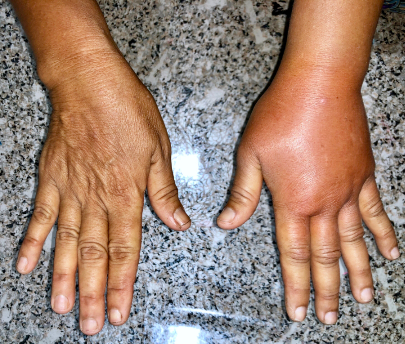
You move on to conduct a focused examination of the lower extremities. You remove ace wraps from both legs, which she says she applies daily. Although she has prescription compression stockings (30-40 mm hg, firm compression) which are about 10 months old, she says they "are all stretched out" as far as they will go (she admits to putting them in the washer and dryer sometimes more than once a week). You also remove a hydrocolloid dressing over the right leg wound; you note some swelling of both lower extremities, but the right lower extremity appears slightly more edematous than the left. Her dorsal and posterior tibial pulses are fairly strong (3+) bilaterally, and you note there is 2+ pitting in the right foot evidence when pressing on the dorsalis pedis pulse in the right foot (and 1+ noted in the left foot).

There is a shallow 2.5cm diameter ulcer with irregular margins over the medial right ankle above the malleolus, which you expected to see as it is consistent with a classic venous ulcer. Mrs. S. complains that it "itches" a lot around the lower edge of the wound, especially at night, and while there is slight maceration and very slight periwound redness that extends 0.5 cm from the inferior wound edge, the wound bed is pink, moist, and otherwise unremarkable.

You also notice something else. The toes of the right foot look like stacked inner tubes, and when you attempt to pinch the skin at the base of the 2nd toe, you are unable to do so; note that this is a positive Stemmers sign suggestive of a lymphedema component that has previously not been identified in the patient's chart. You continue your exam to gather more clinical clues.

Even though Mrs. S. has been wearing her stockings, both feet and lower legs are edematous, and the skin is taut. The dorsum of the right foot appears more "puffy" than the left, giving the contour of her foot a 'box-like' appearance- another indication of lymphedema. Although it is sometimes difficult to discern hemosiderin staining of the gaiter region in people with darker skin, you palpate this area and feel a woody non-compressible quality to the tissues that are also consistent with venous insufficiency. However, you continue palpating proximally and note that the calf areas are also firm or what is often described as 'fibrotic,' and this clue is suggestive of lymphedema. The thigh and inguinal areas are spared.

You ask Mrs. S. if she has ever heard of a 'lymphedema' diagnosis, and she says she has never heard of that before. You explain that longstanding venous insufficiency can be associated with secondary lymphedema, which can make it more difficult to manage but, once properly identified and treated, can end the cycle of re-ulceration or at least minimize recurrence.

You decide that this case could likely be managed in the clinic without referral to a lymphedema specialist since there is not one in the local area, and many will not treat while there is still an active wound. Nevertheless, you talk to Mrs. S about a potential future referral to a lymphedema therapist as an option, should it become necessary.

Since Mrs. S's lower extremity pulses are palpable and her ABI was 0.8, you anticipate compression can be applied at higher levels to reach therapeutic benefits and get the edema under better control while addressing the wound. Over the next few weeks, you evaluate the compression options and after talking to Mrs. S., decide to go with multilayer compression (absorptive contact/base layer, covered with a short stretch bandage and a more elastic top layer) on both legs, verifying the amount of compression at rest, standing and walking the first time it is applied (with a pressure monitor explicitly made for this purpose). Your goal is to reach pressures between 40 mm Hg and 60 mm Hg when walking at the strongest calf muscle contraction. You also evaluate Mrs. S's compression tolerance to ensure she has no discomfort from the compression at rest and her pulses are still palpable (and toenails have good capillary refill) when the legs are elevated and at rest with the bandages intact. You also choose a more breathable, antimicrobial fiber absorptive cover dressing over the wound before applying compression bandages. You schedule twice-weekly visits in your clinic to re-evaluate her progress for the first 2 weeks, with a goal of re-measuring her legs, ordering her new compression stockings every 6 months and giving Mrs. S. instructions to hand wash and air dry to maintain elasticity longer.

It is important to note that some insurance companies may not approve compression therapy on a non-ulcerated, contralateral extremity-so it is important to verify coverage limitations. If this limitation is present, make sure that the diagnosis of lymphedema is mentioned in the justification and refer to any progress made by the Lymphedema Treatment Act submitted to congress in March 2015 (visit source) to see if any states have adopted the stipulations mandating treatment for lymphedema including compression garments. Also, consider referral to a lymphedema therapist as an alternate avenue to initiate earlier in the treatment plan.

You encourage Mrs. S. to limit her salt intake, continue efforts with weight loss (maintaining adequate low-fat protein intake), and consider starting a walking exercise program. You proceed to monitor progress with evidence-based wound care for the open ulcer. Serial circumferential measurements of both lower extremities are taken at each visit along with measurements of the wound(s), and Mrs. S. is delighted to see the measurements steadily decrease.

Finally, the ulcer is closed, and you start the next phase of combined lymphedema/venous insufficiency treatment: maintenance. If there is a short gap of time to await new compression stockings, you realize that if you discontinue compression therapy while the custom stockings are being made, there is a good chance of the ulcer reappearing. Since there is no reimbursement for continued compression wrapping on intact legs, you place two layers of a tubular compression material over each leg and instruct her to remove the second layer at night to avoid compromising the circulation and replace both layers during the day.

Mrs. S. returns to the clinic in about 3 weeks with her new stockings (ordered in a darker brown color to blend with the patient's natural skin tones better). Her legs have increased slightly in circumference, but thankfully the ulcer has not reopened, and you start her on daily wear of her new stockings with instructions to apply them first thing in the morning when she first wakes up but before she has a chance to walk around, and remove them at night just before retiring to bed. You reinforce washing them by hand at night, air drying overnight for wear the next day, and you schedule a return visit in 2 weeks. If possible, you may consider 2 pairs of stockings – the patient can alternate washing one pair and wearing the other.

Select one of the following methods to complete this course.

Take TestPass an exam testing your knowledge of the course material.
OR
Reflect on Practice ImpactDescribe how this course will impact your practice.   (No Test)

Implicit Bias Statement

CEUFast, Inc. is committed to furthering diversity, equity, and inclusion (DEI). While reflecting on this course content, CEUFast, Inc. would like you to consider your individual perspective and question your own biases. Remember, implicit bias is a form of bias that impacts our practice as healthcare professionals. Implicit bias occurs when we have automatic prejudices, judgments, and/or a general attitude towards a person or a group of people based on associated stereotypes we have formed over time. These automatic thoughts occur without our conscious knowledge and without our intentional desire to discriminate. The concern with implicit bias is that this can impact our actions and decisions with our workplace leadership, colleagues, and even our patients. While it is our universal goal to treat everyone equally, our implicit biases can influence our interactions, assessments, communication, prioritization, and decision-making concerning patients, which can ultimately adversely impact health outcomes. It is important to keep this in mind in order to intentionally work to self-identify our own risk areas where our implicit biases might influence our behaviors. Together, we can cease perpetuating stereotypes and remind each other to remain mindful to help avoid reacting according to biases that are contrary to our conscious beliefs and values.

References

  • Bjork, R. (2018). Emerging Paradigms Integrating the Lymphatic and Integumentary Systems: Clinical Implications. Wound Care & Hyperbaric Medicine, 9(2), 50-55.
  • Fife, C. SAWC oral presentation Fall, 2014.
  • Gasbarro V, Michelini S, Antignani PL, Tsolaki E, Ricci M, Allegra C. (2009). The CEAP-L classification for lymphedemas of the limbs: the Italian experience. Int Angiol. 28(4), 315-24.
  • Herbst, K. PhD, MD website (2014). Visit Source.
  • Hettrick, H. and Ehmann, S. (April, 2019). Compression: More Than a Number. Podium presentation at Wound Health Society (WHS) International Symposium for Advancement of Wound Care (SAWC Spring 2019).
  • Lawenda, B. D., Mondry, T. E. & Johnstone, P.A. (2009). Lymphedema: A primer on the identification and management of a chronic condition in oncologic treatment. CA: A Cancer Journal for Clinicians, 59(1), 8-24.
  • Lee BB, Andrade M, Antignani PL , Boccardo F, Bunke N, Campisi C, Damstra R, Flour M, Forner I, Gloviczki P, Laredo J, Piller N, Michelini S, Mortimer P, Rabe E, Rockson S, Scuderi A, Szolnoky G, Partsch H, Villavicencio JL. Diagnosis and Treatment of Primary Lymphedema: Consensus Document of the International Union of Phlebology (IUP)-2013. Visit Source.
  • Nowicki, J. & Siviour, A. (2013). Best practice skin care management in lymphedema. Wound Practice & Research, 21(2), 61-65. Visit Source.
  • Ostler, M. (2018). EDEMA CONFUSION. Physical Therapy and Lymphedema: An Art and A Science. Lecture given at APWCA Conference in Texas on Sept 15, 2018. Visit Source.
  • Partsch, H. (2014). Compression for the management of venous leg ulcers: Which material do we have? Phlebology, 29(15), 140-145.
  • Rabe, E., Partsch, H., Hafner, J., Latimer, C., Mosli, G., Neumann, M., and Carpenter, P. (2018). Indications for medical compression stockings in venous and lymphatic disorders: an evidence-based consensus statement, Phlebology, 33(3), 163-184.
  • Vojackova, N., Fialova, J. & Hercogova, J. (2012). Management of lymphedema. Dermatologic Therapy, 25, 352-357.