Sign Up
You are not currently logged in. Please log in to CEUfast to enable the course progress and auto resume features.

Course Library

Blood Flow Restriction Training and Uses in Rehabilitation

1.5 Contact Hours
Listen to Audio
CEUfast OwlGet one year unlimited nursing CEUs $39Sign up now
This peer reviewed course is applicable for the following professions:
Advanced Practice Registered Nurse (APRN), Athletic Trainer (AT/AL), Certified Nurse Practitioner, Certified Registered Nurse Practitioner, Clinical Nurse Specialist (CNS), Licensed Practical Nurse (LPN), Licensed Vocational Nurses (LVN), Nursing Student, Occupational Therapist (OT), Occupational Therapist Assistant (OTA), Physical Therapist (PT), Physical Therapist Assistant (PTA), Registered Nurse (RN)
This course will be updated or discontinued on or before Friday, May 15, 2026

Nationally Accredited

CEUFast, Inc. is accredited as a provider of nursing continuing professional development by the American Nurses Credentialing Center's Commission on Accreditation. ANCC Provider number #P0274.


BOC
CEUFast, Inc. (BOC AP#: P10067) is approved by the Board of Certification, Inc. to provide education to Athletic Trainers (ATs).

FPTA Approval: CE24-1195500. Accreditation of this course does not necessarily imply the FPTA supports the views of the presenter or the sponsors.
Outcomes

≥ 92% of participants will know the basic concepts of blood flow restriction training (BFRT), patient safety considerations, and patient populations that may benefit from BFRT.

Objectives

After completing this course, the learner will be able to:

  1. Identify principles of muscle strengthening and hypertrophy.
  2. Compare the benefits of blood flow restriction training (BFRT) to traditional resistance training.
  3. Summarize the safety considerations related to BFRT.
  4. Compare training parameters for typical types of exercise used with BFRT.
  5. Outline conditions and patient populations that may benefit from BFRT.
CEUFast Inc. and the course planners for this educational activity do not have any relevant financial relationship(s) to disclose with ineligible companies whose primary business is producing, marketing, selling, re-selling, or distributing healthcare products used by or on patients.

Last Updated:
Restart
Restart
  • 0% complete
Hide Outline
Playback Speed

Narrator Preference

(Automatically scroll to related sections.)
Done
Blood Flow Restriction Training and Uses in Rehabilitation
0:00
0:15
 
To earn of certificate of completion you have one of two options:
  1. Take test and pass with a score of at least 80%
  2. Reflect on practice impact by completing self-reflection, self-assessment and course evaluation.
    (NOTE: Some approval agencies and organizations require you to take a test and self reflection is NOT an option.)
Author:    Cindy Endicott (PT, DPT, FAAOMPT, ATC, Cert Dn)

Introduction to Blood Flow Restriction Training

Blood flow restriction (BFR) training (BFRT) is a rehabilitation intervention that helps promote increased muscle strength and mass with less load than traditional high-load methods. This is accomplished by applying a pneumatic band pressurized at the proximal portion of a limb to alter blood flow to the limb while exercising, creating low levels of hypoxia and leading to muscle strengthening. The cuff is pressurized to a level that creates a partial occlusion of arterial blood flow and a total occlusion of venous return distal to the cuff. According to the American Physical Therapy Association (APTA), BFRT can "help patients make greater strength gains while lifting lighter loads, thereby reducing overall stress placed on the limb" (APTA, n.d.). Blood flow restriction training is becoming increasingly popular as a treatment intervention; it is still relatively unknown by many healthcare professionals, often creating skepticism of efficacy or safety. Many general practice physicians, advanced practice providers, nurses, and other healthcare professionals have either never heard of BFRT or have little understanding of its benefits to patients. Because BFRT creates muscle strength gains with less load on the involved limb, it becomes an excellent intervention for populations who are physically unable or cannot tolerate high-intensity exercise. Populations such as individuals with musculoskeletal injury, which prohibits heavy resistance loads, pre- or postoperative patients in which atrophy is prevalent, bedridden individuals, older adults who cannot lift heavy weights, and other special populations will be discussed later in this course.

While the benefits of strengthening with less physical load are appealing, there are many factors to consider when deciding if this is appropriate for a patient. A professional who is well-trained in safety screening, application, and exercise prescription should closely direct and monitor a session during which BFRT is in use. While many cuff devices are on the market, many are not pneumatic and, therefore, are difficult, if not impossible, to reproduce or document the training parameters between sessions. For this reason, healthcare professionals are encouraged to use a pneumatic/inflatable cuff with a Food and Drug Association (FDA)-approved pressure monitoring device, allowing for accurate cuff inflation pressure monitoring. Five FDA BFR cuff systems are currently on the market (H+cuffs, 2019), all with varying features and price points.

History of Blood Flow Restriction Training

BFR originated in Japan and was developed by Yoshiaki Sato in 1966. Initially, it was intended to be used as a performance training tool for bodybuilding; however, following an injury in 1973, Sato applied his technique to a knee injury he sustained in a ski accident (Sato, 2005). Sato continued his self-experimentation with the method he referred to as KAATSU, with numerous failures, successes, and modifications, until he eventually developed training manuals and parameters, leading to what is known as the KAATSU Training method (Sato, 2005). KAATSU training became available to the general public in approximately 1994, spreading across Europe and eventually to the United States. The United States military adopted BFRT in 2011 (Abadir & DeFrancesco, n.d.) to rehabilitate veterans and aid in strengthening limbs for prosthetics. It was quickly adopted by professional, Olympic, and collegiate athletes and has since been growing in popularity among many healthcare professionals, including Medical Doctors (MD), Doctor of Chiropractic (DC), Physical Therapists (PT), Physical Therapy Assistants (PTA), Athletic Trainers (ATC), and Occupational Therapists (OT). It is increasingly common to see BFRT from Sports Medicine Orthopedic Surgeons in a postoperative protocol.

Muscle Growth and Strengthening

Muscle Strength

The strength of a muscle is the ability to produce a maximal force against an external resistance (Schoenfeld et al., 2021). Skeletal muscles must have enough strength to withstand the external resistance of gravity and our body weight to walk, stand up from a chair, or ascend and descend stairs. We must have enough strength to resist activities throughout our daily living, such as lifting a gallon of milk or pushing open a door. Outside the activities of daily living, our strength needs vary greatly and depend on our activity goals and lifestyle. A weightlifter will have different strength needs than an 80-year-old golfer.

Evidence shows that training load can influence muscular adaptations through metabolic, hormonal, neural adaptions, and cardiovascular responses to resistance training, and the magnitude of the load used (or the amount of weight lifted) is considered one of the most important training variables (Schoenfeld et al., 2021). A low repetition with heavy loads (1-5 repetitions per set with 80%-100% of a one repetition maximum (1RM) has been shown to optimize strength increases (Shoenfeld et al., 2021). Low-intensity resistance training is much less effective in increasing muscle strength and growth than moderate and high-intensity exercise, as outlined above. A real-world example would be as follows: a person can lift 100 pounds (lbs.) during a biceps curl but can only perform this once. 100 lbs. would be this person's 1RM for the biceps curl. According to the traditional model of resistance training for strengthening, this person would need to perform 1-5 repetitions per set at a training load of 80-100 lbs. to increase the strength of their biceps. In a clinical environment, it is difficult to establish a patient's 1RM, as many of our patients are injured or frail, or there is a lack of clinical equipment to establish this number. We modify strength training parameters by altering the amount of weight used and the number of reps and sets performed. Clinically, we tend to rely on manual muscle testing or dynamometry to establish the force produced by a given muscle. Still, some formulas can be used to predict this 1RM value, including bioelectrical impedance (Sue et al., 2022).

Muscle Hypertrophy

Hypertrophy is when the actual size of the muscle increases. Hypertrophy is optimized with moderate loads with 8-12 repetitions per set at 60%-80% of 1RM (Schoenfeld et al., 2021). Muscle hypertrophy is slow and virtually non-existent in the initial stages of resistance training, with most strength changes occurring from neural adaptations. However, within a few months of resistance training, hypertrophy becomes dominant due to increased myofibril and sarcomeres added in parallel and eventually increased diameter of individual fibers (Schoenfeld, 2010).

Physiology Overview of Muscle Growth and Strengthening

If a muscle is never challenged, it is never prompted to become stronger. If the stress on a muscle is confined to resistance well within its strength capabilities, it tells the muscle that it is as strong as it ever needs to be. Resistance is essential to increasing strength for any patient population. Through resistance training, a demand is placed on the muscle; there is neural recruitment of that muscle, the increased stress creates microscopic damage, and then the muscle repairs and recovers, increasing strength and size. Here, we will cover an overview of the physiology of muscle strengthening- this will not be a deep dive into the physiology, such as the Krebs and Cori Cycles, which play a significant role in how muscles are utilized but are outside this course's scope.

The physiology of muscle strength is a factor of five primary mechanisms: tensile strength, metabolic accumulation, muscle cell swelling, neural adaptations, and post-exercise hormones (Schoenfeld, 2010).

  • Tensile stress: Development of strength and hypertrophy depends on progressive tensile loading and stretching of the muscle. This repetitive tensile stress creates micro-damage to the muscle fiber satellite cells. These cells increase in number, eventually fusing with existing cells to develop new myofibers.
  • Metabolic Accumulation: This process is simultaneously facilitated by metabolites that stimulate pathways favoring muscle synthesis over degradation.
    • Lactic Acid- accumulation of lactic acid during exercise promotes muscle hypertrophy.
    • Creatine Kinase- an indicator of excessive muscle damage.
  • Muscle Cell Swelling: During resistance training, intracellular swelling occurs, which is theorized to stretch the cell membranes, further stimulating metabolic signaling.
  • Neural Adaptations: Not all muscle fibers are fired at once during exercise. This constant fluctuation of which myofibrils are stimulated allows for stronger contractions for longer (endurance). During resistance training, more muscle fibers are recruited to increase maximal power.
  • Post-exercise Hormones: Insulin-like growth factor (IGF-1) and growth hormone (GH) are two of the main anabolic hormones in human muscle that have been shown to spike during resistance training and stimulate muscle growth.

There are, however, negative regulators of muscle growth. One significant factor is myostatin, which is crucial in inducing muscle atrophy (Jang et al., 2021). It has also been demonstrated that myostatin inhibition can protect against muscle atrophy and allow skeletal muscle to increase in size (Jang et al., 2021). When considering resistance training, minimizing or inhibiting the effects of myostatin would be desirable. Lactate that is released during exercise can inhibit myostatin.

These five factors will become important when discussing why BFR with low resistance training is highly effective and desirable when building muscle strength and hypertrophy in many patient populations.

photo of rats showing myostatin inhibition

Myostatin Inhibition

Blood Flow Restriction Compared to Traditional Resistance Training

BFRT continues to gain popularity as a strength training method for various patient populations. It has been shown to increase skeletal muscle mass and strength to a similar extent as heavy load resistance training. It demonstrates superior strengthening effects as low load resistance training (Nielsen et al., 2012), thus becoming a highly effective method of strengthening with less tissue and joint stress to the patient. While the exact mechanism behind the efficacy of BFR is unknown, it is believed there is an increased accumulation of metabolites in the muscle created by the hypoxic environment created by the partial occlusion of the cuff; this leads to increased muscular fatigue, neural activation, and anabolic signaling than what would have been made through low-resistance training alone (Lorenz et al., 2021).

Table 1. Muscular and Metabolic Changes in High-Intensity Exercise, BFRT, Low-Intensity Exercise, and Low-Intensity Exercise Alone.
Training Range60-100% 1RM30-40% 1RM30-40% 1RM
Cross-Sectional AreaIncreasedSignificant increaseNo increase
Muscle Damage (Creatine Kinase)PresentNot significantNot significant
LactateIncreasedIncreasedNot present
Neuromuscular RecruitmentFast twitch fiber activation at near-maximal effortFast twitch fiber activation at submaximal effortNo additional recruitment
GHIncreasedSignificant increaseNo increase
IGF-1IncreasedSignificant increaseNo increase
MyostatinInhibitionInhibitionNo change
Mammalian Target of Rapamycin (mTOR)Increased activationIncreased activationNo increase
Satellite CellSignificant increaseSignificant increaseSlow proliferation
(Pearson & Hussain, 2014)
 Med/High IntensityBFRT + Low IntensityLow Intensity

To summarize, there are several theories on why BFR promotes muscle strengthening and hypertrophy. These include activating the cellular swelling mechanism, stimulating intracellular signaling pathways, and cell-to-cell signaling (Cognetti et al., 2022). It has been shown that metabolites that can inhibit muscle growth, such as lactate, GH, IGF-1, satellite cell production, and mTOR, are all increased through low-intensity exercise with BFR, similar to high-intensity exercise. Additionally, metabolites that can inhibit muscle growth, such as myostatin, are inhibited, likely through the increased activation of mTOR. These metabolic signaling pathways are similar to high-intensity exercise but require less load and strain on the patient. They have been shown to occur in less time to adapt than high-intensity exercise.

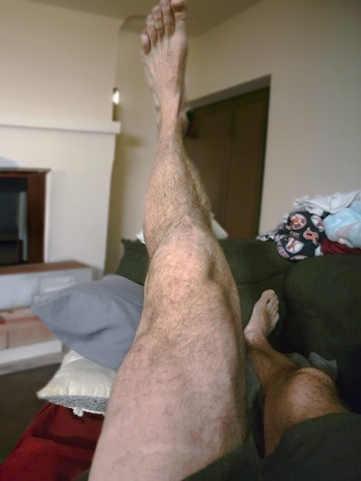
photo of leg quadriceps

Quadriceps/VMO Atrophy Postoperative

photo of mans quadriceps following rehab

Quadriceps/VMO Hypertrophy Following Rehab With BFRT

Proximal vs. Distal Muscle Strengthening

Until recent years, many of the potential benefits of BFR were thought to be limited to tissues undergoing occlusion distal to where the pressure is applied; however, recent findings indicate that there may be some benefit to tissues directly proximal to the occlusion site (Hedt et al., 2022). Increases in rotator cuff strength and limb circumference have been documented via electromyography (EMG) following BFR and low-intensity exercise (Hedt et al., 2022). It does appear, however, that there is a threshold of volume or cumulative time under occlusion for proximal effects to occur. Similar benefits have been demonstrated in the proximal strengthening of the gluteus maximus with occlusion training of the hip and lower extremity (Hedt et al., 2022). Unfortunately, the proximal effects spanning to other regions (chest muscles, back muscles, etc.) are highly understudied.

Safety Considerations

Provider Training

When providing patient interventions, the clinician must be aware of the safety, methods of safe implementation, clinical application, and efficacy. It is recommended that the clinician take appropriate training courses before utilizing BFR devices. Many courses are available, and depending on specific professional state practice, acts can be taken by licensed PTs, ATs, DCs, MDs, Doctors of Osteopathy (DOs), Nurse Practitioners (NPs), PAs, and (OTs). There are usually no prerequisites to these courses, which are available in person and online. Unfortunately, there is no certification process or process to ensure that a clinician completes any coursework before utilization with patients.

Risk of Blood Clot

One of the most common questions about the safety of BFR revolves around clot formation or deep vein thrombosis (DVT). It is known that DVT is a risk of pneumatic tourniquet use during surgery. Most people are less aware that tissue ischemia after inflation stimulates the release of tissue plasminogen activator (TPA), which activates anti-thrombolytic proteins. When the tourniquet is deflated, this causes systematic thrombolysis in the affected limb (Kam et al., 2001). Studies investigating the potential of clot formation following the use of BFRT show no longstanding adverse effects following BFRT with low-resistance exercise. Some studies suggest a slight elevation of TPA following BFR exercise, suggesting a mild anti-thrombolytic effect (Nascimento et al., 2022). Nonetheless, there may be a potential for DVT formation in people with a complex medical history (Nascimento et al., 2022).

General Patient Safety

As with any other therapeutic intervention, especially in physical exercise and rehabilitation, safety considerations must be taken before beginning BFR with any patient. Regardless of age, when performing any resistance training or cardiovascular exercise, the patient must be medically well enough to participate in the exercise safely. If you are concerned about a patient's medical safety when exercising, then you should not consider the application of blood flow resistance to their rehabilitation. Again, as with any form of general exercise, consult with the referring physician if there is any doubt. In direct access states, however, there may not be a referring physician, so diligence is required when implementing any form of resistance or cardiovascular training. The clinician should consider whether BFRT has a clinically relevant benefit to the patient. Consideration should also be taken regarding other potential treatments or interventions that could provide similar results with less risk (Nascimento et al., 2022). Risk vs. benefit is a common consideration for any clinician, and this intervention should be no different.

Safety Screening for Patient Use

Consider a patient's physical appearance before implementing BFR into your treatment plan. How is their circulatory system? Do they have poor capillary refill time, varicose veins, or indications of circulatory loss in their skin or nails? Do they have concerning health conditions in their medical history? Conditions such as uncontrolled hypertension, cardiopulmonary conditions, history of DVT, arterial calcification, diabetes, sickle cell trait, or active infection would serve as indications to take precautions when considering the use of BFRT. While there are no identified contraindications to the use of BFR, there are conditions that have been identified, and point values have been assigned that can serve as a screening to determine the appropriateness of implementation in a given patient. Given that no validated screening system has been developed, the following chart, adapted from Nascimento et al. (2022), can be a good start toward a patient safety screening system. Generally, a point value greater than five (5) should warrant not proceeding with BFR.

Table 2. Patient Safety Screening Based on Medical History.
Each Risk Factor Represents 1 pointEach Risk Factor Represents 2 points
  • Abnormal pulmonary function (COPD)
  • Acute myocardial infarction
  • Age between 41 and 59 years
  • Blood transfusions
  • Chemotherapy
  • Congestive heart failure < 1 month
  • Diabetes requiring insulin
  • History of inflammatory bowel disease
  • History of prior major surgery < 1 month
  • Length of a surgery > 2 hrs
  • Medical patient currently on bed rest
  • Minor surgery planned
  • BMI > 25-39
  • Obstructive pulmonary disease
  • Sepsis <1 month
  • Serious lung disease < 1 month
  • Smoking
  • Currently swollen legs
  • Varicose veins
  • Age 60 to 74 years
  • Arthroscopic surgery
  • BMI > 40
  • Central venous access
  • Immobilized plaster cast < 1 month
  • Laparoscopic surgery > 45 min
  • Major surgery > 45 min
  • Malignancy (present or previous)
  • Patient confined to bed >72 hours
Each Risk Factor Represents 3 pointsEach Risk Factor Represents 5 points
  • Age over 75 years
  • Any acquired congenital thrombophilia
  • Family history of thrombosis
  • History of DVT/pulmonary embolism (PE)
  • Acute spinal cord injury (paralysis) < 1 month
  • Elective major lower extremity arthroplasty
  • Hip/pelvis or leg fracture < 1 month
  • Multiple trauma < 1 month
  • Stroke < 1 month
For Women Only (Each Represents 1 point)
  • Oral contraceptives or hormone replacement therapy
  • Pregnancy or postpartum < 1 month
  • History of unexplained stillborn, recurrent spontaneous abortion > 3 premature birth with toxemia or growth-restricted infant
(Nascimento et al., 2022)

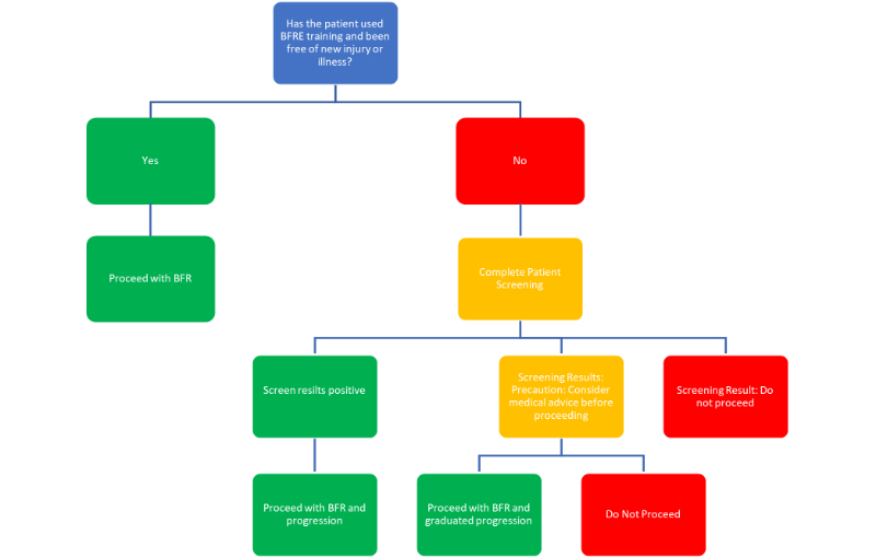
Additionally, the following flow chart can serve as an example of a decision tree to determine when to implement or exclude BFR into your training regime (AIS, n.d.).

flow chart showing decision tree for bfr

BFR Decision Tree

Avoidable Tourniquet Complications

Common complications from tourniquet use include the following (Sharma & Salhotra, 2012):

  • Nerve injury: nerve injury following tourniquet use is likely a result of neural ischemia and mechanical compression. A threefold increase in neurologic complications has been shown for each 30-minute increase in inflation time.
  • Tourniquet pain: pain has been shown to increase with more narrow cuffs (less than 7 centimeters [cm]). Tourniquet pain is shown to develop 30-60 minutes following cuff inflation.
  • Tissue edema: develops if the tourniquet time exceeds 60 minutes. Generally, most surgeons try to limit tourniquet inflation time to 90-120 minutes if possible, with a maximum inflation time of three hours before temporary deflation is needed.
  • Severe injury could occur if excessive pressures are utilized; therefore, there are pressure recommendations for both upper and lower extremities based on the patient's systolic pressure that a surgeon must follow.
  • Massive PE can occur if the tourniquet is applied over a DVT.

Overall Safety Conclusions

First and foremost, the patient's needs must match the clinical benefits provided through BFRT. Once this has been confirmed, patient safety is the next most considerable determination before occlusion training. Based on current literature, including case reports, case studies, and randomized control trials, it has been determined that blood flow restriction provides a tremendous opportunity to accelerate rehabilitation and possibly aid in injury prevention safely. As with any other modality, BFR should be performed by professionals who have undergone appropriate training and education, but overall, it has been shown to be a safe and useful technique (Anderson et al., 2022).

Application Guidelines

Cuff Design

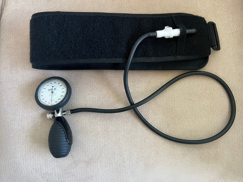
There are many situations in which BFRT is utilized that do not follow evidence-based medical models. You can see these regularly used in gyms or by bodybuilders. Often, you will see people in these settings using narrow cuffs, other forms of straps, or non-pneumatic elastic bands, or where there is a single-size cuff that is cinched to the desired perceived tightness by the wearer. These are often used by lay people, who may not be fully aware of application parameters and can be ineffective or dangerous if utilized improperly. While this equipment is less expensive and easily attainable at sporting goods stores or online shopping sites, it should not be used in a medical setting.

Physical therapists and other medical professionals for whom BFR is within their scope of practice should use inflatable tourniquets that meet FDA standards. Pneumatic cuffs used for BFR fall under FDA Sec 878.5910, which defines a pneumatic tourniquet as "an air-powered device consisting of a pressure-regulating unit, connecting tube, and an inflatable cuff. The cuff is intended to be used to be wrapped around a patient's limb and inflated to reduce or totally occlude circulation" (U.S. Food and Drug Administration [FDA], 2023).

photo of pneumatic cuff and pump

Pneumatic Cuff and Pump

Cuff Placement

Cuff placement for BFRT purposes should be as high proximally on the targeted limb as possible. For the upper extremity, this is between the axilla and the proximal biceps. The lower extremity is in the femoral crease, as proximal to the quadricep as possible; this allows for comfortable muscle contraction, protects superficial nerves, and allows for the lowest pressure required to create occlusion. If the cuff is placed over the thick portion of the muscle belly (quadriceps, for instance), it will take more pressure to push into the muscle before vascular occlusion will occur. The clinician should select a cuff size appropriate to the size of the involved limb, allowing for a small amount of overlap in the cuff ends to create even pressurization around the limb. If a cuff is too small and the ends do not wrap around sufficiently, the patient may experience pinching or discomfort, which may prevent them from participating in exercises. If a cuff is too large for the limb, it may not inflate adequately to provide a reliable reading of the inflation pressure.

Cuff Pressure

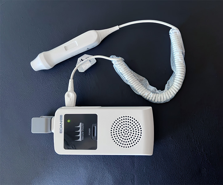
The pressure required to create blood flow occlusion to a limb depends on many individual limb characteristics (Patterson et al., 2019). For maximal safety, reliability, and reproducibility, the pressure should be specific to the patient's limb (left or right), the cuff shape/width/length, and the application location. This is achieved by measuring the limb occlusion pressure (LOP) or the arterial occlusion pressure (AOP); both are accepted terminology. LOP is the minimal pressure to occlude arterial blood flow (Patterson et al., 2019). The gold standard for measuring LOP has been using a Doppler ultrasound technique to measure the exact pressure when arterial blood flow is disrupted. In the lower extremity, this can be measured at the dorsalis pedis or the posterior tibial artery. For the upper extremity, this is typically measured at the radial artery; this pressure is then utilized at varying percentages during BFRT to prevent mild hypoxia and ischemic effects but not to fully occlude arterial blood flow into the limb during rehabilitative exercises (McEwen et al., 2018). Because LOP has been shown to vary significantly between different people, it is suggested that clinicians measure LOP consistently for each patient (Evin et al., 2021). Recently, newer integrated models that automatically calculate LOP have emerged. The automatic pump versions have been shown to accurately measure LOP with no statistical difference from the previous gold standard of the Doppler technique (Abbas et al., 2022). It is generally recommended that pressures between 40-80% of the LOP be utilized during BFR training (Cognetti et al., 2022; Patterson et al., 2019). The cuff inflation should be limited to 40-50% of the LOP for the upper extremity and 60-80% of the LOP for the lower extremity (Smart Tools, n.d.). Exercise prescription depends on whether it is applied passively, with resistance training, or during aerobic exercise.

photo of doppler ultrasound

Doppler Ultrasound

Exercise Prescription

BFR can be used in several capacities, with the most common methods being resistance training, passive exercise, and aerobic training.

Resistance Exercise

BFR use during low-intensity resistance training is the most common and well-studied in the literature. The pressure applied is determined to some degree by the relative load used during the resistance exercise and can be adjusted according to patient effort and comfort/tolerance. As described earlier, cuff inflation is recommended to be 40-50% of the LOP for the upper extremity and 60-80% of the LOP for the lower extremity (Smart Tools, n.d.). It has been found that training loads of 20-40% of the individuals with 1RM consistently produce muscle adaptations and maximize strength and muscle growth (Patterson et al., 2019). If a patient is working on the lower load range (20% 1RM), a higher cuff inflation pressure should be considered (70-80% if performing low-intensity exercise). It has also been suggested that training muscle groups proximal to the cuff may require higher pressure to create maximal adaptation (Patterson et al., 2019).

A training volume utilizing 75 repetitions is common and frequently recommended (Smart Tools, n.d.; Patterson et al., 2019); this repetition scheme is divided into four sets, the first set of 30 repetitions followed by three sets of 15 repetitions with short rests of 30-60 seconds between sets. While other repetitions and set configurations have been looked at, the 30/15/15/15 provides "sufficient volume to lead to adaptations in most people" (Patterson et al., 2019).

Training frequency is typically recommended to perform resistance training 2-4 times per week to produce strength gains and muscle hypertrophy in both traditional strength training models as well as BFR literature, but a high-frequency approach of 1-2 times daily can be used for short periods (Patterson et al., 2019).

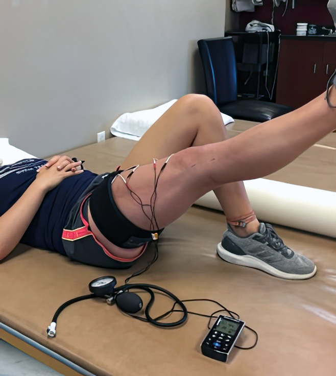
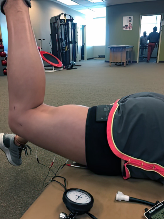
photo of non weight bearing post op patient

photo of non weight bearing post op patient

BFR with Non-Weight Bearing Post-Op Patients

Passive Exercise

Passive BFR involves cuff inflation to a limb or bilateral limbs without the participant performing any exercise; this method can serve as an option for people who are bedridden and unable to exercise, such as following orthopedic surgeries, and can serve as a means of maintaining muscle girth and preventing disuse atrophy. Passive BFR can also prepare the muscle for application during resistance exercise. This passive method is often referred to as the cell swelling protocol or is also known as ischemic preconditioning (Smart Tools, n.d.) and induces cell swelling. As described earlier, intracellular swelling stretches the cell membranes and stimulates metabolic signaling. With passive applications, higher inflation pressures have been shown to provide a stronger stimulus for atrophy prevention (Cognetti et al., 2022). The standard protocol for passive BFR involves cuff inflation up to 100% LOP for 5 minutes, followed by 3 minutes of deflation to all for muscle reperfusion (Patterson et al., 2019). This can be performed for 3-4 cycles once or twice daily for 1-8 weeks. Studies investigating other protocol parameters, different inflation/deflation ratios, and specific cuff pressure have not been performed yet (Patterson et al., 2019).

Electrical stimulation during passive BFR sessions has also been proposed. These protocols suggest using neuromuscular electrical stimulation (NMES) or Russian stimulation settings to perform 10-second isometric contractions followed by 10 seconds of rest throughout the ischemic preconditioning. Very little evidence supports the use of electrical stimulation with BFR to date.

Aerobic Exercise

BFR during aerobic exercise is another common application, typically during walking or cycling. The literature demonstrates no standardization for inflation pressures during aerobic exercise (Patterson et al., 2019); however, Smart Tools (n.d.) recommends a maximum 20-minute exercise session with a minimum of 130 millimeters of mercury (mmHg) pressure on the lower extremities. Exercise can be conducted continuously for up to 20 minutes or at intervals at 50% max effort (Patterson et al., 2019).

Documentation

When documenting the use of BFRT in a patient chart, it is important to capture all of the patient-specific aspects of the BFR session. For example, when documenting the patient onboarding process, it is important to document that the rationale for use, what to expect, and risk screening were reviewed with the patient. LOP does not need to be measured each time BFR is used with a patient, but it is important to document LOP for each limb involved in BFRT. For example, "the cuff was placed at the inguinal crease of the left lower extremity. LOP was measured to be 240 mmHg at the posterior tibial artery with the patient lying supine." Capturing the training load, volume, and rest periods used in each session is important for exercise documentation. For example:

  • Exercise: Ischemic Preconditioning. 5 min 100% LOP x 1 set
  • Exercise: Straight Leg Raises with a one lb. cuff weight load at the ankle. 1x30, 3x15 with 30-second rest between sets at 60% LOP (144 mmHg)
  • Exercise: walking: bilateral lower extremity cuffs at the inguinal crease. Cuff inflation to 130 mmHg x 20 min duration

Documenting in this manner creates an accurate note with treatment parameters that are patient-specific, measurable, and reproducible. This also creates the most defensible documentation should litigation ever occur.

Other Clinical Applications

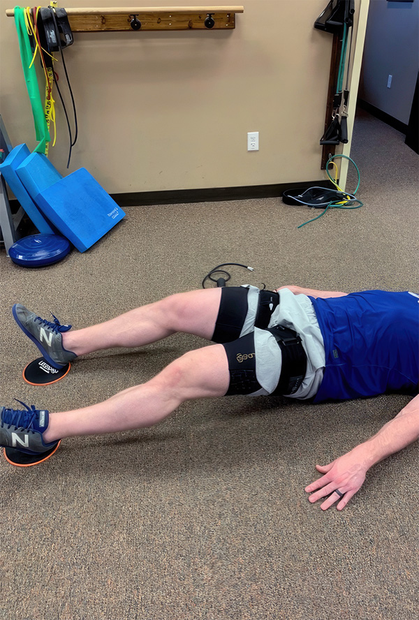
For obvious reasons, BFRT has primarily been studied and utilized in sports medicine. BFRT was once considered an emerging intervention allowing sports medicine professionals to help athletes improve strength for athletic endurance, to aid in the non-operative rehabilitation of injury, to prepare an athlete for surgery through pre-operative "prehab" to limit muscle weakness as a result of injury, and finally postoperatively to prevent atrophy and promote muscle strengthening. Slowly, BFRT has been integrated into similar applications for other teens and adults- not just a treatment for the athlete. Even weekend warriors, tactical professionals, and everyday patients with physical work activities could be treated like Olympians or professional athletes. Studies are emerging now, showing benefits through using BFRT in many other clinical applications and patient populations. There are even studies examining the "application of BFR to optimize exercise countermeasures for human space flight" (Behringer & Willberg, 2019). Other uses include bone healing, neurologic and systemic disorders, and pain modulation.

photo of bfr for sport performance

BFR for Sport Performance

Cardiovascular and Aerobic Capacity

In addition to improvements in cross-sectional area and overall muscle strength, recent studies have demonstrated improvement in the volume or amount of oxygen the body consumes (VO2 max) following BFRT (Bradley et al., 2023). Similarly, low to moderate-intensity aerobic training with BFR significantly improved overall aerobic capacity compared to aerobic exercise without BFR (Formiga et al., 2020). Interestingly, no significant improvement in aerobic capacity was noted with BFR combined with high-intensity aerobic exercise (Formiga et al., 2020). In 2022, Tan et al. (2022) studied the effects of BFR with low-intensity resistance training on microvascular rarefaction in the myocardium of hypertensive rats. He found that BFRT did improve the density and pressure of the microvascular vessels and improved microvascular circulation (Tan et al., 2022). These changes in the microvascular structure led to improved cardiac function and decreased blood pressure, achieving the preventative effect of early hypertension. While not conducted on human subjects, this is an exciting potential for disease prevention in humans, although further testing is warranted.

Bone Healing

Resistance training is a well-known combatant of osteoporosis and a way to improve overall bone density. Studies involving low-intensity resistance training and BFR can increase osteoblastic activity to favor bone formation. Through BFR, this can be accomplished without placing the body under the physical stress of high-intensity resistance training. A meta-analysis conducted by Wang et al. (2023) reviewed 285 studies related to the effects of BFRT on bone metabolism. Ultimately, they included 12 studies in their review, which involved 378 participants. Their review indicated that BFR with low-intensity exercise demonstrated a better impact on bone health than that of low-intensity exercise alone but was less effective (minimally) than high-intensity training. Additionally, they discovered that walking with BFR had a better effect on bone health than without BFR. Wang et al. (2023) concluded that BFR with low-intensity exercise is an effective and efficient alternative to high-intensity training to promote gains in bone health for people who are not able to withstand the higher mechanical loads of high-intensity exercise. This may include "untrained individuals, older adults, or patients undergoing musculoskeletal rehabilitation" (Wang et al., 2023).

Neurologic and Systemic Disorders

BFR has primarily been investigated for use in the treatment of orthopedic conditions. Some emerging studies investigate the outcomes of BFR in treating neurological conditions (Yuan et al., 2023).

BFRT is also being increasingly applied to the rehabilitation treatment of many chronic and systemic diseases. Rodrigues et al. (2020) conducted a study involving rheumatoid arthritis (RA). They deemed that pain experienced during exercise and the health assessment questionnaire scores were improved in the group participating in a 12-week training session with and without BFRT. The outcomes of this study showed that BFRT is a feasible treatment method for RA management. Another study found decreased insulin levels and sensitivity following BFRT training every two weeks for eight weeks (Kambič et al., 2019). In contrast, an additional study suggests improved metabolic control in patients with type II diabetes secondary to improved muscle mass, metabolism, and decreasing insulin levels (Saatmann et al., 2021).

A systematic review investigating the usefulness of blood flow resistance training with various neurologic conditions (Vinolo-Gil et al., 2022) found that BFR could be a beneficial rehabilitation tool for several pathologies, such as Parkinson's disease, stroke, multiple sclerosis, cerebral palsy, and spinal cord injury. Improvements were noted for (Vinolo-Gil et al., 2022)

  • Sensorimotor function
  • Frequency and step length symmetry
  • Perceived exertion
  • Heart rate and gait speed
  • Walking endurance
  • Fatigue
  • Quality of life
  • Muscle thickness
  • Gluteus density
  • Muscle edema

No improvements were found in lower limb balance. Studies involving neurologic conditions have been few; many were case studies or involved low participant numbers, and more clinical trials are needed. Additionally, protocols for using BFR with neurological patients have not been developed.

Pain Modulation

Pain is a common problem for many patients participating in rehabilitation and can be a significant limiting factor in many cases. Many interventions are geared specifically toward pain modulation. Many patients have difficulty performing moderate-intensity exercise because of pain; therefore, their activities are limited and devised in a way to avoid pain. In 2018, Korakakis et al. (2018) found significant pain reduction in anterior knee pain with functional activities following BFRT and low-intensity exercise. Pain reduction was sustained for at least 45 minutes following the occlusion training. This could suggest a clinical application of BFRT as a precursor to other activities within a rehabilitation session, providing clinicians a window of opportunity for knee-loading activities (Korakakis et al., 2018), thus leading to greater compliance and consistency of physical therapy activities (Hedt et al., 2022). The mechanism of pain reduction is still unknown. Still, theories revolve around "opioid and endocannabinoid mediated pain inhibition, recruitment of high threshold motor units, exercise-induced metabolite production and an interaction between cardiovascular and pain regulatory systems" (Hedt et al., 2022).

Older Adults

Similar to findings in younger populations, BFRT has been shown to effectively increase muscle mass and strength, prevent atrophy, and facilitate pre- and postoperative injury rehabilitation in the older adult population when compared to traditional training methods (typically body weight or low-weight resistance exercise) (Centner et al., 2018; Yuan et al., 2023). It has also been shown to effectively improve older adults' cardiovascular function and aerobic capacity (Yuan et al., 2023). In addition to decreased physical capabilities, aging is often associated with cognitive decline. There is substantial evidence that resistance training works to combat both physical and cognitive decline. In 2018, a study was published indicating that BFR can boost the efficacy of resistance training regarding cognitive performance (Törpel et al., 2018). Resistance training is thought to improve cognition through various factors, including increased release of IGF-1 and serum GH. Cognitively, these hormones have been associated with the following (Törpel et al., 2018):

  • Proliferation, differentiation, survival, and migration of neuronal progenitors
  • Synaptic processing
  • Angiogenesis in the brain
  • Neuroprotection
  • Axon growth
  • Dendritic maturation
  • Synaptogenesis

It is also assumed that there is a relationship between IGF-1 levels and neurodegenerative diseases. As outlined in previous sections of this course, one of the metabolic effects of BFRT is a significant increase in IGF-1 and GH production compared to both high and low-intensity resistance training (Törpel et al., 2018).

The existing and continued research proves that BFR with low-intensity resistance training can be applied to many chronic diseases older adults are prone to experience. Additionally, since it is a low-load and low-intensity exercise, it does not produce high mechanical pressure on the often-compromised joints of older adults. This type of training provides an effective treatment intervention and training strategy for some elderly adults.

Case Study

Robert is a 68-year-old male who has been referred to physical therapy for postoperative rehabilitation following a patellar tendon rupture repair. Robert is an active individual with a regular gym routine of cardiovascular exercise five times per week for 30-45 minutes per session and light to moderate weightlifting. He is an avid golfer and tennis player. Robert is presently three days postoperative for his patellar tendon repair and is following a fairly conservative protocol designated by his surgeon. He is presently non-weight bearing (NWB) with a postoperative knee brace locked in full extension. He is not allowed to contract his quadriceps or bend his knee. He can only perform glute squeezes, hip abduction exercises, and ankle pumps for clot prevention for the first ten days following his surgery. He was instructed on the appropriate exercises and asked to schedule his next appointment in 7-10 days, placing him within week two of the postoperative protocol. BFR was discussed as a potential future, given his previous activity level and sports participation.

At the next visit, Robert was doing well; he was frustrated with not being able to be active and starting to see atrophy in his quadriceps muscle. He has been compliant with his home exercise program (HEP) and stated that his pain was well managed. His protocol now allowed submaximal quadriceps tightening (quad sets) and range of motion (ROM) into flexion to 60 degrees, progressing 10 degrees per week until six weeks postoperative. He was to remain in his postoperative brace at all times and must keep it locked into full extension when walking with weight bearing as tolerated. He may unlock his brace for exercises and sitting within the abovementioned parameters for flexion. He was excited to begin working with BFR and be actively involved in his healing. He was instructed in standing calf raises, straight leg raises (SLRs), and short arc quads, and the performance of hip abduction and extension exercises was reviewed. His ROM was measured to be 45 degrees of active flexion. He was also instructed to perform self-patellar mobilizations to maintain the mobility of this joint. Safety screening for BFR was conducted, and since his score was under five points, it was agreed that BFR would begin at the next session later that week. The goals of BFR at this point in his rehabilitation were to promote muscle strengthening and prevent atrophy since he could not utilize external resistance per the surgical protocol. He was instructed to schedule twice weekly for four to six weeks.

At Roberts's next visit, BFR LOP measurements were taken using his posterior tibialis pulse, and three minutes of cell swelling with quad sets were performed. The BFR cuff was inflated to 70% of the LOP, and he proceeded with SLRs, hip abduction, and standing calf raises. Each exercise was performed at 1x45 reps and 3x30 reps per recommended protocols with at least a 1–2 minute pressure letdown break between exercises. At the end of the session, he reported feeling no pain but was quite fatigued with the exercises. BFR continues to be utilized at each subsequent session with similar exercises, adding other exercises as the surgical protocol allowed, using no resistance to the limb.

At Robert's 6-week post-op appointment, the surgeon indicated that he was impressed that Robert had a good quad definition and only minimal atrophy. Robert was informed that he could wean off crutches and begin gait training with his brace on. He was now allowed to move full ROM for flexion. Robert was allowed to progress to body weight and light resistance exercise progressions over the next six weeks, with a final postoperative surgical follow-up at 12 weeks post-op. Robert continued physical therapy sessions at a frequency of one to two times per week over the next four weeks. Frequency was then decreased to one time a week for the last two weeks. His exercises progressed per the protocol to include bodyweight squats, light hip and hamstring resistance training, core stabilization, and proprioception exercises, all while using BFR during the treatment sessions. At his 12-week surgical follow-up, Robert's knee ROM, quad girth, and strength were measured to equal his non-surgical knee, and he has no functional limitations. The surgeon indicated that he is very impressed with how much farther along with strength gains Robert is compared to patients who had not incorporated BFR into their rehabilitation program. He was discharged from physical therapy with instructions on the progressions of his home exercise program, preparing him for a return to recreational sports.

Conclusion

BFRT is a valuable clinical tool to help improve muscle strength and muscle size, prevent hypertrophy, and improve function, with less stress to the joint and surrounding tissue, little to no muscle damage, little to no recovery needed, and little to no soreness or delayed onset muscle soreness (DOMS) (Nichols, n.d.). It is theorized that these benefits can be accomplished by creating mechanical and metabolic stresses that stimulate the body's natural physiologic processes with significantly lower-intensity exercise. Additionally, there seem to be benefits in osteoblastic activity, increased cardiovascular capacity, VO2 max, and a possible decrease in hypertension. BFRT is a great intervention option for pre-op and postoperative patients, geriatric patients, patients who are required to be immobilized or undergo bed rest, generally active patients, athletes, and bodybuilders. Continued research must be conducted to standardize the optimal training parameters and safety screening methods and utilize them in other patient populations.

Select one of the following methods to complete this course.

Take TestPass an exam testing your knowledge of the course material.
OR
No TestDescribe how this course will impact your practice.

Implicit Bias Statement

CEUFast, Inc. is committed to furthering diversity, equity, and inclusion (DEI). While reflecting on this course content, CEUFast, Inc. would like you to consider your individual perspective and question your own biases. Remember, implicit bias is a form of bias that impacts our practice as healthcare professionals. Implicit bias occurs when we have automatic prejudices, judgments, and/or a general attitude towards a person or a group of people based on associated stereotypes we have formed over time. These automatic thoughts occur without our conscious knowledge and without our intentional desire to discriminate. The concern with implicit bias is that this can impact our actions and decisions with our workplace leadership, colleagues, and even our patients. While it is our universal goal to treat everyone equally, our implicit biases can influence our interactions, assessments, communication, prioritization, and decision-making concerning patients, which can ultimately adversely impact health outcomes. It is important to keep this in mind in order to intentionally work to self-identify our own risk areas where our implicit biases might influence our behaviors. Together, we can cease perpetuating stereotypes and remind each other to remain mindful to help avoid reacting according to biases that are contrary to our conscious beliefs and values.

References

  • Abadir, J. & DeFrancesco, J. (n.d.). Blood Flow Restriction Training. University Orthopaedic Associates. Visit Source.
  • Abbas, M. J., Dancy, M. E., Marigi, E. M., Khalil, L. S., Jildeh, T. R., Buckley, P. J., Gillett, J., Burgos, W. D., & Okoroha, K. R. (2022). An automated technique for the measurement of limb occlusion pressure during blood flow restriction therapy is equivalent to previous gold standard. Arthroscopy, Sports Medicine, and Rehabilitation, 4(3), e1127–e1132. Visit Source.
  • Anderson, K. D., Rask, D. M. G., Bates, T., & Nuelle, J. A. (2022). Overall safety and risks associated with blood flow restriction therapy: A literature review. Military Medicine, 187(9–10), 1059–1064. Visit Source.
  • American Physical Therapy Association (APTA). (n.d.). Blood Flow Restriction Training. American Physical Therapy Association. Visit Source.
  • Australian Institute of Sport (AIS). (n.d.). Blood flow restriction training guidelines. Australian Institute of Sport. Visit Source.
  • Behringer, M., & Willberg, C. (2019). Application of blood flow restriction to optimize exercise countermeasures for human space flight. Frontiers in Physiology, 10, 33. Visit Source.
  • Bradley, K. M., Bunn, J., Feito, Y., & Myers, B. J. (2023). Effects of blood flow restriction training on muscle size, power, and V̇O2max in active adults. Translational Journal of the American College of Sports Medicine, 8(1). Visit Source.
  • Centner, C., Wiegel, P., Gollhofer, A., & König, D. (2018). Effects of blood flow restriction training on muscular strength and hypertrophy in older individuals: A systematic review and meta-analysis. Sports Medicine, 49(1), 95–108. Visit Source.
  • Cognetti, D. J., Sheean, A. J., & Owens, J. G. (2022). Blood flow restriction therapy and its use for rehabilitation and return to sport: Physiology, application, and guidelines for implementation. Arthroscopy, Sports Medicine, and Rehabilitation, 4(1), e71–e76. Visit Source.
  • Evin, H. A., Mahoney, S. J., Wagner, M., Bond, C. W., MacFadden, L. N., & Noonan, B. C. (2021). Limb occlusion pressure for blood flow restricted exercise: Variability and relations with participant characteristics. Physical Therapy in Sport: Official Journal of the Association of Chartered Physiotherapists in Sports Medicine, 47, 78–84. Visit Source.
  • Formiga, M. F., Fay, R., Hutchinson, S., Locandro, N., Ceballos, A., Lesh, A., Buscheck, J., Meanor, J., Owens, J. G., & Cahalin, L. P. (2020). Effect of aerobic exercise training with and without blood flow restriction on aerobic capacity in healthy young adults: a systematic review with META-analysis. International Journal of Sports Physical Therapy, 15(2), 175–187. Visit Source.
  • H+ Cuffs. (2019). FDA Listing. H+ Cuffs. Visit Source.
  • Hedt, C., McCulloch, P. C., Harris, J. D., & Lambert, B. S. (2022). Blood flow restriction enhances rehabilitation and return to sport: The paradox of Proximal performance. Arthroscopy, Sports Medicine, and Rehabilitation, 4(1), e51–e63. Visit Source.
  • Jang, J., Park, S., Kim, Y., Jung, J., Lee, J., Chang, Y., Lee, P., Park, B., Wolfe, R. R., Choi, C. S., & Kim, I. (2021). Myostatin Inhibition-Induced increase in muscle mass and strength was amplified by resistance exercise training, and dietary essential amino acids improved muscle quality in mice. Nutrients, 13(5), 1508. Visit Source.
  • Kam, P. C. A., Kavanaugh, R., & Yoong, F. F. Y. (2001). The arterial tourniquet: pathophysiological consequences and anaesthetic implications. Anaesthesia, 56(6), 534–545. Visit Source.
  • Kambič, T., Novaković, M., Tomažin, K., Strojnik, V., & Jug, B. (2019). Blood flow restriction resistance exercise improves muscle strength and hemodynamics, but not vascular function in coronary artery disease patients: a pilot randomized controlled trial. Frontiers in Physiology, 10, 656. Visit Source.
  • Korakakis, V., Whiteley, R., & Giakas, G. (2018). Low load resistance training with blood flow restriction decreases anterior knee pain more than resistance training alone. A pilot randomised controlled trial. Physical Therapy in Sport: Official Journal of the Association of Chartered Physiotherapists in Sports Medicine, 34, 121–128. Visit Source.
  • Lorenz, D. S., Bailey, L., Wilk, K. E., Mangine, R. E., Head, P., Grindstaff, T. L., & Morrison, S. (2021). Blood flow restriction training. Journal of Athletic Training, 56(9), 937–944. Visit Source.
  • McEwen, J. A., Owens, J. G., & Jeyasurya, J. (2018). Why is it crucial to use personalized occlusion pressures in blood flow restriction (BFR) rehabilitation? Journal of Medical and Biological Engineering, 39(2), 173–177. Visit Source.
  • Nascimento, D. D. C., Rolnick, N., Neto, I. V. S., Severin, R., & Beal, F. L. R. (2022). A Useful Blood Flow Restriction Training Risk Stratification for Exercise and Rehabilitation. Frontiers in Physiology, 13, 808622. Visit Source.
  • Nichols, Emily. (n.d.). Blood Flow Restriction Training 101. Performance Health Academy. Visit Source.
  • Nielsen, J.L., Aagaard, P., Bech, R.D., Nygaard, T., Hvid, L.G., Wernbom, M., Suetta, C. and Frandsen, U. (2012). Proliferation of myogenic stem cells in human skeletal muscle in response to low-load resistance training with blood flow restriction. The Journal of Physiology, 590(17), 4351-4361. Visit Source.
  • Patterson, S. D., Hughes, L., Warmington, S., Burr, J., Scott, B. R., Owens, J., Abe, T., Nielsen, J. L., Libardi, C. A., Laurentino, G., Neto, G. R., Brandner, C., Martin-Hernandez, J., & Loenneke, J. (2019). Blood flow restriction exercise: Considerations of methodology, application, and safety. Frontiers in Physiology, 10, 533. Visit Source.
  • Pearson, S., & Hussain, S. R. (2014). A review on the mechanisms of blood-flow restriction resistance training-induced muscle hypertrophy. Sports Medicine, 45(2), 187–200. Visit Source.
  • Rodrigues, R. W. P., Ferraz, R., Kurimori, C. O., Guedes, L. K. N., Lima, F. R., De Sá-Pinto, A. L., Gualano, B., & Roschel, H. (2020). Low‐Load resistance training with Blood‐Flow restriction in relation to muscle function, mass, and functionality in women with rheumatoid arthritis. Arthritis Care & Research, 72(6), 787–797. Visit Source.
  • Saatmann, N., Zaharia, O., Loenneke, J. P., Roden, M., & Pesta, D. (2021). Effects of blood flow restriction exercise and possible applications in type 2 diabetes. Trends in Endocrinology and Metabolism, 32(2), 106–117. Visit Source.
  • Sato, Y. (2005). The history and future of KAATSU training. International Journal of Kaatsu Training Research, 1(1), 1–5. Visit Source.
  • Schoenfeld, B. J. (2010). The mechanisms of muscle hypertrophy and their application to resistance training. The Journal of Strength and Conditioning Research, 24(10), 2857–2872. Visit Source.
  • Schoenfeld, B. J., Grgic, J., Van Every, D. W., & Plotkin, D. L. (2021). Loading recommendations for muscle strength, hypertrophy, and local endurance: A re-examination of the repetition continuum. Sports (Basel, Switzerland), 9(2), 32. Visit Source.
  • Sharma, J. P., & Salhotra, R. (2012). Tourniquets in orthopedic surgery. Indian Journal of Orthopaedics, 46(4), 377–383. Visit Source.
  • Smart Tools (n.d.). What is BFR? Smart Tools. Visit Source.
  • Sue, K., Kobayashi, Y., Ito, M., Midorikawa-Kijima, M., Karasawa, S., Katai, S., & Momose, K. (2022). Bioelectrical impedance analysis to estimate one-repetition maximum measurement of muscle strength for leg press in healthy young adults. Scientific Reports, 12(1), 17142. Visit Source.
  • Tan, Z., Zhao, Y., Zheng, Y., & Pan, Y. (2022). The effect of blood flow-restricted low resistance training on microvascular circulation of myocardium in spontaneously hypertensive rats. Frontiers in Physiology, 13, 829718. Visit Source.
  • Törpel, A., Herold, F., Hamacher, D., Müller, N. G., & Schega, L. (2018). Strengthening the brain—Is resistance training with blood flow restriction an effective strategy for cognitive improvement? Journal of Clinical Medicine. 7. 377. Visit Source.
  • U.S. Food and Drug Administration (FDA). (2023). CFR - Code of Federal Regulations Title 21. U.S. Department of Health and Human Services. Visit Source.
  • Vinolo-Gil, M. J., Rodríguez-Huguet, M., Martín-Valero, R., García-Muñoz, C., Lagares‐Franco, C., & García-Campanario, I. (2022). Effectiveness of blood flow restriction in neurological disorders: a systematic review. Healthcare, 10(12), 2407. Visit Source.
  • Wang, X., Wang, Y., Yang, X., Mohd Nasiruddin, N. J. B., Dong, D., Samsudin, S. B., & Qin, X. M. (2023). Effects of blood flow restriction training on bone metabolism: a systematic review and meta-analysis. Frontiers in Physiology, 14, 1212927. Visit Source.
  • Yuan, J., Wu, L., Xue, Z., Xu, G., & Wu, Y. (2023). Application and progress of blood flow restriction training in improving muscle mass and strength in the elderly. Frontiers in Physiology, 14, 1155314. Visit Source.