Americans make approximately 115 million visits to the emergency department annually. Out of those visits, approximately one quarter or almost 24 million visits are with children (Merrill et al 2005). The pediatric age is 0-18 years, which includes infancy, toddler, pre-school and schoolage children. Rates of pediatric emergency department visits are widely varied with the highest average age being the very young (ages 0-4 years). Boys tend to be seen more frequently than girls, especially the school age, with the mean age being seven years old. The largest portion of emergency room visits are the type where the patient is treated and released, however over a half million pediatric visits result in hospital admissions, especially amongst the youngest children (Merrill et al 2005).
The most common reasons for visits to the emergency department by children are respiratory infections, fever, cough, vomiting, ear infection/ache, skin rash, abdominal pain, sore throat, urinary infections and injuries, including sprains, bruises, fractures, open wounds and lacerations (CDC 2006). Injuries are the main culprit for emergency room visits by older children (6 years or older), whereas in children 0-4 years old, the main culprit is the respiratory system ailments, such as acute bronchitis and upper respiratory infections. (Merrill et al 2005).
The four most common reasons pediatric patients are admitted to the hospital are related to the respiratory system pneumonia, asthma, acute bronchitis and upper respiratory infections (HCUP 2008).
Injuries, including sprains, bruises, fractures, open wounds and lacerations are often accidents but can be related to abuse. If the caregivers or parents account of events does not adequately explain the injury with regard to its nature, distribution or severity then the disparity may be a case of abuse. Health care providers of all types must be alert to the indicators of abuse and must be willing to report their suspicions to the appropriate child protective agencies.
The process of triage for the emergently/urgently ill or injured pediatric patient entails a similar process as that of an adult with special attention required based on the developmental and physiologic differences of the child (Dieckmann 2005). The assessment of the pediatric patient is rapid, simple and duplicable for each and every pediatric patient, despite their injury or illness. A rapid assessment is vital to determine the urgent or emergent treatment modalities necessary.
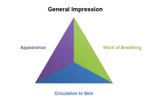
The pediatric assessment triangle (PAT) is a quick, precise, easily adaptable model for the triage assessment of the pediatric patient (Fuzak & Mahar2009). The triangle consists of appearance, breathing, and circulation (ABC approach). This ABC approach allows the triage nurse to use visual clues to swiftly assess the urgency of treatment related to the child's illness or injury. Because of this fast assessment can be done from across the room. It does require an experienced pediatric triage nurse with good judgment and insight in pediatric cardiopulmonary assessment skills to complete the triage process (Dieckmann 2005). The triage nurse must remember that an immediate hands-on approach may cause fear, agitation and/or crying, which as a result, impedes the assessment process. Awareness of developmental age and psychological aspects must be considered for each age group (APLS, 2012). The pediatric assessment triangle has become the cornerstone for pediatric assessment related to emergency medical services.
This rapid approach assessment is a tool for medical professionals to quickly assess a child on sight to determine if the child needs immediate intervention. See the table below for the PAT general impression and first view use.
The appearance of a child is a significant and precise measurement directly related to the degree of distress, illness or injury in a pediatric emergency (APLS, 2012). Most children with mild or moderate distress (and even those moving toward severe distress) are alert and will probably have a normal neurological exam. If the child does not appear to have a normal neurological exam, it is most likely due to such conditions as childhood epilepsy, brain injury in infants and children, neuromuscular diseases, neurogenic and neurodevelopmental disorders (Wong & Kee, 2010). The emergency is then based on what the parent or the caregiver state is abnormal for the child. Another aspect is injuries based on traumatic accidents involving motor vehicles, near drowning, bicycle accidents and falls. In fact, one of the highest causes of death specifically targeting toddler deaths is drowning. Approximately 50% of all pediatric trauma victims will have a head injury with 90% of pediatric deaths from trauma directly related to head injury and/or trauma (Bemis, 2007).
Evaluation of the child's airway and appearance is based on an open and clear airway with normal muscle tone and body position. The pediatric patient should have a normal cry or speech pattern, rate and pitch for their stated age (Bemis, 2007). If the presence of an abnormal cry, absent cry or absent speech is present, the pediatric illness or injury must be considered emergent (Behrman et al, 2004).
Likewise, the pediatric patient should be able to move all of their extremities normally and without problems, responding normally to their parent or caregiver and to the environment around them (i.e. plays with a rattle, toy, stethoscope, name badge and/or lights) (Fleisher et al, 2006).
If the muscle tone is floppy, rigid or nonexistent, in conjunction with decreased response to parents or caregiver (i.e. the parent or caregiver is unable to console the child or wake the child up by their voice or touch) and the child has a decreased response to the environment, this first impression of the patient absolutely should be considered emergent (Roy, 2008).
A simple mnemonic for the appearance assessment is TICLS (tickles) (Behrman et al, 2004):
Breathing is the next part of the triangle. The effort of breathing is a great indicator of oxygenation and ventilation. If the patients breathing pattern appears normal and is without distress, tachypnea, retractions, increased muscle effort, nasal flaring, head bobbing, tripoding, noisy breath sounds (i.e. stridor, wheeze or grunting) and/or absent breath sounds, the patient may be considered a non-urgent patient (Agur & Dalley, 2005). Retractions are usually easily seen in the pediatric patient because of the thin chest wall tissue. Therefore, even if the patient is obese, their ribs are still in the process of maturing and are malleable. Because of this malleable state, the retractions may be more readily seen than in an adult.
Professional Hint: The telltale signs of pediatric respiratory distress are nasal flaring (i.e. light outward/inward flaring of the nostrils when breathing in or out); constant nasal flaring (i.e. the nares are in an outward flared position continuously) and/or retractions of the chest wall (Dieckmann et al, 2005).
They indicate significant effort on the part of the child to breathe easier and move more air. Increased work of breathing will be evident from the doorway or across the room. In the presence of any abnormal findings, the pediatric patient must be considered emergent (Dieckmann et al, 2005).
Circulation is an astute direct observation of color such as mottling, cyanosis, pallor or paleness and bleeding that is significant or overtly obvious. If the skin of the pediatric patient appears white, pale or pasty, it is an indicator for lack of peripheral blood flow and pallor or paleness can be one of the first signs of shock in the pediatric patient (Agur & Dalley, 2005).
The nurse must not accidently confuse vascular instability with being cold as babies can often appear marbled when they are cold, while on the other hand, a bluish discoloration of the skin and mucus membranes is cyanosis. So should the skin have areas of bluish or purple discoloration, these may be patches of cyanosis. Cyanosis and mottling are latent signs of shock and can indicate lack of compensation of vital organs, or be a sign of respiratory failure (Fleisher et al, 2006). Circulation is a direct gauge for core perfusion to vital organs and cardiac output and an alteration in circulation may suggest the patient is in respiratory failure or compensating for shock (Bemis, 2007).
Professional Hint: The decision point for any pediatric emergency is any abnormal finding. Only one leg of the pediatric assessment triangle needs an abnormal assessment finding to consider the patient in need of immediate and/or emergent intervention.
Pediatric vital signs vary significantly based on age. A general guide is the following (Family Practice Note Book, 2012, pg. 1):
Danger Signs (Hazinski et al, 2002)
After completing a rapid assessment from across the room and noting the pediatric patients general appearance, the hands on assessment can begin. Infants and young children should be undressed or at least have their chest exposed for visual assessment including the ABCDE (Airway, Breathing, Circulation, Disability and Exposure) assessment (AHRQ, 2011). The ABCDE assessment will be addressed in detail below:
Airway patency is the first priority. If at any time during the triage or assessment the airway is compromised then immediate treatment is initiated. The sniffing position or head tilt-chin lift or jaw thrust is utilized. In infants, the sniffing position is recommended and for toddlers or school age children, the head tilt-chin lift is suggested (Newberry & Criddle, 2005). If cervical trauma is suspected, do not perform a head tilt, use jaw thrust only. Something as simple as suctioning can result in significant improvement of airway obstruction. However, absent or decreased breath sounds are an indicator of complete airway obstruction and require immediate emergent intervention. (Dieckmann, 2005)
Breathing, along with respiratory rate and effort, is a critical part of the primary assessment. Auscultation should be done with a stethoscope at the midaxillary area anterior to the sternal notch. Respiratory syncytial virus is the most common virus in infants and is a marker for lower respiratory infections.
Types of auscultation:
A Patients respiratory rate and pulse oximetry are useful tools in assessing whether or not the pediatric patient has to work harder at breathing. The nurse needs to remember that children compensate more effectively than adults. It is important to not underestimate the effort of breathing even if the oxygen saturation is greater than 94% (APLS, 2012). One could be easily fooled into believing the patient is in a more stable state than they truly are by ignoring the patients respiratory rate and oxygen saturation. In addition, also note, a rapid respiratory rate could be a sign of fever, fear, anxiety, or pain. Respiratory rate danger zones are >60 respirations/minute or <10 respirations/minute (Fleisher et al, 2006).
The circulation assessment is checking for strength and quality of peripheral pulses, looking at capillary refill (less than 2-2.5 seconds), skin temperature and listening to heart sounds. An abnormal temperature has the potential to mislead the circulation assessment so it is important to determine that the child is not fevered or cold from exposure (James, 2008).
Disability or assessment of the neurological status is the next part of the continual assessment beyond triage. This assessment is done by observation of appearance, pupil response to light, motor activity and response to environment, as well as level of consciousness. The AVPU mnemonic is a widely accepted neurological assessment tool (Fleisher et al, 2006).
Information regarding what kind of exposure (hypothermia or hyperthermia) and how long that exposure was must be inclusive in order to have accurate physiologic findings (APLS, 2012). Any child who has been exposed to extreme outdoor temperature (i.e. cold water drowning, snow or left in hot car) will have an abnormal finding in their assessment. It is important to maintain a warm ambient environment to properly assess and evaluate the pediatric patient (AHRQ 2011). This avoids the ambient temperature of the triage exam room or the emergency room from contributing or altering your assessment of the patient (Dieckmann et al, 2005). It is imperative to avoid confusing the fine marbled appearance often seen in infants due to a cool room with that of mottling due to a lower core temperature due over exposure or vascular instability (Dieckmann, 2005). One can be corrected easily with wrapping the infant or child in a warm blanket (the fine marbled appearance) whereas the other (mottling) may require more intensive interventions (Hazinski et al, 2002). Monitor temperature and treat fever as necessary. The infant will lose body heat rapidly. While exposing them is necessary for a through exam, young children, especially infants less than one year of age, are prone to hypothermia. Although complete exposure should be undertaken in order to get a complete and accurate assessment, care should be taken to preserve the patients body temperature throughout the process (AAP/ACEP, 2004).
In the event of an emergency, the use of the Broselow tape (now more commonly called the resuscitation tape) should be used, if at all possible, to determine the weight and appropriate dosages of life saving drugs and emergency equipment sizes. It is not always convenient to weigh and measure an infant, toddler or child when time is of the essence and more critical. In the event this is the case, the resuscitation tape (or Broselow tape) is a better tool in determining the appropriate weight and drug doses to be utilized due to its color coded system. This color coded system helps clinicians decide what doses of medications are to be used based on height and weight. If a Broselow tape or resuscitation tape is not available, then the following chart will provide guidance (PALS Manual).
Averages:
Example: A 9 year old child would have an average weight of 26 kg. Kg of 8 + (9x2) =26 kg. The 26 kg is used to figure drug dosages.
Example: A 3 year old child would have an average weight 14 kg. Kg of 8 + (3x2) =14 kg. The 14 kg is used to figure drug dosages.
The SAMPLE mnemonic is useful for obtaining information related to the pediatric assessment.
(AHRQ)
The goal of the pediatric triage assessment is to quickly determine acuity level for patients.
Some examples of resources are labs, ECGs, X-rays, IV hydration, IV medications, IM medications, specialty consultation, simple procedure, and complex wound care. (AHRQ)
ESI is a standardized triage system that is supported by both ENA, and ACEP (American College for Emergency Physicians), and provides validity and consistency of a rating system amongst medical institutions across the United States (Briggs & Grossman, 2006).
ESI permits the RN to have a means for quick, effective and accurate sorting of patients, which begins as soon as the nurse sees the patient. This allows the nurse to identify patients who are in immediate need of lifesaving interventions or those who are less critical (Trimarchi, 2012). This further allows the emergency room physician to have a guideline for determination of thresholds for diversions or natural disaster events.
ESI Triage Algorithm (Rockwill, 2005)
ESI levels 3-5 are determined by the number of resources utilized. Resources include:
Count the different type of resources, not the individual tests. (i.e. all labs count as one resource, all x-rays count as one resource) (Eitel et al, 2001). Prediction of number of resources utilized is initiated immediately, starting with the first encounter of the patient by the triage nurse (Trimarchi, 2012). The triage nurse will use information obtained from the subjective and objective aspects of the triage assessment, the patients medical history, medications, age and gender.
The very young (< 3 months) may increase the patients ESI category up a level simply based on their age and (AHRQ, 2011). Co-morbidities may increase the patients ESI category up a level (Gilboy et al).
Level 5: Requires no resources and may be a Nurse only visit.
Example:
Level 4: One resource
Level 3: Two or more resources
Level 2: Does NOT require immediate lifesaving intervention, but is CRITICAL for recovery of the patient and would place the patient on a high risk status.
Level 1: REQUIRES immediate LIFE SAVING intervention
Determining between level 1 and 2:
The patients ESI level may change from the triage time to time of disposition (Briggs & Grossman, 2006). It may increase or decreased depending upon the outcome of the diagnosis and what is revealed during the exam as the nurse and doctor work together to determine the patients needs. The patient may different aspects to different healthcare team member as the patient recalls or is prompted to provide additional information.
Regardless of the ESI level, depending on a patient's specific medical condition, the emergency room physician will admit the patient to the hospital, discharge the patient home or transfer the patient to a more appropriate medical facility (Gilboy et al). Remember, demographics, living arrangements, ability to pay for medications, primary care physician (PCP), must also be considered for final disposition.
ANY high risk situation can rapidly develop into a life threatening situation which requires lifesaving intervention, thereby upgrading a level 2 to a level 1.
Please note: for sake of the case studies, the pediatric assessment triangle (PAT) format will be used. Key points are that the three sides of the PAT tool are: appearance, breathing and circulation.
It is vitally important that after rapidly initiating an across the room assessment using PAT to determine the level of distress, or emergency, move on to the hands on assessment to initiate emergency decision and treatment modalities. When performing vital signs do the least invasive first as crying, fear and anxiety will alter the normal vital signs. Look for danger zone vital signs. Follow the chief complaint and obtain a past medical history if parent or caregiver is available including the events precipitating the emergency room visit. Include immunizations, allergies, and current medications in past medical history (Newberry & Criddle, 2005).
Advance Pediatric Life Support (APLS), The Pediatric Emergency Medicine Resource Jones & Bartlett Learning LLC., American Academy of Pediatrics and American College of Emergency Physicians, 2012
Agency for Healthcare Research and Quality (AHRQ). Emergency Severity Index, Version 4 U.S. Department of Human & Health Services 30 June 2011
American Academy of Pediatrics and the American College of Emergency Physicians. (AAP/ACEP), Textbook for APLS: The Pediatric Emergency Medicine Resource. 4th Edition, 2004 Sudbury, MA. Jones and Bartlett Publishers.
Agur., A, & Dalley, A., Grants Atlas of Anatomy, 11th Edition, Lippincott Williams & Wilkins, 2005.
Bemis, P., Emergency Nursing Bible, 4th Edition, 2007 National Nurses in Business Associations, Inc. Rockledge, Florida
Behrman, R., Kliegman, R., & Jenson, H., Nelson Textbook of Pediatrics 17th Edition 2004 Elsevier Science, Philadelphia, Pennsylvania
Briggs, J & Grossman, V., Emergency Nursing 5-tier Triage Protocols, 2006. Philadelphia, Pennsylvania, Lippincott, Willliams & Wilkins
Centers for Disease Control and Prevention (CDC), National Hospital Ambulatory Medical Care Survey, Emergency Department Summary, Table 9, 2009
Dieckmann, R., Gausche, M., & Brownstein, D., Textbook of Pediatric Education for Prehospital Professionals, Jones and Bartlett, 2005
Fleisher, G., Ludwig, S., & Henretig, F., Textbook of Pediatric Emergency Medicine, 5th Edition, 2006 Lippincott Williams & Wilkins Philadelphia, Pennsylvania
Fuzak, J. & Mahar, P., Emergency Department Triage, 2009. Retrieved 9th July, 2012 from (Visit Source).
Hazinski, M., Zaritsky, A., & Nadkarni, V., et al: PALS Provider Manual, American Heart Association, 2002
Healthcare Cost and Utilization Project (HCUP), U.S. Agency for Healthcare Research and Quality, 2008 Rockville, MD. Retrieved 10th July 2012 from (Visit Source).
Roy, J., Core Concepts of Pediatrics 2008. Retrieved 10th July 2012 from (Visit Source).
Merrill, C, Owens, P and Stocks, C., Pediatric Emergency Department Visits in Community Hospitals from Selected States 2005.
Newberry, L., & Criddle, L., Sheehys Manual of Emergency Care Emergency Nurses Association, 6th Edition, 2005.
Wong, J., & Kee, P., Paediatric Neurology SBCC Baby & Child Clinic, Thomson Paediatric Centre, Singapore 2010. Retrieved 7th July 2012 from (Visit Source).
Zitelli, B, & Davis, H., Atlas of Pediatric Physical Diagnosis, 4th Edition, 2002 Philidelphia, Mosby.