Sign Up
You are not currently logged in. Please log in to CEUfast to enable the course progress and auto resume features.

Course Library

Wound Series Part 3: Pressure Ulcers and Injuries-Risk Factors, Diagnosis, Staging, Management

2 Contact Hours
Listen to Audio
CEUfast OwlGet one year unlimited nursing CEUs $39Sign up now
This peer reviewed course is applicable for the following professions:
Advanced Practice Registered Nurse (APRN), Athletic Trainer (AT/AL), Certified Nurse Midwife, Certified Nurse Practitioner, Certified Registered Nurse Practitioner, Clinical Nurse Specialist (CNS), Licensed Practical Nurse (LPN), Licensed Vocational Nurses (LVN), Midwife (MW), Nursing Student, Occupational Therapist (OT), Occupational Therapist Assistant (OTA), Physical Therapist (PT), Physical Therapist Assistant (PTA), Registered Nurse (RN), Registered Nurse Practitioner
This course will be updated or discontinued on or before Friday, August 1, 2025

Nationally Accredited

CEUFast, Inc. is accredited as a provider of nursing continuing professional development by the American Nurses Credentialing Center's Commission on Accreditation. ANCC Provider number #P0274.


CEUFast, Inc. is an AOTA Provider of professional development, Course approval ID#02147. This distant learning-independent format is offered at 0.2 CEUs Intermediate, Categories: Foundational Knowledge. AOTA does not endorse specific course content, products, or clinical procedures. AOTA provider number 9757.


BOC
CEUFast, Inc. (BOC AP#: P10067) is approved by the Board of Certification, Inc. to provide education to Athletic Trainers (ATs).

FPTA Approval: CE24-649249.Accreditation of this course does not necessarily imply the FPTA supports the views of the presenter or the sponsors.
Outcomes

≥ 92% of participants will be able to identify risk factors for pressure ulcer development, describe pressure ulcer staging, and discuss evidence-informed pressure ulcer prevention and management strategies.

Objectives

After completing this continuing education course, the participant will be able to meet the following objectives:

  1. Describe at least three etiological factors of pressure ulcers/injuries.
  2. Summarize two common pressure ulcer/injury risk assessment tools.
  3. Classify pressure ulcer/injury stages.
  4. Identify at least two evidence-based interventions to consider in managing pressure ulcers/injuries.
  5. Assess research trends in the prevention and management of pressure ulcers/injuries.
CEUFast Inc. and the course planners for this educational activity do not have any relevant financial relationship(s) to disclose with ineligible companies whose primary business is producing, marketing, selling, re-selling, or distributing healthcare products used by or on patients.

Last Updated:
Restart
Restart
  • 0% complete
Hide Outline
Playback Speed

Narrator Preference

(Automatically scroll to related sections.)
Done
Wound Series Part 3: Pressure Ulcers and Injuries-Risk Factors, Diagnosis, Staging, Management
0:00
0:15
 
To earn of certificate of completion you have one of two options:
  1. Take test and pass with a score of at least 80%
  2. Reflect on practice impact by completing self-reflection, self-assessment and course evaluation.
    (NOTE: Some approval agencies and organizations require you to take a test and self reflection is NOT an option.)
Author:    Linda J. Cowan (PHD, ARNP, FNP-BC, CWS)

Background and Significance

Pressure ulcers have been known as "bedsores" for hundreds of years. Historically, the term decubitus ulcer or decubiti has also been used to describe these wounds related to pressure or immobility. The term "pressure ulcer" was replaced by "pressure injury" in the 2016 Pressure Injury Staging System by the National Pressure Injury Advisory Panel (NPIAP) and Pan Pacific Pressure Injury Alliance (PPPIA). The NPIAP defines a pressure injury as "localized damage to the skin or underlying soft tissue usually over a bony prominence or related to a medical or other device. The injury can present as intact skin or an open ulcer and may be painful. The injury occurs due to intense or prolonged pressure or pressure in combination with shear. The tolerance of soft tissue for pressure and shear may be affected by microclimate, nutrition, perfusion, co-morbidities, and condition of the soft tissue" (Edsberg et al., 2016).

Avoidable facility-acquired PUs are recognized internationally as a quality-of-care indicator for healthcare organizations (Office of Disease Prevention and Health Promotion [ODPHP], 2022; Agency for Healthcare Research and Quality [AHRQ], 2023; Centers for Medicare and Medicaid [CMS], 2022). Many healthcare organizations have adopted the new "pressure injury" terminology as well as the updated staging terminology (stages 1-4, unstageable, deep tissue pressure injury [DTPI], medical device-related pressure injury, and mucosal membrane pressure injury). The degree of tissue damage in PUs is typically classified by the NPIAP staging system (JCAHO, 2022). CMS changed its terminology to "pressure ulcers/injuries" for skilled nursing facility (SNF) quality reporting in 2018 (CMS, 2019). However, coding systems such as the International Statistical Classification of Diseases and Related Health Problems – version 10 (ICD-10) continue to use the pressure ulcer term (L89 codes). For this educational course, we will use the term pressure ulcer (PU) without disrespecting the NPIAP.

PUs can have devastating financial implications. Whittington & Briones estimated annual medical costs in the United States associated with treating PUs exceed 5 billion dollars annually. Fogerty et al. (2008) estimated this cost to be higher at $10,845 per patient, exceeding 18.5 billion dollars annually. They also concluded that treating stage 3 and stage 4 hospital-acquired PUs accounts for 58% of national costs (Padula & Delarmente, 2019). Furthermore, studies suggest preventing a PU costs less than treating it (Demarré et al., 2015). Individuals with PUs have higher mortality rates and up to five-fold increased hospital length of stay. Medicare estimated each PU added $43,180 in costs to a hospital stay, while AHRQ estimated the potential cost of individual patient care related to PUs ranging from $20,900 to $151,700 per PU (AHRQ, 2017).

Pressure Ulcer Rates and Reporting

In 2020, AHRQ published Patient Safety Indicators with reported observed rates of hospital-acquired PUs per 1,000 admissions from 47 states in the United States (from hospital discharge data). The overall PU rate for the 2020 report was 0.65 per 1,000 admissions (0.52 per 1,000 admissions for females and 0.79 per 1,000 admissions for males). The overall PU rate for the 2022 report was 0.60 per 1,000 admissions (0.47 per 1,000 admissions for females and 0.73 per 1,000 admissions for males), indicating a slight decrease from the 2020 report to the 2022 report.

CMS reported PUs were present in 3.7 to 7.3% of residents in most long-term care facilities or nursing homes in the United States in 2014 (CMS, 2015). However, the numbers of actual PUs are under-reported. In a study examining the accuracy of nursing home reporting PU rates, the research team stated, "for primary pressure ulcers, 22% were not reported by the nursing homes to CMS; that percentage doubled to 45% when accounting for secondary pressure ulcers" and, "nursing homes substantially under-reported pressure ulcers between 2011 and 2017" (Chen et al., 2022). The 2014 CMS reported rates of PUs in long-term care facilities represent an estimated annual cost of over 3.3 billion dollars (CMS, 2015). More recent estimates of the cost of PUs in United States hospitals could exceed $26.8 billion annually (Padula & Delarmente, 2019). Furthermore, it is important to note a healthcare disparity related to the higher percentages of stage 4 PUs in African American nursing home residents. For example, 50.4% of stage 4 PUs were in African American residents vs. 40.8% in Caucasian residents in short-stay nursing homes. In long-stay nursing home residents, 54.2% of stage 4 PUs were in African American residents vs. 45.6% in Caucasian residents (Chen et al., 2022).

In a large 2018/2019 critical care study of 41,866 critical care patients drawn from a sample of 296,014 patients in United States acute care facilities, investigators found the overall prevalence of PUs in critical care patients was 14.3%, and the hospital-acquired PU prevalence was 5.85%. They compared these rates with 2015 rates; the overall prevalence in critical care patients in 2015 was 14.5%, and the hospital-acquired PU rate was 6.4%. The most common stage reported was DTPI at 33.6% (Cox et al., 2022). The three most common anatomical locations of the PUs in critical care patients were the Coccyx/Sacrum (33.2%), the buttocks (14.8%), and the heels (12.6%). The strongest risk factors for PUs in critical care patients included diabetes mellitus, mechanical ventilation, peripheral vascular disease, and receiving vasopressor agents.

In a national inpatient sample of over six million persons discharged from acute care facilities in the United States, 94,758 individuals had a discharge diagnosis of a PU (Fogerty et al., 2008). However, this number only accounts for patients with a PU diagnosis code documented at discharge. The real numbers of incident PUs (those that start in the facility) are difficult to estimate since studies report widely varied rates across healthcare settings. Most of these rates are prevalence rates (any PU present over a given time) and are under-reported (European Wound Management Association [EWMA], n.d.).

Furthermore, individuals with spinal cord injuries (SCI) are particularly vulnerable to PUs. The National Spinal Cord Injury Statistical Center (NSCISC) 2021 Annual Statistical Report indicates that about 25% of persons under one-year post-SCI have developed a PU within the last 12 months. About 40% have developed at least one PU after 12 months post-SCI (NSCISC, 2021). Shiferaw et al. (2020) conducted an international systematic review and meta-analysis on the global burden of PUs in persons with SCI. They report a global pooled magnitude of PUs among patients with an SCI was 32.36%, and based on the subgroup analysis, the highest magnitude of PU was observed in Africa.

It is evident from the scientific literature that PUs are painful, prone to infection, reduce the quality of life, and create a worldwide economic dilemma (Maklebust, 2005). While healthcare and medical technology have improved in the United States during the past 20 years, facility-acquired PUs have not been eliminated (Rondinelli et al., 2018). Concurrent with this advancing technology, modern-day patients live longer and are typically hospitalized with higher acuity levels than patients hospitalized years ago. Healthcare organizations must continuously improve this important quality of care indicator (National Database of Nursing Quality Indicators [NDNQI], n.d.).

Etiology of Pressure Ulcers

Historically, PUs have been described in medical literature since at least the 1500s, when Fabricius Hildanus first documented his hypotheses of the causes and characteristics of bedsores. He highlighted the role of "internal supernatural" and "external natural" factors that interrupt the supply of blood and nutrients to tissue as causes of bedsores. French surgeon de la Motte identified mechanical pressure and incontinence as key factors in developing PUs in 1722 (Defloor, 1999). Major risk factors for PU development include increased age, impaired mobility, decreased physical activity, poor nutrition, urinary or fecal incontinence, and sensory impairment. Other studies have identified additional risk factors, including smoking status, diabetes mellitus (DM), coronary artery disease (CAD), intensive care unit (ICU) stay greater than three days, ventilator dependency, pneumonia, sepsis, obesity, surgery, female gender, vasopressor agents, and peripheral vascular disease (PVD) (Cowan et al., 2012; Tschannen & Anderson, 2020; Cox et al., 2022).

Most PUs are considered to be avoidable and preventable. An "avoidable" PU means that the patient developed a PU and the facility or healthcare providers did not perform (or document) one or more of the following: evaluate the patient's/resident's clinical condition and PU risk factors; define and implement interventions that are consistent with resident needs, resident goals and recognized standards of practice; monitor and evaluate the impact of the interventions, or revise the interventions as appropriate. There may be a few instances where PUs are unavoidable. "Unavoidable" means the patient/resident developed a PU although the facility and healthcare providers had evaluated (and documented) the resident's clinical condition and PU risk factors; defined and implemented interventions that are consistent with resident needs, goals, and recognized standards of practice; monitored and evaluated the impact of the interventions; and revised the approaches as appropriate – all within a timely manner (Scarborough, 2018).Kennedy ulcers (a specific type of PU often seen at the end of life due to organ/system failure) are one example of "unavoidable" PUs.

graphic showing the breakdown of etiology of pressure sores

Etiology of Pressure Sores

Exploring Risk Factors

Previous studies have identified risk factors for PUs and include the following:

  • A diagnosis of gangrene, septicemia, osteomyelitis, nutritional deficiencies, pneumonitis, urinary tract infection, or paralysis
  • 59-75 years old
  • African American race

The research published by Fogerty et al. (2008), Cowan et al. (2012), and many others have demonstrated how important adequate nutrition and hydration are to maintaining intact skin and facilitating wound healing. Individuals who are nutritionally compromised are at greater risk of a PU.

Chung et al. (2022) conducted a narrative synthesis evaluation of research studies published before December 2020 related to PU risk factors. The total combined sample represented 679,660 individuals. They reported, "in low to moderate risk of bias studies, non-blanchable erythema reliably predicted pressure injury stage 2." Other possible risk factors for the development of a PU identified in this study include the following:

  • Higher interface pressure or BMI < 18.5
  • Male gender
  • Diagnosis of anemia, diabetes, hypoalbuminemia
  • Hypotension
  • Low physical activity
  • Increased length of stay in hospital

Cowan et al. (2012) conducted a retrospective analysis in a Veteran population to identify the strongest PU predictive model, demonstrating that four medical factors (malnutrition, surgery, pneumonia, and candidiasis) were more predictive of a PU. Identifying factors affecting the development of PUs is imperative in the present-day population to select patients for effective prevention interventions. Furthermore, evaluation of the efficacy of existing preventive interventions must be ongoing, and new innovative interventions must be explored to impact PU incidence and prevalence significantly.

Identifying a risk factor is necessary to potentially intervene and lower the associated risk. Identifying the strongest modifiable PU risk factors is important to provide evidence-based interventions and thereby lower the likelihood of someone developing a PU or halting the progression of the PU. PU risk assessment tools provide a tangible way to quantify potential risk so that interventions may be reserved for those at the highest risk and avoid unnecessary interventions and higher financial expenditures on those who may not need them. However, risk assessment tools do not include all risk factors present in an individual, and clinicians are encouraged to use clinical judgment to appropriately account for any additional PU risk factor not addressed by the risk assessment tool their facility is using (Moore & Patton, 2019).

One criticism of existing PU risk assessment tools is that neither risk factors nor the weights attributed to them have been identified using adequate statistical techniques. Risk factors are those factors or conditions that are noted to be most strongly associated with the outcome of interest. To provide evidence-based measures for preventing PUs, an effective means of identifying those at the highest risk is imperative. Another criticism of PU risk assessment tools is the lack of clear evidence that risk assessment tools significantly impact clinical outcomes such as incidence rates. Other criticisms include the subjective nature of some of the assessments, the lack of tools for specific settings such as the perioperative environment, and the fact that no one assessment tool could account for every risk factor (Moore & Patton, 2019). Current risk assessment tools may require further development, improved statistical evaluation, and possible modification to remain applicable to present-day populations.

Thomas posits an explanation for an unchanging incidence of PUs as "a failure of known effective prevention treatment to be applied, or the failure of prevention strategies to be effective despite being applied" (p.298). Effective preventive measures may not be applied if individuals are not appropriately identified as at risk. Risk screening tools are useless if they do not apply to the population being screened, are used inconsistently, or are scored incorrectly. Furthermore, Chung et al. (2022) notes the evidence supports close communication among healthcare givers concerning risk factors to optimize prevention and treatment efficacy.

Risk Assessment Tools

Norton Scale

The PU risk assessment tools most frequently utilized worldwide are the Norton Scale (published in 1962) and the Braden Scale (published in 1987) or modified versions of these. Doreen Norton (along with Rhoda McLaren and Dr. Norman Exton-Smith) developed the Norton Scale in Great Britain (Norton et al., 1989). It is the first of all the PU risk assessment scales; indeed, it is one of the earliest risk assessment scales of any kind. At first, Norton et al. (1989) devised a data collection tool with columns to describe all factors noted in every patient that might be relevant to PU development, such as the patient's weight, build, appetite, medications, preventive measures (14 different skin care products), treatment measures, site and condition of the skin, and skin changes.

They developed a rating 'scale' (when rating scales were uncommon) with five elements with weighted descending values for each element from 4 to 1. The elements or factors in their tool were listed as column headings for general physical condition (including overall nutritional state), mental condition, mobility, activity, and incontinence. Norton reports the tool was scored '4' for a normal or good function in each factor and '1' for very poor or bad function, with a total possible high score of 20 (patient in good overall condition) and a low score of 5 (patient in poor overall condition). A descending scale was selected because it correlated with a decline in the patient's condition.

The Norton conceptual model is a simple model based on their observed factors of general physical condition, mental condition, mobility, activity, and incontinence with a Likert-type scale for each factor totaled as one independent variable and PU risk as the dependent or outcome variable. The model posits that higher total scores strongly correlate with higher PU risk. Norton states her scale was frequently misinterpreted as "over-predicting pressure ulcer lesions" (Norton et al., 1989, p.41) when used incorrectly as a predictive tool. In addition, it was criticized for leaving out a nutritional assessment. However, according to Norton, a nutritional assessment was included in their data collecting form and was intended as an integral part of the overall "general condition" assessment. In retrospect, Norton (1989) regretted not having an explicit user's guide to go along with her original tool and suggested that all future risk assessment tools remain simple and easy to use and have a guide explaining specifically how to utilize the tool (Post, 1963).

Braden Scale

The Braden Scale is today's most widely used PU risk assessment tool. The theoretical framework is based on a physiological model depicting factors contributing to the development of PUs. It includes factors affecting the intensity and duration of pressure (decreased mobility, decreased activity, and decreased sensory perception), which combine with intrinsic factors (age, nutrition, vascular perfusion) and extrinsic factors (increased moisture, increased friction, and increased shear forces) that affect tissue tolerance. The Braden Scale is publicized as the most extensively tested and studied of the assessment tools. The Braden Scale has a potential score ranging from 6 to 23 derived from the total scores of its six subscales (sensory perception, mobility, activity level, moisture/incontinence, nutrition, and friction/shear). Lower scores on the Braden Scale indicate a greater risk for PU development. For example: Very high risk = 9 or below; High risk = 10-12; Moderate risk= 13-14; and Mild risk = 15-18. Subscale scores range from 1 to 4, with lower subscale scores indicating higher risk. So, if the patient were severely nutritionally compromised, the nutrition subscale would have a score no higher than 1 or 2. There is literature by Braden to suggest that if a person has other major risk factors (age, fever, poor nutrition, hemodynamic instability), their score should be advanced to the next highest level of risk (i.e., the next lower score). Yet, observational studies suggest nurses do not routinely do this.

Clinical Pearl:
Nurses tasked with conducting daily PU risk assessments should take them very seriously and score them accurately, taking into account any change in the patient's condition and risk not assessed by the specific tool being used. It is always better to overestimate risk than to underestimate risk! And sometimes, this means assigning a lower score if lower scores indicate higher risk on the risk screening tool you are using (such as the Braden Tool). If the person's sub-score falls between numbers, always select the lower number to try and capture any possible risk. For instance, if the patient's nutritional sub-score falls between a "2" and "3" on the Braden Tool, always mark it a "2".

Diagnosis

Diagnosing PUs depends on assessing possible pressure, shear, and moisture-related etiological factors and the location of the tissue damage. The first question in your mental wound assessment "checklist" should be: "Is it over a bony prominence?" While PUs may develop over any bony prominence on the body, the sacrum, coccyx, buttocks, and heels of the feet are the most prevalent sites. Other common locations may include greater trochanters, ischial tuberosities, ankles, knees, elbows, scapulas, shoulders, and the occiput (IQWIG, 2018). In addition, a pressure-related injury may also be related to devices such as oxygen tubing, cervical collars, endotracheal tubes, oxygen saturation monitor probes, drain tubes, foley catheters, etc. Device-related PUs may be observed over the ears, around the urethral opening of the genitals, or in other soft tissue locations not associated with a bony prominence (NICE, 2015).

Furthermore, this type of device-related injury may cause full-thickness tissue damage, which is difficult to stage because, as in the case of the ears, there is no subcutaneous tissue evident. A very shallow wound on the ears or nose may result in the exposure of cartilage (full-thickness wound) without the involvement of muscle, tendon, or bone. Again, it is important to note that while the onset of PU is associated with unrelieved pressure, it can also be caused by a combination of pressure, friction, and shearing forces. Evaluating these potential forces (head of the bed raised for long periods, patient sliding down in the bed, chair transfers, improper overhead sling positioning, etc.) is imperative when assessing your patient's skin integrity risks.

Pressure Ulcer Stages

PU staging provides a way to communicate the degree of tissue damage in PUs. The staging system was defined by Shea in 1975 and provided a name for the amount of anatomical tissue loss. The original definitions confused many clinicians and led to the inaccurate staging of ulcers associated with or due to perineal dermatitis and those due to deep tissue injury. The original stages ranged from stage I (less obvious and potentially reversible damage) to stage IV (damage extending to muscle, tendon, or bone). "Unstageable and "suspected deep tissue injury" (SDTI) were added as additional 'stages' or descriptors in 2007 (Edsberg et al., 2016). In April 2016, the NPIAP again revised its PU staging system to include a new "pressure injury" terminology to replace "pressure ulcer" terms, deleting "suspected" from the deep tissue injury classification. One key factor to consider when staging PUs is that stage 1 PUs are potentially reversible, so quick intervention is critical.

graphic showing states of pressure sores

Stages of Pressure Ulcers

Stage 1 Pressure Injury: Non-blanchable erythema of intact skin. Intact skin with a localized area of non-blanchable erythema, which may appear differently in darkly pigmented skin. The presence of blanchable erythema or changes in sensation, temperature, or firmness may precede visual changes. Color changes do not include purple or maroon discoloration; these may indicate DTPI.

Stage 2 Pressure Injury: Partial-thickness skin loss with exposed dermis. The wound bed is viable, pink or red, moist, and may also present as an intact or ruptured serum-filled blister. Adipose (fat) is not visible, and deeper tissues are not visible. Granulation tissue, slough, and eschar are not present. These injuries commonly result from adverse microclimate and shear in the skin over the pelvis and shear in the heel. This stage should not be used to describe moisture-associated skin damage (MASD), including incontinence-associated dermatitis (IAD), intertriginous dermatitis (ITD), medical adhesive-related skin injury (MARSI), or traumatic wounds (skin tears, burns, abrasions).

Stage 3 Pressure Injury: Full-thickness loss of skin, in which adipose (fat) is visible in the ulcer, granulation tissue, and epibole (rolled wound edges) are often present. Slough and eschar may be visible. The depth of tissue damage varies by anatomical location; areas of significant adiposity can develop deep wounds. Undermining and tunneling may occur. Fascia, muscle, tendon, ligament, cartilage, and bone are not exposed. If slough or eschar obscures the extent of tissue loss, this is an Unstageable Pressure Injury.

Stage 4 Pressure Injury: Full-thickness skin and tissue loss with exposed or directly palpable fascia, muscle, tendon, ligament, cartilage, or bone in the ulcer. Slough and eschar may be visible. Epibole (rolled edges), undermining, and tunneling often occur. Depth varies by anatomical location. If slough or eschar obscures the extent of tissue loss, this is an Unstageable Pressure Injury.

Unstageable Pressure Injury: Obscured full-thickness skin and tissue loss. Full-thickness skin and tissue loss occur in which the extent of tissue damage within the ulcer cannot be confirmed because it is obscured by slough or eschar. If slough or eschar is removed, a stage 3 or stage 4 pressure injury will be revealed. Stable eschar (dry, adherent, intact without erythema or fluctuance) on the heel or ischemic limb should not be softened or removed (Hess, 2020).

Deep Tissue Pressure Injury: Persistent non-blanchable deep red, maroon, or purple discoloration. Intact or non-intact skin with localized area of persistent non-blanchable deep red, maroon, purple discoloration or epidermal separation revealing a dark wound bed or blood-filled blister. Pain and temperature changes often precede skin color changes. Discoloration may appear differently in darkly pigmented skin. This injury results from intense and prolonged pressure and shear forces at the bone-muscle interface. The wound may evolve rapidly to reveal the actual extent of tissue injury or may resolve without tissue loss. If necrotic tissue, subcutaneous tissue, granulation tissue, fascia, muscle, or other underlying structures are visible, this indicates a full-thickness pressure injury (unstageable, stage 3 or stage 4). 

Additional pressure injury definitions include the following:

Medical Device-Related Pressure Injury: This describes an etiology. Medical device-related pressure injuries result from using devices designed and applied for diagnostic or therapeutic purposes. The resultant pressure injury generally conforms to the pattern or shape of the device. The injury should be staged using the staging system.

Mucosal Membrane Pressure Injury: Mucosal membrane pressure injury is found on mucous membranes with a history of a medical device in use at the location of the injury. Due to the anatomy of the tissue, these ulcers cannot be staged.

It is important to note PUs should never be "back-staged," meaning once a wound is identified as a stage 3 pressure ulcer/injury, it is never referred to as a stage 1 or 2. Rather, the stage 3 pressure ulcer/injury, which is healing would be referred to as a "healing stage 3 pressure ulcer/injury." In addition, only pressure ulcers/injuries are "staged." Diabetic foot ulcers may be "graded" (such as Wagner Grades), but no other wounds are "staged."

Evidence-Based Management of Pressure Ulcers

The approach to managing PUs should always focus on PREVENTION. If the patient develops pressure-related injury (despite preventive measures), then evidence-based practice related to wound healing should be administered but always with PREVENTION in the treatment plan. Healthcare providers should also prevent the PU from deteriorating or worsening to a deeper stage. Evidence suggests that offloading and pressure reduction/redistribution should be a primary goal of treating PUs and eliminating shearing and friction forces. Addressing other key contributing or risk factors is also critical. Knowing what intrinsic and extrinsic factors contribute to skin breakdown is important. Intrinsic factors such as age, immune function, nutrition, and disease states (comorbid conditions) should be considered. Extrinsic factors such as dry skin, friction, transfer equipment, medications, and moisture should also be addressed. The scientific literature suggests PU prevention (PUP) "bundles" may be helpful (Rivera et al., 2020). A PUP or skin bundle is typically a group of interventions or evidence-based PUP strategies that are implemented as a package in a healthcare facility or unit. Components may include but are not limited to standardized staff education, competency check-offs, patient/caregiver education, risk assessment tool(s), preventive interventions by the level of risk, turning schedules, prophylactic dressings, specialized seating interventions, nutrition consults, standardized application of specialty mattresses, documentation templates and checklists and audit tools (Tayyib et al., 2016; Gaspar et al., 2019; Rivera et al., 2020).

Maintaining adequate nutrition and hydration should be essential to PU management plans. Nutritional assessments should include current and usual weight, history of involuntary weight loss, nutrition intake (protein, calories, fluid), appetite, dental health, chewing, swallowing or feeding problems, medical or surgical history affecting intake or absorption, drug/nutrient interactions, psychosocial factors (mood, finances, cooking ability, culture, preferences) and appropriate laboratory values. Other factors to identify potential nutritional impairments include body mass index (BMI), skin fold thickness, arm circumference, etc.

Common laboratory analyses include hemoglobin A1c (HbA1c), blood urea nitrogen (BUN), total lymphocyte count, serum transferrin, serum albumin, and serum pre-albumin. A total lymphocyte count indicates protein-calorie malnutrition (values below 1,800 mm3 indicate risk); serum transferrin is also a malnutrition indicator (values below 200 mg/dl indicate risk). Serum albumin (20 days half-life indicates chronic disease state, values below 3.5g/dl indicate an increased risk of malnutrition); serum pre-albumin has a two to three-day half-life, which some providers feel may provide a more current reflection of protein stores (values below 15 mg/dl indicates increased nutritional risk). However, several nutritional screening recommendations state that serum proteins such as albumin and serum pre-albumin are "not endorsed for nutritional screening," by the evidence presented in the 2019 NPIAP Pressure Injury Prevention Guidelines (Munoz et al., 2020).

Managing excessive moisture (incontinence, sweat, spilled liquids) on the skin surface of patients at risk for PUs is another critical preventive and management goal (Saindon & Berlowitz, 2020). Managing incontinence includes establishing a bowel and bladder program, cleansing the skin after soiling with pH-balanced cleansers, and using incontinence skin barriers (creams, ointments, etc.) to protect and maintain intact skin. Consider a pouching system or collection device to protect from effluence if fecal incontinence is an issue. Indwelling catheters may be indicated for short-term use with severe incontinence-related dermatitis and difficulty managing urinary incontinence. In addition, limit the use of diapers, but if briefs, diapers, or underpads are used, make sure they are the type that wicks moisture from the skin. In addition, temperature plays an important role concerning moisture. Saindon and Berlowitz (2020) systematically reviewed the PU literature. One of their research study conclusions reported that "higher temperatures at the (skin) surface are transferred to deeper tissues, resulting in greater susceptibility to injury. Too much or too little moisture increases the coefficient of friction, with resulting increases in shear deformation of subcutaneous tissues" (and thus, leading to a potential PU).

Addressing impaired mobility includes patient and caregiver education and establishing a turning/repositioning schedule for patients confined to the bed or a chair. This should include offloading direct pressure over bony prominences and repositioning the patient every two to four hours. Keeping the head of the bed raised at 30 degrees or less except for tube-fed patients (but allowing for elevation for 1 hour after meals) is also deemed important. Avoiding "donut" type seating cushions (rings meant for under coccyx areas) and avoiding pulling the patient across the bed or a chair surface is important for reducing friction and shearing forces. Pressure-redistribution surfaces on bed and chair surfaces for "at-risk" patients should also be utilized (Saindon & Berlowitz, 2020).

Pressure Reduction and Redistribution

There are many ways to help reduce or redistribute pressure under bony prominences; the most common are repositioning and the use of support surfaces. Examples of support surfaces include mattresses and mattress overlays, integrated bed systems, seat cushions, seat overlays, and heel floatation devices. Mattresses and mattress overlays include three groups. Group 1 support surfaces do not require electricity, are relatively inexpensive, and are for patients at low or moderate risk of PUs. Group 2 support surfaces may or may not be dynamic-powered devices and are appropriate for patients at moderate or high risk. Group 3 support surfaces include air-fluidized beds and are for patients at very high risk. The goal of these surfaces is to assist in creating an environment that enhances tissue viability and promotes healing. The surface should prevent "bottoming out" when the patient is positioned on it. The surface should also assist in the prevention of the shearing of tissue.

Wound Healing

Open PUs are wounds. Many become chronic wounds, and stage 3-4 PUs have been associated with a higher complication rate, especially infection and sepsis, and a higher mortality rate than other chronic wounds (Saindon & Berlowitz, 2020). We will review some helpful mnemonics to guide wound bed preparation and management of full-thickness open wounds. The first mnemonic is T-I-M-E. There are pivotal articles describing T-I-M-E to guide clinicians in managing full-thickness wounds. (Leaper et al., 2012; Schultz et al., 2003)

  • T stands for tissue: addresses non-viable tissue in the wound bed. Removing dead and necrotic tissue from the wound bed promotes healing and reduces bacterial breeding grounds.
  • I stand for infection and inflammation. Address potential sources of bacterial contamination. Use excellent technique when changing the dressings and providing wound care. Identify early signs of infection and address them. If warranted, use appropriate methods (quantitative tissue cultures) to culture the wound bed. Chronic wounds tend to be stuck in an inflammatory phase of wound healing. High levels of wound enzymes (matrix metalloproteinases [MMPs]) and inflammatory chemical messengers (cytokines) dominate the fluid of these wounds, making a very inhospitable environment for healing processes and cells. Consider wound dressings and topical therapies which address this and help minimize it.
  • M stands for moisture. Maintaining adequate moisture in the wound is necessary for healing cells to migrate and for microcellular processes to continue optimally. A dry wound bed may take almost 50% longer to heal than a moist wound environment. Too much moisture, on the other hand, perpetuates high levels of MMPs and cytokines and may contribute to the inflammatory state of a chronic wound.
  • E stands for the edges of the wound (and surrounding tissue). Healthy wound edges have a "beachfront" look, as if the edges of the wound were the sand on the water's edge of the beach, gently flowing down into the water. If the edge of the wound is rolled under, like a cliff instead of a beach, or if it is undermined as if a cave were hiding under the wound edge, then skin cells (keratinocytes/epithelial cells) cannot migrate easily, and the wound will not contract (get smaller) normally. These problematic wound edges must be addressed and smoothed to facilitate or stimulate wound healing. In some cases, rolled edges may be addressed by chemical cauterization with silver nitrate on a stick by a professional trained to do this within their scope of practice.
Clinical Pearl:
Undermining (an underground cave, "lip," or shelf under the edge of a wound) is often caused by shearing forces (such as when a person slides down in bed or someone slides harshly on a transfer board). Eliminating these forces is the only way to prevent/reduce this undermining in PUs, particularly a sacral, coccyx, and ischial PU.

The following mnemonic illustrates similar principles for topical wound therapy:

  • D – Debride any non-viable tissue (necrotic tissue impedes wound healing)
  • I – Identify and treat infection – infection is a powerful impediment to wound healing (it is recommended to get a biopsy versus a swab culture, if possible)
  • P – Pack dead space LIGHTLY – the wound environment should be friendly to fibroblasts (the white cells that manufacture new granulation tissue in full-thickness wounds). Packing a wound too tightly reduces the fibroblast activity, impairs cellular migration, and may cause tissue ischemia in the wound bed if excessive pressure results from the packing.
  • A – Absorb excess exudate
  • M – Maintain moist wound surface
  • O – Open or excise closed wound edges
  • P – Protect healing wounds from trauma/infection
  • I – Insulate (maintain a normal body temperature to reduce vasoconstriction and enhance cellular activity)

Another published mnemonic, "DIDN'T HEAL," provides a useful way to help remember factors that adversely affect wound healing (Daley, 2020):

  • D = Diabetes: The long-term effects of diabetes impair wound healing by diminishing sensation and arterial inflow. In addition, even acute loss of diabetic control can affect wound healing by causing diminished cardiac output, poor peripheral perfusion, and impaired polymorphonuclear leukocyte phagocytosis.
  • I = Infection: Infection potentiates collagen lysis. Bacterial contamination is a necessary condition but is not sufficient for wound infection. A susceptible host and wound environment are also required. Foreign bodies (including sutures) potentiate wound infection.
  • D = Drugs: Steroids and antimetabolites impede the proliferation of fibroblasts and collagen synthesis.
  • N = Nutritional problems: Protein-calorie malnutrition and deficiencies of vitamins A, C, and zinc impair normal wound-healing mechanisms.
  • T = Tissue necrosis, resulting from local or systemic ischemia or radiation injury, impairs wound healing. Wounds in characteristically well-perfused areas, such as the face and neck, may heal surprisingly well despite unfavorable circumstances. Conversely, even a minor wound involving the foot with a borderline blood supply may mark the onset of a long-term nonhealing ulcer. Because of local oxygen deficits, hypoxia and excessive tension on the wound edges also interfere with wound healing.
  • H = Hypoxia: Inadequate tissue oxygenation due to local vasoconstriction resulting from sympathetic overactivity may occur because of blood volume deficit, unrelieved pain, or hypothermia, especially involving the distal extent of the extremities.
  • E = Excessive tension on wound edges, leading to local tissue ischemia and necrosis.
  • A = Another wound: Competition between several healing areas for the substrates required for wound healing impairs wound healing at all sites.
  • L = Low temperature: The relatively low tissue temperature in the distal aspects of the upper and lower extremities (a reduction of 1-1.5°C [2-3°F] from normal core body temperature) is responsible for the slower healing of wounds at these sites.

Political, Economic, and Other Implications of Pressure Ulcers

Political issues affecting prevention and treatment may include scenarios like the one in Iran, where pressure-reducing equipment is scarce, or situations where a lack of healthcare resources prevents the global implementation of even basic preventative measures. Government-sponsored or subsidized medical care, private insurance, homelessness, drug addiction, war, economic crisis, and limited transportation or food sources are a few huge issues affecting healthcare today. In America, Medicare and Medicaid services come to mind when considering economic, social, and political implications on research findings regarding PU risk. To highlight this point, a roundtable discussion of the International Expert Wound Care Advisory Panel highlights one PU-specific ramification of the Deficit Reduction Act of 2005. The expert panel detailed subsequent changes in the CMS financial reimbursement amounts for long-term and acute care settings such as nursing homes and hospitals. Beginning in October 2008, CMS no longer reimbursed higher rates for patients that develop stage 3 or 4 PUs (full-thickness tissue loss) after admission, representing a potentially substantial economic loss to healthcare facilities. The act was thought to provide additional motivation to acute and long-term care facilities to evaluate and improve their documentation and prevention programs. However, there is a lack of research supporting the reduction in coverage of any improvements in PU prevention or rate reduction. This discussion is significant as it stresses the urgency of a consensus among healthcare providers, particularly the wound care community, in providing effective quality research and evidence-based (and innovative) outcome data related to interventions.

Limitations of Research Evidence and Data

The Limited Use of Randomized Control Trials

There are few (if any) current randomized controlled trials (RCTs) where random vulnerable patients are assigned to receive an intervention suspected of "causing" full-thickness PUs because that would be unethical. Three important documents impact biomedical research worldwide by helping to determine what is ethically right or wrong. These documents are the Nuremberg Code (1947), the World Medical Association's Declaration of Helsinki (originally adopted in 1964), and the Belmont Report of the National Commission for the Protection of Human Subjects in the United States (Beauchamp & Childress, 2001). In a nutshell, these documents came about after tragic medical research practices were disclosed to the world. The Nuremberg Code was written in 1946-1947 after the horrific criminal medical research on Jews and other prisoners in Nazi concentration camps came to light. The Code's purpose is described as providing ten directives for human experimentation, including informed consent and assurance within research protocols that "all unnecessary physical and mental suffering and injury" as a result of the experimentation process is avoided.

The ethical dilemma for PU research arises when you want to do an RCT where the dependent variable of interest is a poor medical outcome, like a stage III PU. It would not be ethical to divide two elderly groups into experimental and control groups, apply some intervention to one group, and withhold preventive interventions to high-risk patients in another group. At the same time, you observed both groups to see who developed a stage 3 PU. Ethically, researchers should desire to prevent harm (PUs) in all subjects, and if something were to cause skin or tissue injury, you would want to intervene immediately. To merely record how bad things got in one group would go against biomedical ethical principles of "not causing harm." However, RCTs can be conducted that investigate potentially positive outcomes. Several RCTs have been reported involving patients deemed at risk for PUs that introduce a theoretically preventative intervention (such as a new support surface) versus standard care (existing mattress) to see which is more effective at preventing PU development (comparative effectiveness studies).

Problems with Data

In particular, concerning PU risk prediction, data collected from research studies on populations twenty years ago poses a problem for application to the current population. Many of the prevention guidelines available today are based on those risk factors identified over twenty years ago, and these may not carry the same relevance today.

Clinical Pearl: Access to Guidelines.

Unfortunately, in the US, AHRQ decided to stop hosting the National Guideline Clearinghouse website in 2018. However, there continues to be national and international guidelines available to healthcare providers on other websites, such as those listed below:

Health information technology (HIT) and patient safety approaches have changed dramatically over the past two decades. These advances will likely meet the increased needs of patients admitted to acute care facilities. Vincent et al. (2006) describe medical technology and clinical procedure advances, as well as process of care (organizational/policy) changes within the emergency medicine and intensive care unit (ICU) arenas over the past 25 years. In addition, more patients are having procedures done on an outpatient basis, so fewer patients with "minor" conditions are being admitted to the hospital. These changes will likely alter acuity levels, numbers of patient transfers within facilities, and length of stays for patients admitted to hospitals. Essentially, these factors are apt to change the "face" of the inpatient population and impact the characteristics of those at risk of a PU. The face of healthcare as we know it is also changing, with technological advances only dreamed about ten years ago. The World Economic Forum describes several advancements, such as virtual reality, artificial intelligence, "wearables," 5G-powered remote visits, etc. It is possible that many of these technologies could be applied to PU prevention, diagnosis, and management in the near future.

Summary

Prevention is paramount!

Address healing impediments, especially nutrition, moisture, temperature, friction, and shear.

Use appropriate mnemonics to help guide wound bed preparation and wound healing approaches.

Any topical wound therapy you select should show expected improvement in two to four weeks. If not, then re-evaluate the wound and evaluate for possible infection (quantitative tissue analysis). Consider measures to address etiologic and systemic factors (nutrition, shear, diabetes, or comorbid disease control). Change to another product if necessary. If the wound worsens or does not improve after doing these things, consider malignancy (biopsy for histopathology) or autoimmune disease(s) as potential contributing factors, such as pyoderma gangrenosum.

As stated before, many changes have occurred in health care over the past 20 years, especially with nursing care delivery, hospital organizational frameworks, quality improvement, financial coverages, not to mention technology (including electronic records), and perhaps the overall face of our present-day patient population. Nevertheless, despite multiple prevention programs implemented nationwide, the incidence and prevalence (and the number of deaths attributed to PUs annually by the CDC) have not changed significantly in many facilities. Researchers need to determine why and develop innovative approaches to solve this dilemma. The effectiveness of prevention and treatment interventions must be determined. Guidelines must be based on current, high-quality scientific evidence, and impediments to implementation must be addressed. There are significant gaps in the scientific literature regarding PU risk assessment in different settings, particularly present-day populations, and how PU risk assessment may impact patient outcomes (Warner-Maron, 2015). New conceptual models of PU development, risk assessment, and effective prevention interventions may need to be developed.

What does the future hold? We already have thermographic scanning for temperature assessment, infrared devices, sub-epidermal moisture detection devices, etc. It will be important to continue to evaluate the efficacy of these devices and technologies, especially regarding future prevention approaches. Assimilating technology such as artificial intelligence (machine learning), automated pressure, temperature, and moisture-sensing devices built into support surfaces, ways to assess the microclimate of skin through clothing or wearable technology may not be too far in our future – and some are already here (Cai et al., 2021; Dweekat et al., 2023; Gefen & Ross, 2020; Saindon & Berlowitz, 2020).

Case Study Scenario

Patient

A 76-year-old African American male, Mr. Smith, presented to the emergency room at 4 pm for a fractured right hip after falling at home. No other complaints were noted. History includes type II diabetes (well-controlled with oral medication and diet), hypertension (no current medication, ran out three months ago), height 5'10", weight 150 lbs (recent weight loss of 25 lbs. over the past three months after wife recently died). Mr. Smith lives alone but has a son who lives two hours away and visits every two weeks. He has been using a walker to ambulate for the past two years since he is "not too steady" on his feet sometimes. Mr. Smith reports he sometimes dribbles urine after urinating but denies any incontinence of urine or stool. The current pain level is eight out of ten. His blood pressure is 170/98. His HbA1c level is 7.2, and his serum glucose is 125.

Risk Assessment

The Braden Scale was used by the admission nurse. Here is how she scored the patient: Sensory Perception: 4 "no impairment"; Moisture: 4 "rarely moist"; Activity: 3 "walks occasionally"; Mobility: 3 "slightly limited"; Nutrition: 3 "Adequate"; Friction & Shear: 3 "No apparent problem" – Total score: 20 (not at risk); Visual skin assessment head-to-toe reveals no redness over any boney prominence. However, a skin tear was noted on the left forearm from a fall at home.

Prevention Intervention Strategies

The patient is placed on a foam mattress (standard for the medical-surgical unit Mr. Smith was admitted to) while awaiting hip surgery. Mr. Smith cannot have anything by mouth (NPO) after midnight, and no nutrition consult has been placed yet. He is on bed rest, and an opioid was ordered for pain relief before surgery. Mr. Smith is using a urinal for voiding during the night. The patient does not want to be turned to the side during the night due to pain.

Initial Clinical Outcomes

Mr. Smith goes to surgery without additional skin assessment, and no one documents any skin warmth at the sacral area or right heel. Perianal moisture is present but not documented by transfer personnel. Mr. Smith is prepped for surgery, and the nurse conducting the perioperative skin assessment notes intact but slightly darker skin over the sacral area. The skin is also warmer to touch over the sacral area than the surrounding skin. The nurse also notes some right hip bruising (diffuse bruising over most of the right lateral hip and thigh – not particularly over trochanter boney prominence), in addition to some right heel bogginess and warmth, and the left forearm skin tear.
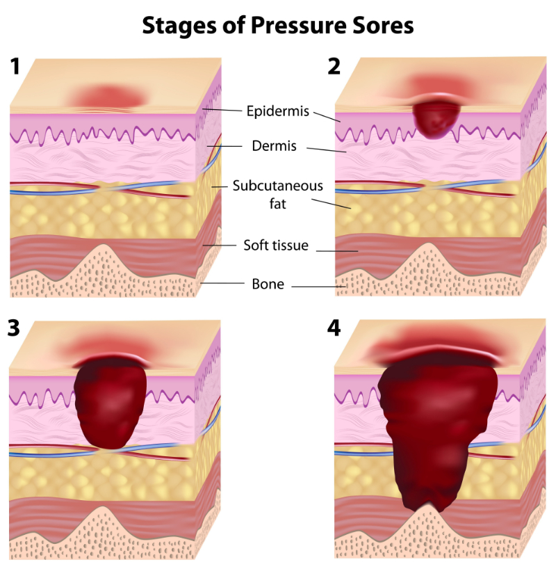
Staging

Perioperative nurse documents right hip bruising likely due to fracture injury, a stage 1 sacral PU, and stage 1 right heel PU. The left forearm skin tear is not staged because it is not a PU, and the right hip is not staged due to the nature of bruising consistent with fall injury and fracture.

Management Strategies

Perioperative staff implement specialty gel operating table padding and carefully apply foam padding to the non-operative extremities, head, etc. They also take care to float heels. In recovery, alternating pressure redistribution surface is used, small adjustments to the patient's position are made hourly to relieve pressure to the sacral area, and heels continue to be floated. Vital signs reveal high blood pressure after surgery (200/110) and elevated heart rate (110/min). Labs are unremarkable. The patient is transferred to surgical intensive care for observation. The beds have alternating pressure mattresses and beds with a "turn to assist" capability. When the patient awakes, he reports pain as four out of ten. Post-operative opioids are ordered for pain. The patient remains NPO. The perioperative staff points out all skin findings to the intensive care staff. When the patient was admitted to intensive care, the Braden risk assessment score was: Sensory Perception: 2 "very limited"; Moisture: 2 "often moist"; Activity: 1 "bedfast"; Mobility: 2 "very limited"; Nutrition: 1 "very poor"; Friction & Shear: 1 "problem" – Total score: 9 (severe risk). A nutrition consult and a wound consult by a wound specialist are ordered.

Outcomes

The patient stabilizes within one day and is transferred to a regular medical-surgical floor (on an alternating pressure specialty mattress). The staff now documents the sacral PU as stage 2 superficial open blister but no purple discoloration, and the right heel is now a deep purple color, intact, and still boggy (documented by staff as stage 1). The interdisciplinary team discusses the PU management plan and steps to initiate a care plan to prevent the progression of the existing PU and any new PU. Physical therapy initiates a mobility and strengthening care plan, and the patient starts to ambulate with assistance. A regular turning schedule is implemented at least every two hours, and sips of fluid are offered with a turning schedule. A moisture barrier cream is ordered for the perineum, and the head of the bed is kept at 30 degrees or below (when in bed) except for meals and 30 minutes after meals. Pain that may be present with turning is addressed by medicating Mr. Smith 30 minutes before the turn as needed, and nutrition interventions have been initiated. The social worker and team discuss the best location to discharge the patient, the potential need for home assistance, and the safety assessment of the home environment. Mr. Smith is discharged 12 days after the resolution of the stage 2 sacral PU with no further evolution of the heel PU. Right hip bruising is resolved, and the left forearm skin tear is closed. Mr. Smith's vital signs, including his blood pressure, pulse, and labs, are all unremarkable at discharge.

Strengths of the Case

Perioperative skin assessment was thorough and accurately noted PUs over the sacral area and right heel. The healthcare staff correctly noted right hip bruising due to injury and not a PU (diffuse bruising over most right lateral hip and thigh – not particularly over trochanter boney prominence) and the left forearm skin tear (not a PU). Perioperative staff also implement prevention strategies during surgery (specialty gel operating table padding and careful foam padding to non-operative extremities, head, etc., and take care to float heels). Alternating pressure redistribution surface is appropriately used, with small adjustments to the patient's position regularly made to relieve pressure to the sacral area, and heels continue to be floated. ICU staff correctly score the risk assessment as" 9" (very high risk): 

  • Sensory Perception: 2 "very limited" – because the patient is over 65 years old, and some sensory decline is normal in ages over 65 as well as people with diabetes (peripheral neuropathy present in feet), and the patient is on pain medication (decreases sensory perception, and pain may also distract the patient from feeling more minor discomforts).
  • Moisture: 2 "often moist" (patient using urinal was noted to frequently spill a little, and perineal moisture was noted even pre-operatively, which could be sweat).
  • Activity: 1 "bedfast" (patient was bedfast for several days).
  • Mobility: 2 "very limited" (mobility was impaired severely for more than three days).
  • Nutrition: 1 "very poor" (NPO for > 24 hours, 25 lb. weight loss prior to admission, and potential appetite changes due to grieving since wife recently died).
  • Friction & Shear: 1 "problem" (the nurse felt the score was between a 1 or 2 because the patient was slipping down in bed frequently, needing staff to lift him back up – he was also moist in the perineal area, increasing friction, but selected 1 to capture the most risk possible, to intervene if possible).

Total score: 9 (severe risk) was accurate at admission to intensive care. 

Appropriate interventions based on the level of risk considered for a Braden Scale score of 9 include: Frequent turning; maximal remobilization (early mobility, PT/OT involvement if feasible and patient's condition allows); protect heels (float heels but take care to not cause increased focused pressure to Achilles tendon – if using pillows, use one lengthwise behind each leg at calf and with enough height that heels do not touch bed surface); manage moisture (may include moisture barrier creams, avoid drying the skin, bed pads that wick moisture away from body, address cause of moisture if possible, etc.), manage nutrition and hydration (nutrition consult, increase protein intake, supplement if needed, offer liquids with turn schedules if patient is able to take oral liquids), and friction and shear (maintain head of bed below 30 degrees when not eating if condition is stable, use trapeze when indicated, use lift sheet to move patient, protect elbows and heels during movement if exposed to friction); pressure relieving support surface; turning schedule (low air loss beds or mattress overlays do not substitute for turning schedules); use of foam wedges for 30 degree lateral positioning (if medical condition allows for this); and supplementing turn schedules with small shifts in position more frequently. 

The actions of the staff in the case scenario addressed some of these. Physical therapists initiated a mobility and strengthening plan of care, a regular turning schedule was implemented at least every two hours, and sips of fluid were offered with a turning schedule. A moisture barrier cream was ordered for the perineum, and the head of the bed was kept at 30 degrees or below (when in bed) except for meals and 30 minutes after meals. Pain that may be present with turning was addressed, and nutrition interventions have been initiated. Nutrition and wound consults were placed. An interdisciplinary team and social worker were involved with discharge planning, and a home safety assessment was ordered (to prevent further falls). Self-care needs at home were also evaluated/addressed.

Weaknesses of the Case

The initial Braden score obtained on admission to the hospital facility did not account for the real measure of PU risk or Mr. Smith's age as a risk factor (not accounted for by the Braden tool). The admission assessment documented: 

  • Sensory Perception: 4 "no impairment" (this is not accurate)
  • Moisture: 4 "rarely moist" (this is not accurate)
  • Activity: 3 "walks occasionally"
  • Mobility: 3 "slightly limited"
  • Nutrition: 3 "Adequate" (this is not accurate)
  • Friction & Shear: 3 "No apparent problem" (this is not accurate)
  • Total score: 20 (not at risk) was documented on admission.

However, this would have been scored more accurately if these sub-scores were marked in this way: 

  • Sensory Perception: 2 (between 2 "Very limited" and 3 "slightly limited," select the lower score) due to the patient's age, diabetes diagnosis, peripheral neuropathy, and pain.
  • Moisture: 3 "occasionally moist" (especially at the perineum, since he did dribble urine and didn't realize it).
  • Activity: 1 "Bedfast" (the patient had a newly fractured hip and would not be up walking until at least after surgery, and he was in severe pain, which impaired his mobility even in bed – so at the time of the assessment, he should have been considered bedfast).
  • Mobility: 2 "very limited" (for the same reasons as activity).
  • Nutrition: 1 (between 1 "very poor" and 2 "probably inadequate" for NPO status that would start after midnight and recent significant weight loss).
  • Friction & Shear: 1 (between 1 "problem" and 2 "potential problem" due to the patient's severe immobility at the start of hospitalization due to hip fracture and severe pain.

The total admission score should have been between 10 and 12 (both are high-risk scores). Appropriate prevention interventions might have been initiated earlier if this level of risk had been communicated, and the PU he developed may have been avoided.

Additionally, there was room for improvement for the medical-surgical staff related to PU staging of the right heel, which was incorrectly staged as a stage 1 but was a deep purple color, intact, and still boggy (should have been staged as a deep tissue injury). Quicker implementation of powered pressure redistribution support surfaces and better attention to offloading heels pre-operatively may have helped prevent the PUs that developed (AHRQ, 2023; NPIAP, n.d.).

Select one of the following methods to complete this course.

Take TestPass an exam testing your knowledge of the course material.
OR
No TestDescribe how this course will impact your practice.

Implicit Bias Statement

CEUFast, Inc. is committed to furthering diversity, equity, and inclusion (DEI). While reflecting on this course content, CEUFast, Inc. would like you to consider your individual perspective and question your own biases. Remember, implicit bias is a form of bias that impacts our practice as healthcare professionals. Implicit bias occurs when we have automatic prejudices, judgments, and/or a general attitude towards a person or a group of people based on associated stereotypes we have formed over time. These automatic thoughts occur without our conscious knowledge and without our intentional desire to discriminate. The concern with implicit bias is that this can impact our actions and decisions with our workplace leadership, colleagues, and even our patients. While it is our universal goal to treat everyone equally, our implicit biases can influence our interactions, assessments, communication, prioritization, and decision-making concerning patients, which can ultimately adversely impact health outcomes. It is important to keep this in mind in order to intentionally work to self-identify our own risk areas where our implicit biases might influence our behaviors. Together, we can cease perpetuating stereotypes and remind each other to remain mindful to help avoid reacting according to biases that are contrary to our conscious beliefs and values.

References

  • Agency for Healthcare Research and Quality (AHRQ). (2017). Pressure Injury Prevention in Hospitals Training Program. Agency for Healthcare Research and Quality. Visit Source.
  • Agency for Healthcare Research and Quality (AHRQ). (2023). Preventing Pressure Ulcers in Hospitals. Agency for Healthcare Research and Quality. Visit Source.
  • Beauchamp, T. L., & Childress, J. F. (2001). Principles of Biomedical Ethics (5th ed.). Oxford University Press.
  • Cai, F., Jiang, X., Hou, X., Wang, D., Wang, Y., Deng, H., Guo, H., Wang, H., & Li, X. (2021). Application of infrared thermography in the early warning of pressure injury: A prospective observational study. Journal of clinical nursing, 30(3-4), 559–571. Visit Source.
  • Centers for Medicare and Medicaid Services (CMS). (2015). Nursing Home Data Compendium 2015 Edition. Centers for Medicare and Medicaid Services. Visit Source.
  • Centers for Medicare and Medicaid Services (CMS). (2019). Skilled Nursing Facility Quality Reporting Program Measure Calculations and Reporting User’s Manual. Centers for Medicare and Medicaid Services. Visit Source.
  • Centers for Medicare and Medicaid Services (CMS). (2022). ICD-10 HAC List. Centers for Medicare and Medicaid Services. Visit Source.
  • Chen, Z., Gleason, L. J., & Sanghavi, P. (2022). Accuracy of Pressure Ulcer Events in US Nursing Home Ratings. Medical care, 60(10), 775–783. Visit Source.
  • Chung, M. L., Widdel, M., Kirchhoff, J., Sellin, J., Jelali, M., Geiser, F., Mücke, M., & Conrad, R. (2022). Risk Factors for Pressure Injuries in Adult Patients: A Narrative Synthesis. International journal of environmental research and public health, 19(2), 761. Visit Source.
  • Cowan, L. J., Stechmiller, J. K., Rowe, M., & Kairalla, J. A. (2012). Enhancing Braden pressure ulcer risk assessment in acutely ill adult veterans. Wound repair and regeneration : official publication of the Wound Healing Society [and] the European Tissue Repair Society20(2), 137–148. Visit Source.
  • Cox, J., Edsberg, L. E., Koloms, K., & VanGilder, C. A. (2022). Pressure Injuries in Critical Care Patients in US Hospitals: Results of the International Pressure Ulcer Prevalence Survey. Journal of wound, ostomy, and continence nursing : official publication of The Wound, Ostomy and Continence Nurses Society, 49(1), 21–28. Visit Source.
  • Daley, B. J. (2020). Wound Care. Medscape. Visit Source.
  • Defloor, T. (1999). The risk of pressure sores: a conceptual scheme. Journal of clinical nursing, 8(2), 206–216. Visit Source.
  • Demarré, L., Van Lancker, A., Van Hecke, A., Verhaeghe, S., Grypdonck, M., Lemey, J., Annemans, L., & Beeckman, D. (2015). The cost of prevention and treatment of pressure ulcers: A systematic review. International journal of nursing studies, 52(11), 1754–1774. Visit Source.
  • Dweekat, O. Y., Lam, S. S., & McGrath, L. (2023). Machine Learning Techniques, Applications, and Potential Future Opportunities in Pressure Injuries (Bedsores) Management: A Systematic Review. International journal of environmental research and public health, 20(1), 796. Visit Source.
  • Edsberg, L. E., Black, J. M., Goldberg, M., McNichol, L., Moore, L., & Sieggreen, M. (2016). Revised National Pressure Ulcer Advisory Panel Pressure Injury Staging System: Revised Pressure Injury Staging System. Journal of wound, ostomy, and continence nursing : official publication of The Wound, Ostomy and Continence Nurses Society, 43(6), 585–597. Visit Source.
  • European Wound Management Association (EWMA). (n.d.). Pressure ulcer monitoring: a process of evidence-based practice, data sharing and joint efforts. European Wound Management Association. Visit Source.
  • Fogerty, M. D., Abumrad, N. N., Nanney, L., Arbogast, P. G., Poulose, B., & Barbul, A. (2008). Risk factors for pressure ulcers in acute care hospitals. Wound repair and regeneration : official publication of the Wound Healing Society [and] the European Tissue Repair Society, 16(1), 11–18. Visit Source.
  • Gaspar, S., Peralta, M., Marques, A., Budri, A., & Gaspar de Matos, M. (2019). Effectiveness on hospital-acquired pressure ulcers prevention: a systematic review. International wound journal, 16(5), 1087–1102. Visit Source.
  • Gefen, A., & Ross, G. (2020). The subepidermal moisture scanner: the technology explained. Journal of wound care, 29(Sup2c), S10–S16. Visit Source.
  • Hess, C. T. (2020). Classification of Pressure Injuries. Advances in skin & wound care,33(10), 558–559. Visit Source.
  • The Joint Commission (JCAHO). (2022). Quick Safety Issue 25: Preventing pressure injuries (Updated March 2022). The Joint Commission. Visit Source.
  • Leaper, D. J., Schultz, G., Carville, K., Fletcher, J., Swanson, T., & Drake, R. (2012). Extending the TIME concept: what have we learned in the past 10 years?(*). International wound journal, 9 Suppl 2(Suppl 2), 1–19. Visit Source.
  • Maklebust, J. (2005). Pressure ulcers: the great insult. The Nursing clinics of North America, 40(2), 365–389. Visit Source.
  • Moore, Z. E., & Patton, D. (2019). Risk assessment tools for the prevention of pressure ulcers. The Cochrane database of systematic reviews, 1(1), CD006471. Visit Source.
  • Munoz, N., Posthauer, M. E., Cereda, E., Schols, J. M. G. A., & Haesler, E. (2020). The Role of Nutrition for Pressure Injury Prevention and Healing: The 2019 International Clinical Practice Guideline Recommendations. Advances in skin & wound care, 33(3), 123–136. Visit Source.
  • National Database of Nursing Quality Indicators (NDNQI). (n.d.). Pressure Injury Training. National Database of Nursing Quality Indicators. Visit Source.
  • National Institute for Health and Care Excellence (NICE). (2015). Pressure ulcers. National Institute for Health and Care Excellence. Visit Source.
  • National Pressure Ulcer Advisory Panel (NPIAP). (n.d.). Pressure Ulcer Staging and Terminology Press Release. National Pressure Ulcer Advisory Panel. Visit Source.
  • National Spinal Cord Injury Statistical Center (NSCISC). (2021). 2021 Annual Report – Complete Public Version. National Spinal Cord Injury Statistical Center. Visit Source.
  • Norton, D. (1989). Calculating the risk: reflections on the Norton Scale. Decubitus, 2(3), 24–31.
  • Office of Disease Prevention and Health Promotion (ODPHP). (2022). Older Adults. Office of Disease Prevention and Health Promotion. Visit Source.
  • Padula, W. V., & Delarmente, B. A. (2019). The national cost of hospital-acquired pressure injuries in the United States. International wound journal,16(3), 634–640. Visit Source.
  • Post, F. (1963). An Investigation of Geriatric Nursing Problems in Hospital. By A. N. Exton-Smith, Doreen Norton, Rhoda McLaren. Published by the Corporation for the Care of Old People, Nuffield Lodge, Regent’s Park, London, N.W.1. pp. 238. Price 12s. 6d. 16.7.62. British Journal of Psychiatry, 109(458), 152–153. Visit Source.
  • Rivera, J., Donohoe, E., Deady-Rooney, M., Douglas, M., & Samaniego, N. (2020). Implementing a Pressure Injury Prevention Bundle to Decrease Hospital-Acquired Pressure Injuries in an Adult Critical Care Unit: An Evidence-Based, Pilot Initiative. Wound management & prevention, 66(10), 20–28.
  • Rondinelli, J., Zuniga, S., Kipnis, P., Kawar, L. N., Liu, V., & Escobar, G. J. (2018). Hospital-Acquired Pressure Injury: Risk-Adjusted Comparisons in an Integrated Healthcare Delivery System. Nursing research, 67(1), 16–25. Visit Source.
  • Saindon, K., & Berlowitz, D. R. (2020). Update on Pressure Injuries: A Review of the Literature. Advances in skin & wound care, 33(8), 403–409. Visit Source.
  • Scarborough, P. (2018). Unavoidable Pressure Ulcer/Injury, Kennedy Terminal Ulcer, Skin Failure: The Clinical and Regulatory Perspectives as We Know It Today. Oregon Health Care Association. Visit Source.
  • Schultz, G. S., Sibbald, R. G., Falanga, V., Ayello, E. A., Dowsett, C., Harding, K., Romanelli, M., Stacey, M. C., Teot, L., & Vanscheidt, W. (2003). Wound bed preparation: a systematic approach to wound management. Wound repair and regeneration : official publication of the Wound Healing Society [and] the European Tissue Repair Society, 11 Suppl 1, S1–S28. Visit Source.
  • Shiferaw, W. S., Akalu, T. Y., Mulugeta, H., & Aynalem, Y. A. (2020). The global burden of pressure ulcers among patients with spinal cord injury: a systematic review and meta-analysis. BMC musculoskeletal disorders, 21(1), 334. Visit Source.
  • Tayyib, N., Coyer, F., & Lewis, P. A. (2016). Implementing a pressure ulcer prevention bundle in an adult intensive care. Intensive & critical care nursing, 37, 27–36. Visit Source.
  • Tschannen, D., & Anderson, C. (2020). The pressure injury predictive model: A framework for hospital-acquired pressure injuries. Journal of clinical nursing, 29(7-8), 1398–1421. Visit Source.
  • Vincent, J. L., Fink, M. P., Marini, J. J., Pinsky, M. R., Sibbald, W. J., Singer, M., Suter, P. M., Cook, D., Pepe, P. E., & Evans, T. (2006). Intensive care and emergency medicine: progress over the past 25 years. Chest, 129(4), 1061–1067. Visit Source.
  • Warner-Maron, I. (2015). The Risk of Risk Assessment: Pressure Ulcer Assessment and the Braden Scale. HMP Global Learning Network. Visit Source.