Sign Up
You are not currently logged in. Please log in to CEUfast to enable the course progress and auto resume features.

Course Library

Angina

1.5 Contact Hours including 1.5 Pharmacology Hours
Listen to Audio
CEUfast OwlGet one year unlimited nursing CEUs $39Sign up now
This peer reviewed course is applicable for the following professions:
Advanced Practice Registered Nurse (APRN), Certified Nurse Midwife, Certified Nurse Practitioner, Certified Registered Nurse Practitioner, Clinical Nurse Specialist (CNS), Licensed Practical Nurse (LPN), Licensed Vocational Nurses (LVN), Midwife (MW), Nursing Student, Registered Nurse (RN), Registered Nurse Practitioner
This course will be updated or discontinued on or before Monday, May 17, 2027

Nationally Accredited

CEUFast, Inc. is accredited as a provider of nursing continuing professional development by the American Nurses Credentialing Center's Commission on Accreditation. ANCC Provider number #P0274.


Outcomes

≥ 92% of participants will know what angina is, its types, common causes, diagnostics, and management.

Objectives

By the end of this course, the learner will be able to:

  1. Define the different types of angina and their underlying pathophysiology.
  2. Identify common signs and symptoms associated with angina, including atypical presentations.
  3. Differentiate between stable, unstable, and variant (Prinzmetal's) angina.
  4. Describe evidence-based nursing assessments and interventions for patients presenting with angina.
  5. Summarize pharmacologic and non-pharmacologic management strategies for angina.
CEUFast Inc. and the course planners for this educational activity do not have any relevant financial relationship(s) to disclose with ineligible companies whose primary business is producing, marketing, selling, re-selling, or distributing healthcare products used by or on patients.

Last Updated:
  • $39 Unlimited Access for 1 Year
    (Includes all state required Nursing CEs)
  • No Tests Required
    (Accepted by most states & professions)
  • Instant Reporting to CE Broker
  • Instant Access to certificates of completion
Logo Audio
Now includes
Audio Courses!
Learn More
Restart
Restart
  • 0% complete
Hide Outline
Playback Speed

Narrator Preference

(Automatically scroll to related sections.)
Done
Angina
0:00
0:15
 
To earn a certificate of completion you have one of two options:
  1. Take test and pass with a score of at least 80%
  2. Attest that you have read and learned all the course materials.
    (NOTE: Some approval agencies and organizations require you to take a test and "No Test" is NOT an option.)
Author:    Krystle Maynard (DNP, RN, SANE-A)

Introduction to Angina

The term "angina" does not always specifically refer to the heart. Rather, it is a word used to characterize a particular type of sensation, irrespective of its underlying cause. Angina may be applied to a variety of conditions that produce a painful, intermittent, strangling sensation, often likened to suffocative constriction or spasms caused by oxygen deprivation. Differentiating the underlying causes of angina-like symptoms can be a complex process (Virani et al., 2021).

In the clinical setting, angina is most commonly used to describe chest pain caused by ischemia or decreased blood flow to the heart. Angina pectoris is another word used to describe angina, and it is the most common symptom of coronary heart disease (National Heart, Lung, and Blood Institute, 2023a).

Angina is a fairly common condition, affecting more than 10 million Americans. As serious as angina can be, having chest pain may not always be related to cardiovascular disease (CVD). Other common potential causes of chest pain may include indigestion, cardiac arrhythmias, and more. Though there could be multiple reasons for someone to experience chest pain, it should not be discredited or downplayed. All chest pain should be acknowledged and evaluated by a medical professional. In the event a person experiences angina that truly has an increased risk for an acute cardiac event, time is of the essence for evaluation and intervention (National Heart, Lung, and Blood Institute, 2023a).

Table 1: American Heart Association (AHA) Heart Disease and Stroke Statistics - 2024 Update
  • The death rate of CVD and coronary heart disease decreased by 15% from 2011 to 2021, with the actual number of deaths increasing by 0.05%.
  • CVD was the leading cause of death in Americans in 2021, ranking around 40%.
  • An estimated nine million American adults have angina related to CVD.
  • The average age for a first heart attack was around 65 years for males and over 70 years for females.
  • More than 375,000 Americans died from CVD in 2021.
  • One American adult dies approximately every 40 seconds from CVD.
(AHA, 2024a)

How Serious Can Angina Be?

Acute coronary syndrome (ACS) is a cardiac emergency, necessitating quick interventions to re-establish adequate blood flow to the ischemic tissue. Studies show that delays in cardiac interventions in true myocardial infarctions result in higher mortality risk (Lukitasari et al., 2023).

Pathophysiology

photo of man with chest pain

Angina pectoris is a well-recognized symptom of ischemic heart disease. Derived from the Latin words angere (to choke) and pectus (chest), angina translates to "the choking of the chest," an accurate reflection of the experience. When blood flow through the coronary arteries is reduced, the resulting oxygen deficit in the myocardium typically produces sensations described as a vice-like squeezing, burning, crushing pressure, or the feeling of an elephant sitting on the chest. This pain is usually centered behind the mid-sternum and may radiate into the arms, neck, or jaw (Alaeddini, 2018).

Anginal equivalents often signify other symptoms that may indicate the same underlying myocardial oxygen deficiency. Common anginal equivalents reported by patients include nausea, profound fatigue, back pain, upper abdominal discomfort, and shortness of breath.

At present, the exact physiology behind angina pectoris is not fully understood. It is thought that ischemic myocardial tissues release chemical mediators such as bradykinin and adenosine, which stimulate sympathetic and vagal nerve fibers, resulting in the perception of pain. Advancing our understanding of these processes could significantly improve the speed and accuracy of differential diagnosis in cases of suspected angina (Alaeddini, 2018).

Angina typically occurs when there is a mismatch between oxygen supply and demand. In other words, the blood flow is not adequate to reach the tissues to ensure adequate oxygen exchange occurs. This is often due to plaque buildup or other blockage mechanisms, resulting in reduced blood flow (Klabunde, 2023).

Types of Angina

  • Stable angina 

Stable angina is the result of long-term vessel narrowing, often due to atherosclerosis (plaque buildup). Clinically, people who have been diagnosed with stable angina will typically experience chest pain during periods of activity or exertion, with pain improving upon rest (Klabunde, 2023).

  • Unstable angina 

 Unstable angina is characterized by a sudden and significant reduction in coronary artery blood flow. When evaluating this condition, it is essential to consider factors that may contribute to compromised coronary perfusion. Unstable angina can occur unpredictably and is not necessarily triggered by physical exertion. Due to its potential severity, it should always be regarded as a medical emergency.

This reduction in blood flow is often associated with the presence of atherosclerotic plaque. These vulnerable plaques are prone to rupture or dislodgement, which can lead to the release of embolic debris that partially blocks a coronary artery. Such partial, non-occlusive obstructions are often detected through coronary angiography or angioscopy and are believed to be the primary cause of unstable angina in most patients (Klabunde, 2023).

  • Variant (Prinzmetal’s) angina 

Prinzmetal's angina occurs when the vessels undergo vasospasm, resulting in a short-lived reduction in blood supply to the cardiac tissues. The typical treatment for this type of angina is medication (Klabunde, 2023).

  • Microvascular angina 

Individuals with microvascular angina will often have symptoms of stable angina (relieved by nitroglycerin and/or rest), yet all diagnoses return normal. A portion of these individuals may experience an abnormal stress test, but this is not the case for every person. Those with a diagnosis of microvascular angina are at a higher risk for coronary artery disease (CAD) (Sweis & Jivan, 2024).

  • Silent ischemia 

Silent ischemia is somewhat self-explanatory. Individuals with silent ischemia will have reduced function and damage to the cardiac tissues but may not experience obvious symptoms (AHA, 2024b).

Differential Diagnosis: Cardiac vs. Noncardiac Causes of Anginal Symptoms

Angina-like chest pain can arise from both cardiac and noncardiac origins. Recognizing and differentiating these potential causes is essential, particularly when distinguishing between life-threatening cardiac events and other serious but less urgent conditions. It is equally important to acknowledge that several noncardiac factors can increase myocardial oxygen demand or mimic true angina pectoris in presentation.

Noncardiac Chest Pain (NCCP) accounts for approximately half of chest pain-related visits to the emergency department and up to 80% of cases seen in primary care settings. These cases often present with symptoms that closely resemble angina, potentially complicating the diagnostic process (Li et al., 2024).

Awareness of these alternative causes enhances clinical judgment, allowing for more accurate diagnoses and timely, appropriate interventions.

Table 2: Possible Noncardiac Causes of Angina or Anginal-Type Pain
Chest Wall-Related Causes
  • Musculoskeletal pain
  • Isolated musculoskeletal chest pain syndromes
  • Costochondritis
    • Costovertebral joint dysfunction syndrome
  • Rheumatic diseases
    • Rheumatoid arthritis
    • Ankylosing spondylitis
    • Psoriatic arthritis
    • Fibromyalgia
  • Nonrheumatic systemic diseases
  • Stress fractures
  • Neoplasms, including pathologic fractures
  • Sickle cell anemia
  • Infections such as septic arthritis or osteomyelitis
  • Skin and sensory nerves
    • Herpes zoster (shingles)
    • Postherpetic or post-radiation neuralgia
Gastrointestinal-Related Causes
  • Gastroesophageal reflux disease
  • Esophageal hyperalgesia
  • Esophageal spasm
  • Esophageal rupture
  • Medication-induced esophagitis
  • Radiating or referred visceral pain
    • Peptic ulcer disease
    • Cholecystitis
    • Pancreatitis
    • Kidney stones
    • Appendicitis
Pulmonary-Related Causes
  • Acute pulmonary thromboembolism
  • Pulmonary hypertension, cor pulmonale
  • Pneumonia
  • Cancer
  • Sarcoidosis
  • Pneumothorax
  • Pleuritis
  • Pleural effusion
Psychogenic-Related Causes
  • General anxiety disorder
  • Panic disorder
  • Depression
  • Hypochondriasis
  • Munchausen syndrome
  • Phobias
(Teragawa et al., 2020) (AMBOSS, 2025)

Chest pain originating from cardiac sources can arise from a variety of conditions beyond CAD. One example is aortic valve disease, in which obstruction of the aortic orifice impairs normal function. This impairment can lead to elevated systolic pressure and a subsequent decrease in myocardial oxygenation. Over time, aortic valve dysfunction often contributes to myocardial hypertrophy, resulting in a sustained increase in oxygen demand and the development of chest pain.

Another cardiac-related source of chest pain is mitral valve prolapse, characterized by excess mitral valve leaflet tissue compromising normal valve function. Individuals with mitral valve prolapse are more prone to experiencing chest pain due to an associated oxygen supply imbalance.

Table 3: Cardiac Associated Causes of Angina
  • Coronary heart disease
  • CAD
  • Stable angina pectoris
  • Unstable angina
  • Non-ST elevation myocardial infarction
  • ST-elevation myocardial infarction
  • Aortic dissection
  • Valvular heart disease
  • Aortic stenosis
  • Mitral stenosis
  • Pulmonic stenosis
  • Pericarditis
  • Myocarditis
  • Stress-induced cardiomyopathy
  • Cardiac syndrome X
  • Coronary vasospasm
  • Idiopathic hypertrophic subaortic stenosis
(Rao et al., 2025)

Clinical Presentation

Anginal symptoms may vary from person to person. Typical symptoms will often include chest pain or pressure; this may or may not radiate to other locations within the body. Along with the potential for chest pain, angina can also be felt in the arms, jaw, back, and even the neck. Symptoms that may occur with the pain or discomfort may include shortness of air, dizziness, diaphoresis, nausea, and even fatigue (Mayo Clinic Staff, 2024).

Women experiencing anginal symptoms could vary even more from the typical symptoms, which is often a reason why some women may have delayed onset to seek care. Women could experience any variety of the typical symptoms, but they may also experience no symptoms or less common ones, such as stomach aches and nausea (Mayo Clinic Staff, 2024).

Nursing Assessment

As nurses perform their assessment, there are many key pieces of data that can be obtained simply by talking to patients and their loved ones. It is important that nurses perform a focused history and symptom evaluation to evaluate for any key data collected or to watch for known risk factors for angina/chest pain. Risk factors may include any of the following (Mayo Clinic Staff, 2024):

  • Age: Adults over the age of 60 are at an increased risk for developing angina
  • Family history: Heart disease is commonly seen in family systems
  • Substance use: Smoking cigarettes, alcohol, and other substances
  • Comorbid conditions: Diabetes, hypertension, hyperlipidemia, kidney disease, metabolic syndrome, and more
  • Sedentary lifestyle
  • Overweight or obese

Diagnostic Evaluation of Chest Pain and Cardiac Ischemia

Cardiac causes of ischemic angina can often be ruled out by obtaining a thorough patient history, conducting a physical examination, and assessing for basic laboratory tests. A complete blood count, along with electrolyte panels that include blood urea nitrogen (BUN), creatinine, and glucose levels, can help identify infectious or metabolic causes of chest discomfort. In individuals with true angina pectoris, these laboratory findings, along with chest X-rays, can be within normal limits. A chest x-ray may be beneficial in revealing cardiomegaly or other abnormalities in patients with a history of myocardial infarction, ischemic cardiomyopathy, pericardial effusion, or acute pulmonary edema.

Nuclear Heart Scan

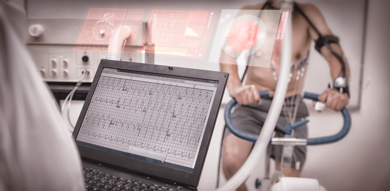
To further assess myocardial perfusion, a cardiac stress test may help determine whether the heart receives sufficient blood flow during physical exertion or pharmacologic stress. For patients unable to exercise due to arthritis or other conditions, medications can be administered to simulate the effects of physical stress on the heart—this is known as a pharmacologic stress test.

Exercise stress tests (also known as nuclear heart scans) remain one of the most commonly used diagnostic tools for evaluating chest pain. It is especially valuable in patients with stable angina, as it can help determine the severity of CAD. This testing can be ordered independently or along with other diagnostics, such as echocardiography or myocardial perfusion imaging. A stress echocardiogram can assess cardiac structure and ventricular function and look for valvular abnormalities. It is especially useful for patients with baseline electrocardiogram (ECG) abnormalities or those with suspected aortic stenosis or hypertrophic cardiomyopathy (National Heart, Lung, and Blood Institute, 2022b).

photo of man taking stress test on bike

Electrocardiogram

Despite its frequent use, a resting ECG has limitations in diagnosing acute angina. Many patients with angina due to cardiac ischemia present with normal ECG findings at rest. Nevertheless, ECGs remain a useful diagnostic component, often revealing prior infarction, conduction abnormalities, arrhythmias, or ST-T wave changes. It is important not to rule out cardiac causes of chest pain based solely on a normal ECG, as some patients with known CAD may exhibit seemingly normal or transiently normalized readings even during episodes of chest discomfort (National Heart, Lung, and Blood Institute, 2022b).

Cardiac Catheterization: Diagnostic and Therapeutic Applications

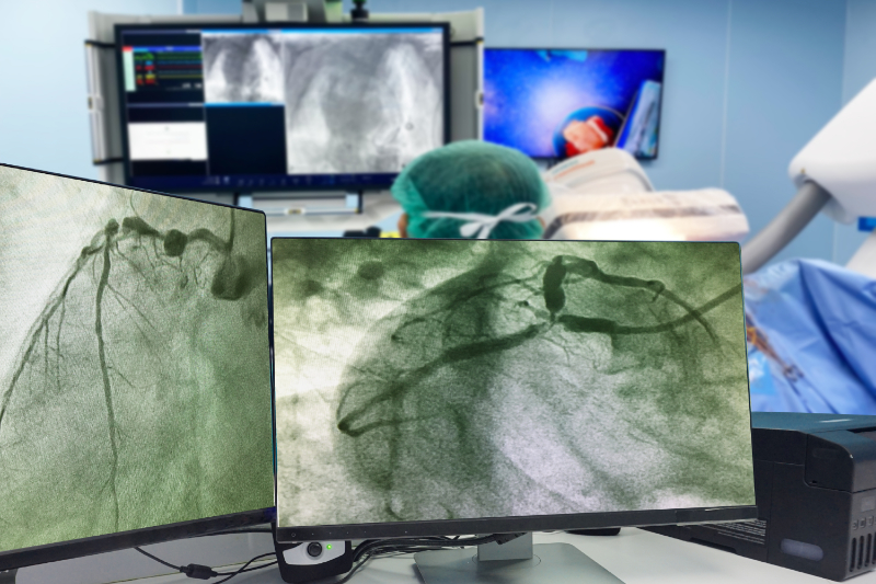
Cardiac catheterization is an invasive procedure that can be utilized as a diagnostic and therapeutic intervention for certain ischemic heart conditions. It plays a critical role in evaluating angina by directly assessing coronary circulation and cardiac function. Typically performed in a specialized catheterization lab under sterile conditions and local anesthesia, the procedure involves the insertion of a flexible catheter into a vein or artery, most commonly in the groin (femoral artery) or wrist (radial artery), and advancing it toward the heart (National Heart, Lung, and Blood Institute, 2022a).

photo of cardiac cath x-ray

Once the catheter reaches the appropriate location, contrast dye is injected into the coronary arteries or cardiac chambers. Real-time X-ray imaging captures the flow of the dye, allowing providers to have a clear visualization of the heart chambers and vessels and assess valvular function. This provides detailed insights into the presence and severity of any abnormalities (National Heart, Lung, and Blood Institute, 2022a).

Cardiac catheterization is indicated for a range of clinical purposes, including:

  • Confirming or ruling out CAD
  • Determining if there is cause for an intervention
  • Detecting valvular heart disease
  • Evaluating left ventricular performance and hemodynamics
  • Identifying congenital heart anomalies
  • Confirming the presence of tumors, cardiomyopathies, or abnormal cardiac anatomy

Cardiac CT Scan

A computed tomography (CT) scan is a noninvasive diagnostic study that allows providers to view advanced images within the body. A cardiac CT scan is specifically utilized to allow for advanced imaging of the heart and coronary vasculature. A CT scan consists of X-ray imaging in combination with computerized methods to create 3D images and models of the cardiac structures. This diagnostic study can often be used to help providers identify diseases or defects within the blood vessels that supply the heart and surrounding structures (National Heart, Lung, and Blood Institute, 2022b).

Coronary Calcium Scan

A coronary calcium scan is a cardiac CT that measures the amount of calcium buildup in the cardiac wall and structures. If there is a significant buildup of calcium, the diagnosis may point toward CAD (National Heart, Lung, and Blood Institute, 2022b).

Cardiac MRI

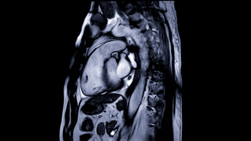
Cardiac magnetic resonance imaging (MRI) is another noninvasive diagnostic study that is often used if the results of previous diagnostics are unclear. This study uses magnets, radio waves, and computers to develop detailed imaging of the heart and its surrounding structures. Cardiac MRIs are especially beneficial in detecting inflammation or scarring of the heart.

photo of cardiac mri

Cardiac Biomarkers

Cardiac biomarkers are chemicals released into the bloodstream when the heart muscles are under stress or damaged. These biomarkers are often utilized to help determine risk for and manage ACS, which is known to be life-threatening. One of the most commonly known cardiac biomarkers is troponin, which is used to diagnose cardiac ischemia. Creatine Kinase (CK) is another biomarker, but this level does not typically begin to elevate until hours after symptoms begin, making it less specific (Patibandla et al., 2023).

Table 4: Canadian Cardiovascular Society Angina Scale
The Canadian Cardiovascular Society grading scale is commonly used for the classification of angina severity:
  • Class I – Angina only during strenuous or prolonged physical activity
  • Class II – Slight limitation, with angina only during vigorous physical activity (walking uphill, climbing stairs rapidly, etc.)
  • Class III – Symptoms with everyday living activities, i.e., moderate limitation (walking 1-2 blocks on a level surface, climbing one flight of stairs under normal conditions)
  • Class IV – Inability to perform any activity without angina or angina at rest, i.e., severe limitation
(Canadian Cardiovascular Society, 2020)

Nursing Interventions

Nursing interventions for angina/chest pain symptoms have been pretty standard for many years, with little deviation. Nurses should always remember to ASSESS THE PATIENT, not just the monitor or labs found in the chart. Monitoring and assessments are key to establishing a baseline, assessing changes in clinical status, and trending findings along the way to help determine a person's trajectory. Patients with symptoms of chest pain should always be on a cardiac monitor with frequent vital signs and regular ECGs (Mechanic et al., 2023).

Providers may order routine labs, such as serial troponins and cardiac enzymes, to assess for any changes in clinical status. Medication and supplemental oxygen may also be ordered for symptom management. For individuals whose oxygen saturations are less than 90%, supplemental oxygen may be indicated. For complaints of acute pain, pharmaceutical interventions may be initiated, such as nitroglycerin or morphine (Mechanic et al., 2023).

In addition to assessment and diagnostics, healthcare providers should continuously monitor for clinical changes and notify providers as necessary.

Table 5: Acute Management of Angina of Unknown Origins
Initial Management:
  • Monitoring of vital signs and oxygen saturation
  • Establishing intravenous access
  • Draw blood specimens: Complete blood count (CBC), chemistry, coagulation studies, cardiac markers (CK-MB, myoglobin, troponin)
  • 12-lead ECG
  • Take a targeted history 
Medical management may include:
  • Administering oxygen
  • Aspirin
  • Nitroglycerin or morphine to relieve chest pain
  • Assessing for thrombolytics 
Diagnostic workup:
  • Cardiac stress testing
  • Cardiac catheterization
(Rao et al., 2025)

Pharmacologic Management

Analgesic Options

  • Nitroglycerin: Sublingual or intravenous
  • Morphine: Intravenous

Antiplatelet Options

  • Aspirin: 325 milligrams (mg) loading dose, followed by 81 mg daily dose
  • Clopidogrel (Plavix®): 300 mg loading dose, followed by 75 mg daily
  • Prasugrel: 60 mg loading dose, followed by a maintenance dose of 5 mg or 10 mg (based on weight) daily
  • Ticagrelor: 180 mg loading dose, followed by a maintenance dose of 90 mg twice daily
Table 6: Medications Used in Treatment of Angina Pectoris
Antianginal and anti-ischemic drugs
  • Aspirin - Inhibits platelet aggregation, maintaining blood flow through narrowed arteries.
  • Clopidogrel (Plavix®) - Selectively inhibits adenosine diphosphate (ADP) binding to platelet receptors, preventing aggregation.
  • Nitroglycerine - Dilates blood vessels, improving blood flow to the heart.
  • Beta-adrenergic blockers - Slow the heart rate and decrease overall oxygen demands.
    • Metoprolol
    • Atenolol
    • Propranolol
  • Angiotensin-converting enzyme (ACE) inhibitors - These drugs allow blood to flow from the heart more easily. For instance, a provider may prescribe ACE inhibitors if a person has had a moderate to severe heart attack, reducing the heart's overall pumping capacity.
    • Ramipril
  • Calcium channel blockers - When treating coronary artery spasms, heart medications such as calcium channel blockers work to relax the coronary arteries and prevent the spasms. They can also aid in Prinzmetal angina (vasospastic angina).
    • Amlodipine
    • Diltiazem
    • Verapamil
  • Ranolazine (Ranexa®) - Selectively inhibits late sodium current, reducing myocardial wall tension and oxygen consumption.
(Rao et al., 2025)

Lifestyle Modifications and Patient Education

  • Smoking cessation

Smoking has been proven to damage blood vessels, making cardiovascular risk factors, such as angina, even more life-threatening (National Heart, Lung, and Blood Institute, 2023b).

  • Diet, exercise, and stress management

Adopting a healthier diet, staying active, and managing stress are all solid interventions to reduce the incidence and threat of anginal symptoms. When a poor diet is at play, risk factors increase for conditions such as CAD, hyperlipidemia, hypertension, diabetes, heart disease, and more. Physical activity is often recognized as a contributor to angina, but a sedentary lifestyle poses an even greater risk (National Heart, Lung, and Blood Institute, 2023b).

  • Medication adherence

All medications serve a specific purpose, so it is important to provide education on what medications are intended for, risks, and side effects, as well as instruct patients not to abruptly stop any medication without speaking to their prescribing provider.

  • Warning signs of worsening angina or myocardial infarction

Angina and chest pain can look and feel different to everyone, but this does not make it any less serious. Any time there is a new or worsening aspect of angina symptoms, patients should be instructed to contact their provider and/or present to the closest emergency department.

Case Studies

Kami – A Veteran Nurse with Recurrent Chest Pain

Patient Profile:

Kami is a 51-year-old nurse and military veteran who completed two tours in the Middle East. During her deployments, she began smoking as a coping mechanism to manage chronic stress and the constant threat of explosive devices. Though she considers herself physically fit and does not have diabetes, she has continued smoking since returning from duty.

Presenting Complaint:

For the past two weeks, Kami has experienced nighttime episodes of chest discomfort described as a crushing sensation radiating to her left arm. The most recent episode lasted approximately 30 minutes and extended to her shoulder and jaw. In response, she smoked a cigarette to alleviate her anxiety. She reports that the chest pain never occurred during menstruation, though she has not yet undergone menopause. Kami has no known allergies, is not pregnant, and her medical history is otherwise unremarkable.

Family History:

  • Father: Deceased at age 71 due to myocardial infarction; had diabetes.
  • Mother: Survived a stroke at age 70; currently diabetic but has no known history of CAD.

Initial Evaluation:

Upon presentation to the emergency department, Kami's vital signs were within normal limits:

  • Oxygen saturation: 98%
  • Heart rate: 82 beats per minute (bpm)
  • Blood pressure: 128/70 millimeters of mercury (mmHg)

A comprehensive workup was initiated, including:

  • ECG: Normal
  • Chest X-ray: Normal
  • Cardiac enzymes and lab work: Within normal limits
  • Additional testing scheduled: Glucose tolerance test and cardiac stress test

Initial Clinical Impression and Management:

Despite unremarkable initial findings, Kami's symptoms and clinical history prompted her physician to suspect Prinzmetal angina (variant angina), a condition involving transient coronary artery vasospasms often occurring at rest and frequently during the night or early morning.

Initial Treatment Plan:

Sublingual Nitroglycerin: For immediate relief during acute vasospastic episodes.

Long-acting nitrate and calcium channel blocker: To improve coronary blood flow and prevent recurrence. Medication adjustments may be necessary based on therapeutic response.

Lifestyle Modification: Kami was strongly advised to quit smoking. A smoking cessation class was recommended, though pharmacologic support for cessation was deferred at this time.

Contingency Plan: If Kami fails to respond to first-line therapy, her physician may introduce alpha-adrenergic blockers to lower blood pressure and counteract the vasoconstrictive effects of stress hormones. Should pharmacologic measures prove inadequate or if coronary obstruction develops, coronary angioplasty or other interventional procedures may be considered.

Prognosis and Follow-Up:

Kami was counseled that Prinzmetal angina is a chronic yet manageable condition. Symptoms often follow a cyclical pattern, and many patients see improvement with medical therapy over time. If well-controlled, medication tapering may be considered after 6–12 months. Regular follow-up and adherence to lifestyle modifications, especially smoking cessation, will be critical in preventing progression and maintaining cardiac health.

John – A Car Salesman

Patient Profile:

John is a 54-year-old car salesman who works 50+ hours per week and has little time for exercise or partaking in a healthy diet. John works hard to provide for his family but spends very little time taking care of himself, as evidenced by eating fast food multiple times weekly, poor exercise habits, and long, stressful work hours.

Presenting Complaint:

John presents to his local emergency department complaining of a heavy, crushing chest pain that began while he was mowing his lawn. He described the pain as a tight, squeezing sensation in the center of his chest, radiating to his left shoulder, arm, and jaw. He rates the pain as 9/10. The pain improved slightly when he stopped activity and rested, but it did not completely go away.

Past Medical History:

  • Hypertension
  • Hyperlipidemia
  • Smoker
  • Positive family history of early CAD (father had a heart attack at age 52)

Medication History:

  • Lisinopril
  • Simvastatin
  • As needed (PRN) ibuprofen for joint pain

Initial Evaluation:

Upon presentation to the emergency department, John's vital signs were as follows:

  • Oxygen saturation: 96% on room air
  • Heart rate: 98 bpm
  • Blood pressure: 164/94 mmHg

Physical Exam:

  • Alert, anxious, diaphoretic
  • No murmurs, rubs, or gallops
  • Lungs clear to auscultation

Diagnostics:

  • ECG: ST-segment depression
  • Troponin: mildly elevated

Plan:

  • Administer aspirin 325 mg
  • Sublingual nitroglycerin was given with partial relief of symptoms
  • Started on a heparin drip and metoprolol
  • Admitted for cardiac catheterization and observation

Maria- 58-year-old Female with Type 2 Diabetes

Patient Profile:

Maria is a 58-year-old female who works as a medical assistant in a busy primary care clinic. Maria has had intermittent chest discomfort for a few days, thought to be indigestion, along with back pain and nausea.

Presenting Complaint:

Maria presents to an urgent care clinic with complaints of profound fatigue, nausea, and mild upper back discomfort for the past 24 hours. She denies significant chest pain but describes a "pressure" across her upper back between her shoulder blades. She reports feeling "off" and short of breath when climbing stairs. Symptoms worsened this morning after walking her dog.

Past Medical History:

  • Type 2 Diabetes Mellitus
  • Obesity (body mass index [BMI] 34)
  • Depression
  • Gastroesophageal reflux disease (GERD)

Medication History:

  • Metformin 1000 mg twice daily
  • Sertraline 50 mg daily
  • Omeprazole 20 mg daily

Initial Evaluation:

Upon presentation to the emergency department, Maria's vital signs were as follows:

  • BP: 142/88 mmHg
  • HR: 104 bpm
  • RR: 22/min
  • O2 Sat: 95% on room air

Physical Exam:

  • Diaphoretic and tired-appearing
  • Tachycardia, no murmurs
  • Lungs are clear to auscultation
  • Mild tenderness in upper thoracic spine on palpation

Diagnostics:

  • ECG: Some ST-T wave changes that are nonspecific
  • Troponin: Elevated

Plan:

  • Administered aspirin 325 mg
  • Initiated nitroglycerin and oxygen therapy
  • Admitted for observation and further cardiac workup
  • ECG revealed hypokinesis in the inferior wall, suggesting ischemia

Conclusion

Angina is most commonly associated with cardiovascular causes, though that is not always the case. As healthcare providers, it is important to have a generalized knowledge base of what angina is, potential causes, and how to identify and treat it properly. Some cases of angina can often present as a sign of something much worse, with a need to intervene rapidly. Anytime angina is suspected, it must be thoroughly evaluated to uncover its root cause. Health professionals need to stay alert not only to the classic signs of angina but also to less obvious, related symptoms to quickly recognize heart-related ischemia and act fast to restore proper oxygen supply to affected tissues. Through better understanding and thorough observation, healthcare providers can more effectively manage this challenging condition (symptom) that continues to impact so many lives.

Select one of the following methods to complete this course.

Take TestPass an exam testing your knowledge of the course material.
OR
No TestAttest that you have read and learned all the course materials.

Implicit Bias Statement

CEUFast, Inc. is committed to furthering diversity, equity, and inclusion (DEI). While reflecting on this course content, CEUFast, Inc. would like you to consider your individual perspective and question your own biases. Remember, implicit bias is a form of bias that impacts our practice as healthcare professionals. Implicit bias occurs when we have automatic prejudices, judgments, and/or a general attitude towards a person or a group of people based on associated stereotypes we have formed over time. These automatic thoughts occur without our conscious knowledge and without our intentional desire to discriminate. The concern with implicit bias is that this can impact our actions and decisions with our workplace leadership, colleagues, and even our patients. While it is our universal goal to treat everyone equally, our implicit biases can influence our interactions, assessments, communication, prioritization, and decision-making concerning patients, which can ultimately adversely impact health outcomes. It is important to keep this in mind in order to intentionally work to self-identify our own risk areas where our implicit biases might influence our behaviors. Together, we can cease perpetuating stereotypes and remind each other to remain mindful to help avoid reacting according to biases that are contrary to our conscious beliefs and values.

References

  • AMBOSS. (2025). Chest pain. AMBOSS. Visit Source.
  • Alaeddini, J. (2018). Angina pectoris in emergency medicine. Visit Source. Medscape.
  • American Heart Association. (2024a). 2024 Heart disease and stroke statistics update fact sheet.  American Heart Association. Visit Source.
  • American Heart Association. (2024b). Ischemic heart disease and silent ischemia. American Heart Association. Visit Source.
  • Canadian Cardiovascular Society. (2020). Canadian Cardiovascular Society grading of angina pectoris. Canadian Cardiovascular Society. Visit Source.
  • Klabunde, R. (2023). Cardiovascular physiology concepts. Cvphysiology. Visit Source.
  • Li, T., Jawish, M. A., Badurdeen, D., & Koop, A. H. (2024). Diagnosis and management of noncardiac chest pain. Gastroenterology & hepatology, 20(9), 533–541. Visit Source.
  • Lukitasari, M., Apriliyawan, S., Manistamara, H., Sella, Y. O., Rohman, M. S., & Jonnagaddala, J. (2023). Focused chest pain assessment for early detection of acute coronary syndrome: Development of a cardiovascular digital health intervention. Global Heart, 17(1), 18. Visit Source.
  • Mayo Clinic Staff. (2024). Diseases and conditions: Angina. Mayo Clinic. Visit Source.
  • Mechanic, O. J., Gavin, M., & Grossman, S. A. (2023). Acute myocardial infarction. In StatPearls. StatPearls Publishing. Visit Source.
  • National Heart, Lung, and Blood Institute. (2023a). What is angina? National Heart, Lung, and Blood Institute. Visit Source.
  • National Heart, Lung, and Blood Institute. (2023b). Living with angina. National Heart, Lung, and Blood Institute. Visit Source.
  • National Heart, Lung, and Blood Institute. (2022a). What is a cardiac catheterization? National Heart, Lung, and Blood Institute. Visit Source.
  • National Heart, Lung, and Blood Institute. (2022b). Heart tests. National Heart, Lung, and Blood Institute. Visit Source.
  • Patibandla, S., Gupta, K., & Alsayouri, K. (2023). Cardiac biomarkers. In StatPearls. StatPearls Publishing. Visit Source.
  • Rao, S. V., O'Donoghue, M. L., Ruel, M., Rab, T., Tamis-Holland, J. E., Alexander, J. H., Baber, U., Baker, H., Cohen, M. G., Cruz-Ruiz, M., Davis, L. L., de Lemos, J. A., DeWald, T. A., Elgendy, I. Y., Feldman, D. N., Goyal, A., Isiadinso, I., Menon, V., Morrow, D. A., Mukherjee, D., … Williams, M. S. (2025). 2025 ACC/AHA/ACEP/NAEMSP/SCAI guideline for the management of patients with acute coronary syndromes: A report of the American College of Cardiology/American Heart Association Joint Committee on Clinical Practice Guidelines. Circulation, 151(13), e771–e862. Visit Source.
  • Sweis, R. N. & Jivan, A. (2024). Microvascular angina. Merck Manual Professional Version. Visit Source. Visit Source.
  • Teragawa, H., Oshita, C., & Orita, Y. (2020). Is noncardiac chest pain truly noncardiac?. Clinical Medicine Insights. Cardiology, 14, 1179546820918903. Visit Source.
  • Virani, S. S., Alonso, A., Aparicio, H. J., Benjamin, E. J., Bittencourt, M. S., Callaway, C. W., Carson, A. P., Chamberlain, A. M., Cheng, S., Delling, F. N., Elkind, M. S. V., Evenson, K. R., Ferguson, J. F., Gupta, D. K., Khan, S. S., Kissela, B. M., Knutson, K. L., Lee, C. D., Lewis, T. T., Liu, J., … American Heart Association Council on Epidemiology and Prevention Statistics Committee and Stroke Statistics Subcommittee (2021). Heart Disease and Stroke Statistics- 2021 Update: A report from the American Heart Association. Circulation, 143(8), e254–e743. Visit Source.