Sign Up
You are not currently logged in. Please log in to CEUfast to enable the course progress and auto resume features.

Course Library

HIV/AIDS: Current Evidence-Based Practice (Two Hour)

2 Contact Hours
Listen to Audio
CEUfast OwlGet one year unlimited nursing CEUs $39Sign up now
This peer reviewed course is applicable for the following professions:
Advanced Practice Registered Nurse (APRN), Certified Nurse Practitioner, Certified Registered Nurse Practitioner, Clinical Nurse Specialist (CNS), Licensed Practical Nurse (LPN), Licensed Vocational Nurses (LVN), Nursing Student, Physical Therapist (PT), Physical Therapist Assistant (PTA), Registered Nurse (RN), Registered Nurse Practitioner, Respiratory Care Practitioner, Respiratory Therapist (RT)
This course will be updated or discontinued on or before Monday, November 17, 2025

Nationally Accredited

CEUFast, Inc. is accredited as a provider of nursing continuing professional development by the American Nurses Credentialing Center's Commission on Accreditation. ANCC Provider number #P0274.


Outcomes

≥ 92% of participants will know the pathology, management, and current clinical guidelines of HIV/AIDS.

Objectives

By the end of this activity, the participant will be able to:

  1. Outline the human immunodeficiency virus (HIV) genome and structural biology.
  2. Characterize the HIV life cycle.
  3. Identify the different symptomatic presentations of people living with HIV and acquired immunodeficiency syndrome (AIDS).
  4. Generalize the different accepted guidelines on exposure prophylaxis for HIV.
  5. Create management plans for people living with HIV/AIDS.
CEUFast Inc. and the course planners for this educational activity do not have any relevant financial relationship(s) to disclose with ineligible companies whose primary business is producing, marketing, selling, re-selling, or distributing healthcare products used by or on patients.

Last Updated:
  • $39 Unlimited Access for 1 Year
    (Includes all state required Nursing CEs)
  • No Tests Required
    (Accepted by most states & professions)
  • Instant Reporting to CE Broker
  • Instant Access to certificates of completion
Logo Audio
Now includes
Audio Courses!
Learn More
Restart
Restart
  • 0% complete
Hide Outline
Playback Speed

Narrator Preference

(Automatically scroll to related sections.)
Done
HIV/AIDS: Current Evidence-Based Practice (Two Hour)
0:00
0:15
 
To earn a certificate of completion you have one of two options:
  1. Take test and pass with a score of at least 80%
  2. Attest that you have read and learned all the course materials.
    (NOTE: Some approval agencies and organizations require you to take a test and "No Test" is NOT an option.)
Author:    Jassin Jouria (MD)

Virus Genome and Structural Biology

Research compilations and epidemiological surveys tracing the origin of human immunodeficiency virus (HIV) have prominently recognized the last century as the biological point frame of the first report. According to multiple reports, the virus arose from several independent zoonotic transmissions of the simian immunodeficiency virus. Currently, the HIV epidemic is globally discussed concerning the spread and healthcare burden of the virulent HIV-1 and the less widespread HIV-2. The HIV viruses belong to the Retroviridae family, subfamily Orthoretrovirinae (Justiz Vaillant & Gulick, 2022). In epidemiological concepts, this family has a prominent relationship with the mammalian genome, responsible for a host of ancient diseases in this animal class and currently making up about 8% of the mammalian deoxyribonucleic acid (DNA) today. By encoding only 16 proteins in the mammalian genomes, the HIV viruses can subvert the mammalian innate and adaptive immune responses, making the biological systems vulnerable to opportunistic infections and end-organ damage. It invades the cell's surface receptors, reprograms the cellular functions, and matures into infectious virions. During these steps, the evolutionary mechanism deployed by the virus makes it escape the mammalian immune defenses and ultimately subvert the body's ability to suppress viral replication (Waymack & Sundareshan, 2022).

HIV is categorized into two types – type 1 and type 2. HIV type 1 (HIV-1) is further divided into four subgroups – groups M, N, O, and P. According to phylogenetic comparisons, some retroviruses in the chimpanzee population can be grouped into the N and O of the human retroviruses; this explains why early structural classifications suggest that HIV was probably transmitted to humans from chimpanzees or gorillas. On further classification, the HIV-1 group M is subcategorized into the subtypes A, B, C, D, F, G, H, J, and K. Based on evolutionary studies, subgroups A and D seem to be the oldest, with subtypes B and D showing common architecture suggestive of a close relation. HIV type 2 (HIV-2) is categorized into groups A-H, with group A further subdivided into A1 and A2. Morphologically, both HIV-1 and HIV-2 are indistinguishable. However, distinct antigenicity differences exist between both types, prompting the need to develop HIV-2-specific diagnostic methods. In 2012, the first such diagnostic tool – the nucleic acid test (NAT) was developed to quantify HIV-2 nucleic acids directly.

The HIV genome comprises two identical single-stranded ribonucleic acid (RNA) molecules enclosed within the core of the virus particle. By the reverse transcription of the viral RNA genome, the provirus HIV particles are generated. The process of provirus DNA generation also includes the degeneration of the single-stranded RNA molecules and the subsequent integration into the human DNA makeup. The arrangement of the DNA genomes impacts different stages, from viral invasions of cells to the maturation and replication of infectious virions; for instance, the 5' long terminal repeat region codes for the transcription of viral genes while the 'Gag gene' encodes the proteins of the outer core membrane. Although the overall genome of HIV appears simple, the complexities involved in cell entry and reprogramming are championed by a host of high-specific proteins in its envelope and core.

graphics showing hiv virion structure

HIV Virion Structure

The mature HIV virion has these proteins functioning consistently to aid viral cells' sustenance and survival. At the mature stage, the virions attain a round shape and roughly measure about 100 nanometers (nm) in diameter. The proteins and other structural components of the mature virion are completely enclosed in an outer lipid membrane envelope. The outer envelope comprises 72 knobs functioning as trimers of the proteins resident in the envelope. In mature virions, the envelope composition is doubled-layered and prominently features the envelope proteins surface glycoprotein (SU) and transmembrane protein (TM). Many oligopeptides have also been identified in the mature virus particles. These peptides are generated by the proteolytic processing of the precursor proteins during viral maturation and replication (Van Heuvel et al., 2022).

Beyond merely ensuring virus cell survival, the regulator proteins play significant roles in the different stages of viral replication, propagation, and virulence. Tat, a transactivator protein, makes viral RNA molecules available for virus production by speeding up the processes involved in viral RNA production. It binds to the trans-activation response (TAR) sequence of viral RNA and transactivates any additional HIV genomes in the cell. Rev, an RNA splicing regulator protein, splices the newly formed virus RNAs. This process accelerates the production of regulatory proteins during viral replication and also aids the translation of structural proteins in the later stages of viral replication. Nef, a negative regulating factor protein, negatively affects the presentation of the CD4 cell population in the host cell; this effect depopulates and inactivates the CD4 cells in a process that culminates in enhanced pathogenicity and impairment of the host systems' immunity response (Pai et al., 2021). Increases in pathogenicity as triggered by Nef protein are prominent in the life cycle of HIV-1 compared to HIV-2. Other regulatory proteins directly and indirectly involved in virus-cell production, maturation, and propagation include the Vif, Vpu, and Vpr proteins.

Case Study

Rolia resides in a quiet suburb of Uganda, a third-world country battling a surge of HIV cases in recent years. With a rapidly increasing population, the primary healthcare system in this country is stretched too thin as self-care campaigns could only capture residents in the large metropolis. Rolia and other middle-aged women in the suburbs could only make do with local medicines or visit the metropolis in emergency cases. At 42, Rolia was diagnosed with chronic HIV after a brief illness characterized by intermittent fever, unexplained weight loss, upper abdominal pain, and non-resolving watery diarrhea. Initial examination identified Crysporidium parvum in her stool, and subsequent cultures identified other microbes implicated in HIV-related opportunistic infections, including Pneumocystis carinii and cytomegalovirus (CMV) retinitis.

She lost 21 kilograms (kg) in the past seven months and had also developed episodic pains in the joints of the lower limbs. On examination, Rolia's temperature was 39.5 C, her pulse was 97/min, and her blood pressure was 154/87 millimeters of mercury (mmHg). The reported laboratory examination is as follows:

  • Hemoglobin (Hgb): 14.0 g/dl
  • White blood cell count (WBC): 3,100
  • Neutrophils: 78
  • Lymphocytes: 14
  • Monocytes: 20
  • Platelets: 230
  • Sodium: 142
  • Potassium: 3.5 meq/L
  • Chloride: 157 meq/L
  • CD4 lymphocyte count 85/uL
  • CO2: 21 meq/L
  • Creatinine: 2.7 mg/dl

Endoscopic examinations revealed a normal ilium with minimal gastrointestinal inflammation. A colonoscopy revealed clusters of Cryptosporidium in the colon and the upper ampulla. Gram staining of blood culture showed no organisms; however, acid-fast bacilli (AFB) blood cultures showed Mycobacterium avium complex. Rolia was immediately started on ethambutol and clarithromycin with a nursing plan to check her vitals every hour. About three days later, Rolia's condition improved as she admitted to having impressive symptomatic relief from the joint pains and the feverish condition. A week after conducting genotyping, she was started on tenofovir, lamivudine, and lopinavir/ritonavir. Rolia was then referred to an HIV adherence monitoring center in her hometown for therapy monitoring. A quarterly visit to the HIV therapy center in the metropolis was also scheduled to better evaluate Rolia's response to therapy and any drug side effects.

Global Epidemiology Statistics

The global distribution of HIV cases, regardless of region and cultural affiliation, seems to follow a common pattern. New cases are mostly recorded in a population group with similar risk factors; this population mostly includes men who have sex with men, intravenous drug users, prisoners and those confined to a close setting, the transgender community, sex workers, and immunocompromised individuals living in regions with inadequate primary healthcare systems (Damtie et al., 2022). The widespread practices of unsafe sex and unhealthy reuse of needles by drug abusers have been classified as behaviors that increase the likelihood of contracting this disease. In regions with poor prenatal healthcare systems, mother-to-child transmission also contributes largely to new cases of HIV. HIV-positive mothers with no access to antiretroviral therapy, especially in sub-Saharan Africa, contribute largely to the steadily increasing new cases of HIV in the infant population. Globally, the volume of new cases contributed by men who have sex with men has remained disturbingly high over the last decade. Receptive anal sexual intercourse and exposure to infected needles in a subpopulation of this group that abuses drugs have relatively increased the rate of transmission.

On a global scale, the healthcare burden of HIV was at 36.9 million cases in 2019, a count corresponding to an estimated 0.5% of the world's population (Govender et al., 2021). Cluster analysis showed that the global burden as of 2019 translates to about 476 cases per every 100,000 group in the population. Epidemiological estimates for new cases and cases under treatment peaked in 2005 and steadily declined for the next five years; this trend has been attributed to the wide circulation of antiretroviral (ART) therapy regimens and their efficacy in the human population. Since 2010, however, the incidence rate has reportedly been increasing steadily. As of 2021, the highest prevalence rates were recorded in South Africa, Portugal, the United States, Mexico, Brazil, Peru, Spain, and Germany. Portugal reported a very high prevalence rate, with an estimated increase from 86 per 100,000 people to 370 per 100,000. However, between 1999 and 2019, South Africa reported a significantly increased prevalence rate from 354 per 100,000 people to 14,251 per 100,000. In sub-Saharan Africa, regional epidemiological studies showed that new cases are particularly concentrated in Lesotho, Mozambique, South Africa, Zimbabwe, and Namibia.

map of Africa showing prevalence of hiv and aids

Prevalence of HIV-AIDS in Africa

Trends in the age distribution of new cases show an irregular distribution, with the most peaks appearing in infancy and young adulthood. Infancy peaks correspond to increased perinatal transmission rates due to inadequate primary healthcare systems. In contrast, peaks at the adolescent stage correspond to an increase in adolescent risky behaviors, including needle sharing and drug abuse. Population clusters in the 20 – 39-year-old groups show the highest incidence rates. The Joint United Nations Programme on HIV/AIDS (UNAIDS) data on prevalence trends also attribute 500 in every 5,000 new cases to children. Young women residents in sub-Saharan Africa also appear to show a high level of susceptibility to the virus. Country-level epidemiological studies have identified the worst cases in Africa. Data from these studies identify South Africa as the worst affected country in the world today. In 2017 alone, about 7.9 million HIV cases across all age groups were reported, with the prevalence rate among men at 14.8% and 26.3% among the female population (Simbayi et al., 2019).

Referencing statistics published by the UNAIDS for 2022, the global burden of HIV was at 38.4 million in 2021, with 1.5 million new cases in the same year. The key at-risk population (gay men, sex workers, those who are transgender, and drug addicts) reportedly accounted for about 70% of all cases documented globally as of 2021. Regional distribution studies place 94% of new HIV infections outside of sub-Saharan Africa and 51% in sub-Saharan Africa. Risk estimates showed that the risk of new cases is 35 times higher in people who abuse injectable drugs, 30 times higher in female sex workers, 28 times higher among men who have sex with men, and 14 times higher in transgender women (Simbayi et al., 2019). Age-specific risk studies suggest a weekly count of about 4,900 young women aged 15-24 reporting new cases of infection. In sub-Saharan Africa, young women and girls aged between 15-24 years appear to be twice as likely to have an active HIV infection compared to men of the same age range.

Pathophysiology, Life Cycle, and Clinical Staging

Understanding the pathophysiology of HIV involves carefully examining the CD4 cells and their interaction with viral envelope proteins. The CD4+ helper T cells are key regulators of humans' innate and humoral immune response systems. These cells are prominently expressed on the surface of T-lymphocytes, monocytes, macrophages, and dendritic cells. HIV virions recognize and specifically target these cells, depopulating them and effectively impairing their defense functions. The destruction of CD4 cells significantly increases the risk of opportunistic infections, resulting in a syndrome state of low immunity and infection vulnerability. Using a series of complex processes, HIV invades healthy calls, hijacks their functionalities, and reprograms its transcriptions and translational abilities to initiate viral replication (Biyazin et al., 2022). Beyond merely reprogramming the cells, HIV also counteracts the innate antiretroviral tendencies of the healthy cells in a calculated attempt to eliminate the cell's defensive structure and ensure self-sustenance. In addition to CD4+ helper T cells, HIV directly impacts the functionalities of monocytes, macrophages, immature dendritic cells, and resting T cell subsets. Although these cells play little to no role in viral replications, they are important in the body's immune response.

HIV Life Cycle

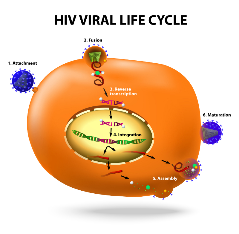
The HIV life cycle is complex, involving multiple series of carefully coordinated attacks on the host cell and an eventual hijacking of cell function. In the early life cycle phase, HIV virions attach to the cell surface and attempt cell entry. If successful, this phase usually ends with integrating proviral DNA into the host cell genome. In the late phase, viral replications start with the transcription of proviral particles and end with the release of virulent virions produced with the infrastructure of the host cell. Lasting between two to three days, viral invasion of healthy cells culminates in the death of infected host cells and other bystander cells. A widely studied phenomenon in this regard is the high virus cell variation rate. With a variation rate estimated at one mutation at every few complete replication cycles, HIV produces an extensive number of variants. However, the survival rates for each variant reportedly depend on the selective combined forces of the body's immune system and ART therapy. The combined forces of massive virion production (estimated as 2 x 10⁹ virions per day) and the short generation time allow the virus to rapidly adapt to the host cell environment and survive against multiple odds. To properly capture the life cycle of HIV, this course will discuss the interaction between the host cell and the virus when discussing viral attachments, binding and fusion, viral entry, reverse transcriptions, uncoating and nuclear entry, integration, transcription, translation and assembly, maturation, and release.

graphic showing hiv life cycle stages

Overview of the HIV Life Cycle Stages

Viral Attachment

In infected humans, HIV virions that are cell-free (yet to infect a cell) die out in about one hour. Cell-free virions must infect a healthy cell within a short time frame to survive. The process usually starts with recognizing and attaching to healthy cells with a CD4 receptor. The co-receptors C-C chemokine receptor type 5 (CCR5) and C-X-C motif chemokine receptor 4 (CXCR4) also play important roles in this early stage of infection. CCR5 co-receptors are primarily expressed on the surfaces of monocytes, macrophages, memory, and CD4+ T cells. CXCR4 co-receptors, on the other hand, are primarily expressed on both memory CD4 cells and native cells. Studies have shown that the densities of the envelope glycoprotein (Env) trimers on the virions and the CD4 receptors on the host cells are usually low, causing significant inefficiencies in the process of viral attachment to the host cell (Negi et al., 2022).

As a rate-limiting step in viral infection, the pharmacological effects of agents that can lower the densities of the Env trimers or temporarily block the CD4 receptor sites have been extensively studied in infected individuals. To aid viral attachment, several receptors, including lectins and polyglycans, can also bind virions in an unspecific manner, ultimately increasing the viral infection rate. These additional receptors also appear as concentrated virions in the cells, aiding improved interaction between virions and cells with active CD4 receptors on their surfaces. Variant features may also determine the rate of viral attachment. For instance, individual virus strains with an envelope glycoprotein or gp120 variation express increased affinity for the co-receptors CCR5 and CXCR4.

Virion Binding and Fusion

Virion binding and fusion specifically initiate the infection cycle with the interaction of cell surface CD4 receptors with the external gp120 on the virion. The binding of the virion's external glycoprotein on the CD4 helper cells initiates a conformational cellular configuration in the Env trimer. The change in conformation further allows the secondary interaction of gp120 with either cofactors - CCR5 and CXCR4. Configurational changes of this nature do not end with the gp120-cofactor binding. Instead, cofactor binding further promotes a new type of binding that involves the gp41 transmembrane protein resident on the gp120 on the virion and the host cell membrane. Micro-imaging observations studying this interaction suggest that the gp41 inserts its hydrophobic fusion peptide apparatus into the host cell membrane in a final interaction that establishes transmembrane contact between the invading virion and the host cell environment (Bruxelle et al., 2021). Following this interaction, the gp41 complex forms a helical bundle that bridges the distance between the virion membrane and the cellular content – effectively pulling both environments together. The resulting complex is a fusion reaction as the virion's contents are released into the invaded cells. CCR5 receptor blockers and fusion inhibitors indicated for the management of HIV exert their pharmacological actions at this stage of infection.

Reverse Transcriptase-catalyzed Reverse Transcription

After membrane fusion, reverse transcriptase interactions follow. The genetic components of the HIV genome, including its double-stranded RNA and nucleocapsid envelope, are fused with the host cell environment. Double-stranded RNAs are transcribed stepwise into double-stranded DNAs using the host cell machinery; this process is described as 'reverse transcriptase.' An enzyme-reverse transcriptase, commonly found in retroviruses, is essential for a successful reverse transcription of the viral genome. The enzyme catalyzes the reaction that reverses the order of a normal transcription process by generating nuclear DNA components from messenger RNA components, followed by export into the cytoplasm and the generations of proteins. Inhibiting the actions of reverse transcriptase is expected to truncate virion survival and halt the life cycle of HIV, especially in cases of early infection (Singh & Das, 2022). The two classes of ART therapy medications, nucleoside and nucleotide analog reverse transcriptase inhibitors and non-nucleoside reverse transcriptase inhibitors, are expected to inactivate this enzyme by clogging its active binding site.

Virion Uncoating and Nuclear Entry

The viral capsid is disrupted during this stage as the virus is uncoated, and the viral genome is released into the cell nucleus. Uncoating is timed and executed at a point of maximum transmembrane fusion to ensure virion survival. Researchers studying this stage have recognized point mutation in the capsid proteins causing premature uncoating and complex interaction with the tripartite motif 5-alpha protein (TRIM5a). These interactions, although uncommon, reportedly impair viral infectivity and halt the virion's continued parasitic survival on the cell machinery (Müller et al., 2021). Depending on the viral variations and virion level of infectivity, capsids may remain intact for several hours until conditions are favorable for uncoating; this is particularly important for the survival of HIV-1. During this stage, the reverse transcription complexes (RTCs) are transitioned to the pre-integration complex (PIC) – a cell component required during the later integration stage.

Integration

Following the successful generation of linear DNAs from the reverse transcription of viral double-stranded RNAs and its release into the cell nucleus, the viral genome is inserted into the cell genome; this is essential for cellular invasion, the expression of proteins, and the initiation of active viral infection. Insertion of viral DNA into the host cell's DNA is catalyzed by the enzyme integrase. The proviral DNA is completely integrated as part of the host cell chromosome, influencing cellular activity and, most importantly, the synthesis of proteins. Active viral infection is achieved by the continued infection of new healthy cells or the generation of new cells containing proviral DNA (Mbhele et al., 2021).

Interrupting the integration or inhibiting the actions of integrase have been proposed as valid pharmacological methods of halting proviral cell survival. Drug compounds inhibiting integrase, integrase inhibitors – are currently used. The immediate effect of viral integrations may be delayed over time. For instance, in some T cells, the proviral genome may remain silent for many years, evading viral eradication and surviving within the host cell structure; this constitutes a major problem for viral eradication as these cells remain undetected. If ART is abruptly discontinued, silent proviral DNA in long-living T cells may become active and initiate active infection. The type and state of activation of the host cell and subsequent exogenous stimulation are factors that reportedly determine if a cell is productively infected or silently infected after viral fusion.

Transcription

In actively infected host cells, the cellular polymerase II (Pol II) initiates the transcription of genomic RNA and viral messengers using the integrated provirus as a biological template. Although it takes a short time, proviral transcription includes a series of complex stages involving the viral promoter and multiple cellular transcription factors (Dutilleul et al., 2020). These factors, including nuclear factor kappa B (NF-kB), nuclear factor of activated T cells (NFAT), and the viral transactivator protein Tat, are essential in viral gene expression (Hokello et al., 2021). The influence of these factors on viral infectivity and transcription is still largely unknown. However, recent research studies suggest that Tat binds to a specific sequence of the TAR element. Model studies have reported increased transcription processivity directly linked to this binding and efficient synthesis of long-length HIV transcripts. Not only does Tat increase the success rate of viral transcription, but it also reportedly exerts huge roles in producing an estimated 25 different mRNAs by splicing. These mRNAs are classified into three size classes and include the following:

  1. Unspliced RNA of size 9 kb serves as a template for producing Gag and Gag-Pol precursors or as a simple genomic RNA.
  2. Single-spliced RNA of size 4 kb responsible for encoding Env, Vif, Vpu, and Vpr.
  3. Fully-spliced RNA of size 2 kb responsible for Nef, Tat, and Rev expression.

Translation and Assembly

Rev, Nef, and Tat are essential in the translation and assembly stages. Tat significantly increases viral infectivity by boosting viral transcription and inducing RNA elongation. On the other hand, Rev moves the partially spliced and unspliced viral RNA to the cytoplasm. Nef down-modulates the surface receptors, making the cells more effective in producing infectious viral particles and, most importantly, making infected cells evade the host immune system. The Gag and Gag-Pol precursors initially produced from unspliced RNA are concentrated in lipid rafts of the plasma membrane and are finally processed into structural and enzymatic proteins. Following the interaction of the Env glycoproteins with the structural proteins, two different copies of genomic viral RNAs are recruited. The complex interaction between the different proteins at this stage of HIV infection ultimately results in the formation and assembly of membrane-coated spherical viral particles (Lerner et al., 2022).

Budding and Maturation of Viral Particles

During the budding phase, progeny virions initially produced and assembled are released from the infected cells into the host's biological environment. The cellular protein tumor susceptibility gene (TSG101) and the Gag proteins are implicated in this stage. Cellular interactions between these proteins and the infected cell membrane allow newly generated HIV virions to pinch off the actively infected cells and enter the host body circulation. At the late stage of budding, the restriction factor tetherin (bone marrow stromal antigen 2 or BST-2) attaches the assembled viral particles to the cell surface. Viral particles tethered to the cell's surface are released in an immature form characterized by a thick-layered coating of Gag and Gag-Pol precursors (Roos, 2022). Shortly after viral budding is completed, a viral protease cleaves the precursors as the released virions attain maturation. Subsequent reconfiguration of the viral proteins renders the particles highly infectious as the matured virions infect new healthy cells in an infectious cycle that starts with viral fusion and ends with the release and maturation of infectious virions. Drug compounds that inactivate the viral protease during viral maturation have reportedly reduced the infectivity of released virions.

Symptomatic Observations and Other Objective Findings

HIV infections advance in stages, getting worse with time, especially in cases of poor management. Without adequate viral suppressant therapy or symptomatic management, HIV attains a stage of massive replication characterized by active protein synthesis and the symptoms. At first, the immune response produces a huge volume of antibodies in the initial stage of 'seroconversion.' The most important stages of symptomatic expression as clinical presentation of HIV are often categorized into three different stages – acute HIV infection, chronic HIV infection, and acquired immunodeficiency syndrome (AIDS).

Acute HIV Infection

Acute HIV infection is usually characterized by the presence of viremia and the lack of antibodies against the invading virus. In less than six months after infection, anti-HIV antibodies may become detectable, and the presence of viremia may be confirmed by a p24 antigen test or by an HIV RNA test. The most reported clinical presentation of the acute HIV infection stage includes high fever, skin rashes, myalgia, pharyngitis, lymphadenopathy, and arthralgia. Depending on the level of viral infectivity, the infected individual may also report sore throat, extreme fatigue, chills, and malaise. Referencing epidemiological studies, an estimated 30% of the population with an acute HIV infection are usually asymptomatic but highly infectious (Cowan et al., 2021). Symptom presentation may occur later than six months and may be without a pattern. In the lymphatics system, lymph nodes in the groin, head, and neck may become swollen as the body's immune response rapidly surges. Night sweats, anorexia, gradual unexplained weight loss, and fatigue are also commonly documented in symptomatic individuals.

Diarrhea, abdominal disturbances, emesis, and nausea constitute the most reported gastrointestinal symptoms at the acute infection stage. Myalgia, joint tenderness, and asymmetric joint swelling are the most reported impairments of the musculoskeletal system. At this stage of infection, the clinical presentation of symptoms may also closely mimic other infections of a viral origin. However, a few distinctive presentations may help medical professionals accurately suspect acute HIV infection. For instance, the first presentation of symptoms, including anemic pallor and lymphadenopathy, may resolve spontaneously without medical intervention. Maculopapular rashes on the proximal extremities (and/or on the trunk) dominate, and the overstressed lymph nodes become discrete and freely mobile. In this case, oral thrush presents as an erythematous mucosa with white exudates. Thrush presentations are commonly found on the soft palate and along the gingival border. In severe cases, thrush might extend to the esophagus, resulting in odynophagia and difficulty swallowing.

Studies aggregating symptomatic presentation at this stage of HIV infection have also independently reported oral hairy leukoplakia presenting as filamentous white lesions found along the lateral borders of the tongue. On the posterior oropharynx, shallow and painful aphthous ulcers may also be present. Oral, genital, and dermal presentation of lesions caused by the herpes simplex virus (HSV) may also be present. In severe cases, perianal and periungual presentation of these lesions may also be noticed. Skin manifestations are commonly reported in severe cases of viral infectivity. Reactivation of herpes zoster virus in previously infected individuals may occur with a general presentation of lesions that extend over several dermatomes. Cutaneous dissemination of lesions may occur. Thrombocytopenia occurring as bleeding gums and easy bruising may be reported, especially in infectious cases where platelet count rapidly falls below 10,000 cells/uL (Shi et al., 2023).

Photophobia, headache, and encephalitis are the common presentations of aseptic meningitis reported during acute infection stages. The involvement of cranial nerves, predominantly nerves V and VII, explains the host of neurologic manifestations reported in acute infection stages. Recent studies have also described acute inflammatory demyelinating polyneuropathy presentations at the early stages of HIV infection. Nerve lesions and myopathy characterized by muscle weakness and laryngeal palsy may also be reported (Shao et al., 2022).

Chronic HIV Infection

At the chronic stages of HIV infection, the immune response to invading virus cells further depreciates as symptomatic manifestations worsen. Viral load count during this stage progresses slowly as the virus particles replicate slowly in the cellular environment. Depending on the level of viral infectivity, this stage of clinical latency may or may not present with a host of HIV-related symptoms (Ellwanger et al., 2023).

The World Health Organization (WHO) symptomatic description for the chronic HIV infection stage (also described as the moderately symptomatic stage) primarily focuses on unexplained body weight loss of greater than 10 percent of the total body weight. In many patients, weight loss occurs rapidly and uniformly across the body. There are prolonged unexplained bouts of diarrhea lasting for more than one month. In many cases, the symptomatic profile in chronic HIV infection also includes empyema, pyomyositis, bone and joint infection, bacteremia, pyelonephritis, and meningitis. In soft tissues and the trunk extremities, mucocutaneous conditions predominate. The most commonly reported infections include acute necrotizing ulcerative stomatitis, oral candidiasis, dysphagia, oral hairy leukoplakia, gingivitis, and soft palate infections.

If ART is delayed or not started at this stage of HIV infection, the condition usually progresses to AIDS within ten years or longer. Patients on ART may remain at the chronic infection stage as viral count decreases significantly. A proper ART regimen may eliminate the risk of transmission by crashing the viral load count to a clinically undetectable level. At this level of viral latency, the HIV-positive patient carries no risk of sexual transmission of the virus to an HIV-negative individual.

AIDS

Chronic HIV infection leads to AIDS in cases of a failed ART regimen or inadequate provision of ART. At CD4 cell counts lower than 200 cells/mm3, AIDS is diagnosed. Knowing the patients' recent CD4 cell count helps the clinician understand the symptomatic presentation of this disease at every stage of progression. Particularly, HIV wasting syndrome helps clinicians better evaluate when a patient develops AIDS as a result of a failing therapy or poor ART adherence. Conditions that have been mostly reported at the AIDS stage of HIV infection include chronic HSV infection, Kaposi sarcoma, HIV encephalopathy, toxoplasmosis, orolabial manifestation of HSV, leukocytopenia, pneumonia, and extrapulmonary cryptococcosis.

AIDS significantly increases the risk of cardiovascular impairment in HIV-positive patients. Symptoms commonly presented in this regard include chest pain, fatigue, dyspnea, and unexplained palpitations (Choi et al., 2021). Clinicians can assess HIV-related cardiovascular complications by examining the jugular veins for distension, checking for peripheral edema, listening to heart murmurs, and checking for signs of pulmonary edema. In most cases, the presenting cardiovascular symptoms in AIDS patients include muffled heart sounds, heart murmurs, pericarditis, point distensions of the jugular veins, and cardiac tamponade secondary to a Mycobacterium tuberculosis infection.

In the pulmonary system, the clinical presentation of AIDS directly influences the rate and manner of airflow. A compromised immune system alters the biological defense of the pulmonary architecture, exposing the upper and lower respiratory tracts to opportunistic infections. Upper respiratory tract infections and bronchitis are the most reported pulmonary complications in these patients. Others of varied levels of expression in the AIDS patient population may include lung cancer, non-Hodgkin's sarcoma, Kaposi sarcoma, sarcoidosis, and emphysema. Clinical evaluations of these symptoms should be prioritized and scheduled daily. Clinicians may assess tripoding, posturing, and other signs of respiratory distress, cyanosis, and tachypnea. Focal adventitious lung sounds may be diagnosed using proper auscultation techniques.

AIDS-related symptoms of the gastrointestinal tract are secondary to opportunistic infections, biological complications, and adverse effects of ART. Lower CD4 counts have been linked to increased susceptibility of the hepatobiliary system, altering normal digestion and upsetting the mechanics of enzyme secretion and action. In addition to this, the daily dosage regimen of ART medications may cause multiple side effects, including steatosis, pancreatitis, and hepatotoxicity (Verma et al., 2022). Other commonly diagnosed gastrointestinal complications of AIDS include esophagitis, herpes simplex infection, and frequent, unexplained bouts of diarrhea secondary to Cryptosporidium infection.

In the central nervous system (CNS), AIDS complications directly impair memory, cognition, and social functioning. Meningitis, cerebral malignancies secondary to immunosuppression, and focal demyelinating lesions are the most commonly presented CNS complications of AIDS (Zenebe et al., 2022).

Depending on the recent CD4 cell count, complaints, as presented, may include frequent headaches, seizures, migraines, cognitive disability, slurred speech, and visual impairments. To properly track symptom development or response to additional symptomatic therapy, clinicians may document the time of symptom onset, severity scale, symptoms' daily frequency, and associated disorders. Reports can also include symptom details on other common CNS presentations, including fevers, neck pain, and any signs of neurologic impairment. Maculopapular rash, morbilliform rash, and oral ulcers are the common symptoms of the dermatologic system in AIDS infection. Others include Kaposi sarcoma, nodules, and vascular neoplasm. In severely immunocompromised patients, dermatologic dissemination of fungal skin infections may also be observed. Clinical observations of dermatological manifestation should prioritize inquiries such as the onset of symptoms, recent skin infections, medical history, allergy profile, and ART side effects (Ramaswami et al., 2021).

Symptomatic Presentation and Viral Load Correlations in HIV-positive Individuals

A transient reduction in the CD4 cell count is perhaps the most important hallmark of acute HIV infection. During this stage of primary infection, immune system compromise is noticed until the CD4 cell count rebounds to near-normal levels as the primary infection resolves. Although peripheral CD4+ T lymphocyte counts are the first to be impacted, recent research findings suggested a slow decline in circulating and tissue-cased CD4+ T lymphocytes. Based on further research findings, HIV replication and the resultant CD4 cell depletions occur primarily in the gut-associated lymphoid tissues. In these tissues, many memory T lymphocytes are also resident. With increased depletion of gut-associated CD4 cells, the intestinal lining becomes weak, permeable, and overly susceptible to opportunistic infections. Ultimately, the biological immune response is activated as rapid CD4 cell depletion occurs simultaneously in other body systems (Mazzuti et al., 2023).

Several theories have been proposed to explain the mechanics of HIV-related CD4 cell depletion. Many researchers hold that HIV replication may be primarily cytopathogenic – directly invading and destroying healthy cells of the immune system. HIV virion infections may also terminate biological cellular replication by generating incomplete reverse transcripts, leading to an inflammatory reaction and cell death. The HIV virion also negatively impacts the biological functions of the thymus and stem cells in a move to significantly impair the production of new CD4+ T cells (Luo et al., 2022).

The level of CD4 cell depletion has been linked to specific symptomatic presentations in people living with HIV; this explains how the continued suppression of the biological immune defense may precipitate different symptoms in these individuals. The sections below highlight the correlation between symptom presentation and CD4 cell count at different stages of HIV infection.

All CD4 Counts in Acute Primary Infection

Mycobacterium tuberculosis

In acute CD4 cell suppression, Mycobacterium tuberculosis may attack the respiratory opportunistic infection documented in HIV-positive patients. The bacilli persist in the system for several years, impairing the alveoli function and causing latent tuberculosis infection. Epidemiological studies in this regard showed that up to 16% of untreated HIV cases have an increased annual risk of active tuberculosis disease; this is a sharp contrast to only 5% in HIV-negative individuals with latent tuberculosis (Gill et al., 2022). Symptom presentation may include fever, unexplained weight loss, chronic cough, dyspnea, fatigue, and spitting of blood. In severe cases of immune compromise, miliary tuberculosis is characterized by the dissemination of bacilli in the bloodstream and subsequent infections in many organ systems.

CD4 Count of Less Than 250 cells/mm3

Coccidioidomycosis-related infection

Coccidioidomycosis-related infections at this level of CD4 depletion are mainly of two types – Coccidioides immitis and Coccidioides posadasii. These microorganisms are found in large deposits in the soil. Contamination with food or water sources may facilitate host-system infections in immunocompromised HIV patients. During infection, the most commonly reported symptomatic presentations include focal pneumonia characterized by fever, pleuritic chest pain, and cough; meningitis characterized by persistent headache, lethargy, and fatigue; positive serology test with no symptomatic presentation; and diffuse pneumonia characterized by hypoxemia, fever, and dyspnea (Agarwal et al., 2022).

CD4 Count of Less Than 200 cells/mm3

Mucocutaneous candidiasis

Candida albicans are considered the causative organism of this infection. Oropharyngeal presentations of candidiasis at this level of CD4 depletion occur as plaque-like lesions with white, creamy appearances in the oral mucosa. Surface distribution of these lesions may extend to the soft palate, esophagus, gums, and tongue surface. In the esophagus, these lesions present with a particular burning sensation, resulting in painful swallowing and discomfort in mastication. Endoscopic examinations may reveal extensions of these white plaque-like presentations down the gut. In females living with HIV, candidiasis infections may precipitate vulvovaginal manifestations characterized by frequent urination, smelly discharges, itching, and mucosal burning sensations. Symptomatic therapy and ART may help relieve these manifestations.

Pneumocystis jiroveci pneumonia (PCP)

The PCP fungus is widely studied in connection to the symptomatic presentation of CD4 depletion in HIV-positive patients. Generally, the most commonly reported manifestations include high fever, dry cough, irregular breathing patterns, and hypoxemia. Chest examinations confirm pulmonary involvement with the development of bilateral, symmetrical infiltrates appearing in a ground glass pattern (Liebenberg et al., 2021).

CD4 Count of Less Than 150 cells/mm3

Histoplasma capsulatum 

At this stage of CD4 cell depletion, AIDS is diagnosed. Histoplasma capsulatum infections primarily affect the pulmonary system in conditions characterized by fever, dyspnea, headache, chills, and myalgia. In HIV-positive patients with an impressive response to ART, CD4 cell rebound may lead to a complete resolution of these symptoms. However, in chronic CD4 cell depletion, severe cases of histoplasmosis infections may develop; this is a common case in elderly HIV-positive patients with a rapidly depleting CD4 cell population. Disseminated histoplasmosis fungal clusters in different organ systems through the lymphatic and circulatory system may manifest as unexplained weight loss, fever, lymphadenopathy, ulcers and lesions of the oral and nasal cavity, hepatosplenomegaly, and ulcerative erosions of the gastrointestinal system.

CD4 Count of Less Than 100 cells/mm3

Herpes simplex virus

Clinical examinations such as viral culture, HSV antigen tests, and HSV DNA polymerase chain reaction (PCR) are often recommended to confirm active HSV in HIV patients. These examinations detect even traces of HSV in the cells. Genital lesions are often recommended for typing to distinguish between active infections of both HSV-1 and HSV-2 viruses. If confirmed in HIV-positive patients, HSV infections at this stage of CD4 depletion manifest as oropharyngeal lesions, genital herpes, encephalitis, and eczema herpeticum. In male patients, HSV proctitis characterized by deep, non-healing ulcers is commonly reported.

Cryptosporidiosis

This infection is caused by a protozoon – Cryptosporidium; this microbe affects the cell population along the gastrointestinal tract and the small bowels. In the acute phases of the infection, Cryptosporidium causes several bouts of unexplained diarrhea and severe abdominal pains. Reports of nausea, gastrointestinal disturbance, and frequent vomiting are common. In people with continued CD4 depletion, Cryptosporidium may cause a severe case of extraintestinal cryptosporidiosis characterized by extensive gut ulcerations, perforations, and watery diarrhea. Clinical examinations that detect Cryptosporidium oocytes in the stool or gut tissue samples are required to confirm active Cryptosporidium infection. Active infection can also be confirmed by enzyme-linked immunosorbent assays (ELISA) and immunofluorescence.

Cryptococcus neoformans

A Cryptococcus neoformans infection may manifest as an acute presentation of meningitis or meningoencephalitis. The most common symptoms of these disease states include fever, malaise, headache, and vomiting. As a yeast characterized by a thick, usually drug-resistant capsule, eliminating Cryptococcus neoformans presents a peculiar difficulty in immunocompromised patients. In severe cases, the yeast causes pulmonary cryptococcosis, upsetting the pulmonary systems and prompting the development of symptoms such as dyspnea, chest pain, and cough. Testing from cryptococcal antigen in the cerebrospinal fluid is considered an accurate clinical examination to confirm an active infection; this is particularly important in patients with meningitis or meningoencephalitis. Blood cultures are also useful in the clinical diagnosis of Cryptococcus neoformans.

John Cunningham virus

Clinical manifestation of an active John Cunningham virus or JC virus (JCV) in immunocompromised individuals presents as progressive multifocal leukoencephalopathy (PML). Patients may suddenly develop PML lesions complicated by edema, often referred to as inflammatory PML (Lauver & Lukacher, 2020). Depending on the part of the CNS affected, the presentation of an active infection may vary significantly. Symptoms may range from cognitive disability to hemiparesis and aphasia. Lesions on the cortex may cause frequent seizures. In many patients, the virus avoids the optic nerve and the spinal cord. However, expert clinical advice is needed to confirm an active JCV infection since immune reconstitution prompted by ART may produce symptoms similar to JCV infection. Finding neuroradiological clues suggestive of JCV infection is the first step in the diagnosis. Magnetic resonance imaging (MRI) may reveal clusters of white matter lesions. In the cerebrospinal fluid, PCR may also detect JCV DNA.

Microsporidia

These organisms occur in nature as zoonotic or waterborne microbes, harmless in immunocompetent individuals but causing opportunistic infections in HIV-positive patients. Symptomatic manifestation of microsporidiosis depends solely on the causative organism. In active infection caused by Encephalitozoon intestinalis, the most commonly presented symptoms include diarrhea, vomiting, nausea episodes, and superficial keratoconjunctivitis. Encephalitozoon cuniculi causes impairment of the hepatobiliary system, with presenting symptoms ranging from hepatitis to encephalitis. Encephalitozoon hellem, on the other hand, causes sinusitis, breathing difficulties, and prostatic abscesses. As expected, these opportunistic infections are more pronounced in HIV-positive individuals with a pronounced depletion of CD4 cells. Microsporidiosis is detected by light microscopic methods employing stains such as Unitex 2B and Chromotrope CR. Sample collection for microscopy can be done with stool or small bowel biopsy in severe cases of infection.

CD4 Count of Less Than 50 cells/mm3

Mycobacterium avium complex (MAC)

MAC infections are perhaps one of the most studied opportunistic infections in the AIDS patient population. In the United States, it is considered the most diagnosed opportunistic infection. Primarily, MAC infections are characterized by a reduced lymphocyte count, bacteremia, and bacterial infiltration into organ systems. In moderate and severe infection cases, MAC ultimately infiltrates the organ tissues, prompting the destruction of cell biology and organ failure. Patients with an active MAC infection may also report bone infections, enlargement of the lymph nodes, ulcerative skin lesions, and CNS lesions. MAC cultures can be grown from the blood cultures of AIDS patients to ascertain the level of infection. Samples from the lymph node, body fluids, and sterile tissues can also be used for culture growth (Simon et al., 2023).

Toxoplasma gondii encephalitis

Toxoplasma gondii is a coccidian protozoan globally distributed in soil samples, water samples, or on the skin surfaces of soil-burrowing animals. This microbe is harmless in immunocompetent humans but constitutes a major health risk in people with a CD4 count of fewer than 50 cells/mm3. The most common clinical presentation of Toxoplasma gondii is focal encephalitis with other symptoms such as headache, altered mental state, motor weakness, muscle weakness, and fatigue. Clinical evaluation of Toxoplasma gondii infection involves the detection of anti-plasma immunoglobulin G antibodies. Imaging investigation may detect cluster lesions, and brain biopsy may provide a definitive biopsy.

Bartonellosis

In HIV-positive patients, Bartonella henselae and Bartonella quintana have been reported as the causative organisms of Bartonellosis. The infectious components from these organisms are spread through the blood, invading soft tissues and forming lesion masses in different tissue organs. Lesions may also appear on the skin as quiet breakouts that populate over time. Symptomatic observations may depend on the specific causative microbe. For instance, Bartonellosis caused by Bartonella quintana has osteomyelitis and endocarditis as its most common symptoms in advanced infection stages. On the other hand, bacillary peliosis hepatitis is considered the most common symptom of Bartonella henselae in the advanced stages of infection.

Pre- and Post-Exposure Prophylaxis

Pre- and post-exposure prophylaxis regimens are designed to significantly reduce the risk of active primary infection in individuals exposed to HIV. Pre-exposure prophylaxis is used before a potential exposure, and post-exposure prophylaxis provides substantive biological protection against the virus after a possible exposure. Today, the most widely studied and recommended pre-exposure prophylaxis is the daily oral co-formulation of tenofovir disoproxil fumarate and emtricitabine (TDF/FTC). Based on the WHO and Centers for Disease Control and Prevention (CDC) recommendations, this ART combination should be administered daily in individuals at risk of contracting HIV through sexual contact or injection drug use. These antiretrovirals disrupt viral replications by antagonizing stages of the HIV virion life cycle. In immunocompetent men who have sex with men, pre-exposure prophylaxis was reported to reduce the risk of HIV infection by 86% and by an estimated 76% in heterosexual couples. However, there have been controversial survey results on the effectiveness of this regimen in other populations of HIV-positive individuals, as efficacy seems to depend on drug adherence (Murchu et al., 2022).

Early studies assessing options for HIV therapy show that when administered shortly after exposure, ARTs could significantly reduce the risk of acute HIV infection. Subsequent findings backing this claim further demonstrated that the efficacy of these regimens is higher when ART is administered shortly after exposure and not longer than 72 hours after exposure. Post-exposure prophylaxis is expected to last four weeks on a daily use schedule. In its 2013 review of post-exposure prophylaxis therapy, the United States Public Health Service recommended using three to four ARTs in a post-exposure prophylaxis regimen. The efficacies of pre-and post-exposure prophylaxis regimens depend on the level of drug adherence and the drugs' pharmacokinetics. For instance, pharmacokinetic reviews on exposure to prophylaxis drugs revealed TDF concentrates in high levels in the vaginal tissues and relatively lower levels in rectal tissues in women. Hence, strict adherence significantly affects the efficacy of pre-exposure prophylaxis in women. Currently, the research on ART medications focuses on exploring other options for pre-and post-exposure prophylaxis. In the future, injectable options and vaginal rings offering long-term biological protection against HIV virion may be available.

HIV Diagnostic Testing and Recommendations

Over the years, HIV testing algorithms have improved. The current recommendations are designed to increase the chances of viral load suppression and also reduce the chances of false positives. The CDC guideline on HIV testing recommends initial screening with an antigen-antibody assay. All positive assay results are further confirmed with a combination immunoassay to differentiate between HIV-1 and HIV-2 infection cases. Detection of a primary HIV infection is considered important to the disease's prognosis and patients' survival. According to the CDC guidelines on acute HIV infection diagnoses, the following scenarios must be considered by clinicians during HIV screening routines.

  1. Acute HIV infection is confirmed by the detection of HIV RNA or p24 antigen in a sample even after a negative or indeterminate HIV antibody test result.
  2. An indeterminate or negative antibody test result in at-risk patients with a reactive antigen/antibody test result or at-risk patients with a high possibility of acute infection should undergo plasma HIV RNA testing to ascertain infection status.
  3. A positive quantitative or qualitative plasma HIV RNA screening after a negative or indeterminate HIV antibody test confirms a high risk of acute HIV infection. In this case, infection status should be confirmed by subsequent documentation of HIV antibody seroconversion.
  4. Acute HIV infection is confirmed in cases of a positive HIV antigen/antibody result or a positive HIV RNA result after a negative antibody test in patients taking daily pre-exposure prophylaxis regimens. New blood specimens can be collected for diagnosis confirmation before initiating an ART schedule (Justiz Vaillant & Gulick, 2022)

Rapid diagnosis can also be done using blood samples from a finger stick or from oral fluids; this option provides a testing result within 30 minutes. In cases when finger-stick tests are considered not accurate enough, the popular fourth-generation antigen/antibody tests or NATs are recommended. These tests show a high accuracy rate and can easily detect HIV virion in the acute infection stages. Globally, routine HIV testing campaigns are widely publicized to catch HIV infection cases in the early stages of infection and increase the reach of ART distribution. In Europe and Africa, these campaigns are targeted at population clusters with poor access to primary healthcare, including HIV prevention programs. Injection drug users, men who have sex with men, and prisoners are specifically targeted in these campaigns. In the United States, the United States Prevention Services Task Force (USPSTF) recommends quarterly HIV screening tests for individuals at high risk of contracting the virus and at least once for immunocompetent individuals. In the same vein, the WHO recommends HIV screening and testing for all children and adults presenting for medical care in areas of generalized HIV epidemics.

A complete blood cell count profile has also been incorporated into the HIV screening algorithm in different parts of the globe; this count helps clinicians quickly evaluate patients' risk of leukopenia, anemia, and thrombocytopenia. If the result suggests an active infection, viral and CD4 counts may be ordered. With a low absolute lymphocyte count, the CD4 count is likely low, and the risk of immunosuppression increases. Clinical surveys have consistently linked low lymphocytes and CD4 cell count to an increased risk of opportunistic infections. Confirmatory tests for HIV may be ordered, especially in cases when the differential diagnoses are many. Cardiac biomarkers and an electrocardiogram (EKG) may be ordered in patients with signs suggestive of a cardiac problem. Ultrasound and echocardiography may be ordered in patients with low lymphocyte counts and a strong differential for acute valvular pathology and pericarditis. Chest radiography may also be useful in those presenting with symptoms indicative of pulmonary infections. In patients presenting with metabolic anomalies as a side effect of pre-/post-exposure prophylaxis, a complete metabolic profile may be ordered to obtain baseline renal and hepatic function and also to evaluate the risk of acute HIV infection.

HIV Management

In the management of HIV infection, the goal of therapy includes viral suppression and increasing CD4 cell count to achieve symptomatic relief. Today, therapies are based on combining two or more ARTs to reduce viral load. The clinical success of this therapy is widely considered one of the most impressive feats of modern medicine in the last decade. About 25 different types of antiretroviral drugs have been developed so far, with each class uniquely targeting different stages of the HIV life cycle. As expected, ARTs generally suppress viral replication and boost the chances of survival of healthy CD4 cells. Primarily, these combined forces reduce the risk of transmission of AIDS and improve the immune system. Although these drugs have demonstrated high efficacy against the virus, the development and transmission of drug resistance within the HIV-positive patient population remain a great threat to the continued use of these drugs. The most widely prescribed classes of ARTs today will now be discussed.

Integrase Strand Transfer Inhibitors (INSTIs)

ARTs in this class interrupt viral replication by disrupting the integration of the HIV genome into the host genome. Dolutegravir, a popular ART in this class, has been found to exert superior viral suppression effects compared to other first-line ARTs. Administered once daily in individuals with a confirmed diagnosis of HIV infection, dolutegravir has shown an impressive safety profile and limited drug-drug or drug-food interactions. Raltegravir, another INSTI, has shown comparable efficacy to dolutegravir. Unlike dolutegravir, raltegravir is administered twice daily and also demonstrated an impressive safety profile in HIV-positive patients. Elvitegravir is another commonly prescribed member of this class. Elvitegravir is co-administered with cobicistat, a pharmacological booster that reduces the rate of eltegravir's hepatic clearance. Fixed ART combinations, including cobicistat, are not recommended. The United States Department of Health and Human Services (HHS) guideline for ART is an INSTI as a first-line regimen of HIV therapy (Foster et al., 2022).

Entry Inhibitors

Entry inhibitors exert pharmacological action against the HIV virion by directly disrupting how the virus invades healthy human cells. Drugs in this class of ART act on the cofactors and other entry apparatus explored by the virus. For instance, maraviroc, an entry inhibitor, directly antagonizes the R5 virus strain from binding to the outer membrane of healthy CD4 cells. Before maraviroc is recommended for use in HIV-positive patients, highly technical tests confirm the absence of virus strains using the CXCR4 cofactor for cellular binding. Enfuvirtide, another entry inhibitor, exerts pharmacological action by binding directly to HIV. Enfuvirtide is rarely used in the general population as it is expensive and must be administered parenterally twice daily (Orkin et al., 2022).

Protease Inhibitors

Protease enzymatically facilitates virion budding and maturation by cleaving long polypeptide chains from the virion, effectively turning it into an infectious functional protein. Protease inhibitors block virion cell budding and maturation by inhibiting the enzymatic actions of protease. Most guidelines on the use of protease inhibitors prescribe their co-administration with two nucleoside analogs; this combination is considered highly effective in progressively reducing the viral load, improving the survival of CD4-bearing cells. As with integrase strand inhibitors, the liver rapidly metabolizes protease inhibitors. Cobicistat is co-administered with protease inhibitors to inhibit the metabolic pathway for their increased elimination (Gandhi et al., 2023).

Nucleoside Reverse Transcriptase Inhibitors (NRTIs)

This class of ARTs disrupts the viral life cycle by blocking the actions of the reverse transcriptase enzyme. They are designed as analogs of natural nucleosides and nucleotides to be incorporated into viral DNA during transcription. On incorporation, the analogs terminate the further extension of the DNA genome, shutting down its transcription process. Today, NRTIs are included in many three-drug ART therapy regimens in combination with an ART from another class. Globally, only two first-line NRTI-based ART regimens are recommended for use in HIV-positive patients; they are abacavir and lamivudine or tenofovir and emtricitabine. Tenofovir, a potent NRTI, has demonstrated impressive results in reducing viral load in HIV-positive patients, with the only downside of causing subtle declines in renal and bone function. To make tenofovir safer in HIV-positive patients with renal impairment, a prodrug of tenofovir, tenofovir alafenamide fumarate, was recently developed. In contrast to tenofovir, this prodrug demonstrates less renal toxicity and little to no interaction with bone metabolism. It also penetrated deeper into the lymphoid tissues, impacting drug action in proviral cells resident in the tissues (Amblard et al., 2022).

Zidovudine and stavudine are other nucleoside analogs not currently prescribed in the HIV population. Although these drugs demonstrate potent drug actions against the HIV virion, they have been linked with a few life-threatening side effects that significantly limit their clinical use. Some of the commonly reported side effects of these drugs include anemia, hepatic stenosis, lipoatrophy, lactic acidosis, and neuropathy. In regions of the world with low investment in ART programs, these drugs are still widely used as second-line regimens. Abacavir is another NRTI linked with life-threatening allergies in HIV-positive patients with the HLA-B*5701 allele. Epidemiological surveys have linked abacavir with a high risk of cardiovascular diseases, especially in HIV-positive patients with multiple comorbidities.

Non-nucleoside Reverse Transcriptase Inhibitor (NNRTIs)

Similar to NRTIs, this class of ARTs also inhibits the action of the reverse transcriptase enzyme, although in a different manner. NNRTIs induce a conformational change in the enzyme by actively binding to a region near the active site of the enzyme; this conformational change inactivates the enzyme, truncating the transcription process and reducing viral load in HIV-positive patients. ARTs in this class are safe, potent, and cheaply produced. Efavirenz and nevirapine are perhaps the most widely prescribed ARTs in this class. Although considered safe and widely tolerated, NNRTIs have been linked to a few CNS toxicities (Patel & Zulfiqar, 2022). Efavirenz has been linked with a high risk of suicidality and depression in patients on a three-drug regimen that includes this medication. Nevirapine, on the other hand, has been linked with rash and severe hepatotoxicity in the HIV-positive patient population. Rilpivirine, a well-tolerated NNRTI, has demonstrated effective antiretroviral action in HIV-positive patients with a viral load of fewer than 100,000 cells/mm3.

Complementary and Alternative Medicine in HIV Management

Complementary and alternative medicine are care practices not generally considered conventional therapy in HIV management. In addition to the conventional practices of prescribing ARTs in HIV management, other alternative medicine systems are becoming popular in the HIV-positive global community. Herbal medications, osteopathy, homeopathy, acupuncture, and massage are considered the most widely prescribed alternative medicine in this regard (Yunihastuti et al., 2022). Unlike conventional ART regimens, complementary medical practices are designed to offer symptomatic relief and not to reduce viral load. Herbal medications such as ginseng, Echinacea or St. John's wort, and the African Potato are reportedly effective in boosting the immune profile of the host cells, an action linked to a significant decrease in the incidence and risk of opportunistic infections in HIV-positive patients.

Homeopathic therapies and Chinese medicine practices, including acupuncture and acupressure, offer improved blood flow and reduce the severity of dyspnea, cardiovascular complications, and renal decline in HIV patients. Supplements such as melatonin, whey protein, and chondroitin help combat unexplained weight loss, bone degeneration, and insomnia. Reflexology also reportedly helps with improved joint movement, decreasing the risk of bone degeneration. Meditation, therapeutic touch, yoga, and aromatherapy are reportedly effective in managing suicide ideation and depression linked with some ARTs. Exercise, cognitive therapy, and hypnosis have also been reported to improve the prognosis of opportunistic infection in HIV-positive patients.

Conclusion

The management of HIV and AIDS is a lifelong process requiring optimum adherence to therapy and continued evaluation of therapy. Complementary medical practices can be combined with conventional clinical care to improve the outcome of therapy. Medical personnel collaboration is essential for tracking therapy outcomes and side effects. In the long run, the primary aim of HIV therapy is the reduction of viral load while improving the population of CD4-bearing cells.

Select one of the following methods to complete this course.

Take TestPass an exam testing your knowledge of the course material.
OR
No TestAttest that you have read and learned all the course materials.

Implicit Bias Statement

CEUFast, Inc. is committed to furthering diversity, equity, and inclusion (DEI). While reflecting on this course content, CEUFast, Inc. would like you to consider your individual perspective and question your own biases. Remember, implicit bias is a form of bias that impacts our practice as healthcare professionals. Implicit bias occurs when we have automatic prejudices, judgments, and/or a general attitude towards a person or a group of people based on associated stereotypes we have formed over time. These automatic thoughts occur without our conscious knowledge and without our intentional desire to discriminate. The concern with implicit bias is that this can impact our actions and decisions with our workplace leadership, colleagues, and even our patients. While it is our universal goal to treat everyone equally, our implicit biases can influence our interactions, assessments, communication, prioritization, and decision-making concerning patients, which can ultimately adversely impact health outcomes. It is important to keep this in mind in order to intentionally work to self-identify our own risk areas where our implicit biases might influence our behaviors. Together, we can cease perpetuating stereotypes and remind each other to remain mindful to help avoid reacting according to biases that are contrary to our conscious beliefs and values.

References

  • Agarwal, P., Gami, R., Osman, A. F., Khor, S. Y., & Haddad, I. (2022). Disseminated Coccidioidomycosis in an Immunocompetent Male Who Lived in an Endemic Region in the Remote Past: A Case Report. Cureus, 14(5), e25249. Visit Source.
  • Amblard, F., Patel, D., Michailidis, E., Coats, S. J., Kasthuri, M., Biteau, N., Tber, Z., Ehteshami, M., & Schinazi, R. F. (2022). HIV nucleoside reverse transcriptase inhibitors. European journal of medicinal chemistry, 240, 114554. Visit Source.
  • Biyazin, Y., Wondwossen, K., Wubie, A. B., Getachew, M., & Gebremichael, B. (2022). Survival and predictors of mortality among HIV-positive children on antiretroviral therapy in public hospitals. Journal of pharmaceutical policy and practice, 15(1), 48. Visit Source.
  • Bruxelle, J. F., Trattnig, N., Mureithi, M. W., Landais, E., & Pantophlet, R. (2021). HIV-1 Entry and Prospects for Protecting against Infection. Microorganisms, 9(2), 228. Visit Source.
  • Choi, H., Dey, A. K., Sharma, G., Bhoite, R., Burkholder, G., Fedson, S., & Jneid, H. (2021). Etiology and pathophysiology of heart failure in people with HIV. Heart failure reviews, 26(3), 497–505. Visit Source.
  • Cowan, E. A., McGowan, J. P., Fine, S. M., Vail, R. M., Merrick, S. T., Radix, A. E., Hoffmann, C. J., & Gonzalez, C. J. (2021). Diagnosis and Management of Acute HIV. Johns Hopkins University.
  • Damtie, Y., Kefale, B., Yalew, M., Arefaynie, M., Adane, B., Muche, A., Dewau, R., Fentaw, Z., Amsalu, E. T., Bitew, G., Ayele, W. M., Kassa, A. A., Chanie, M. G., Melaku, M. S., & Adane, M. (2022). HIV risk behavior and associated factors among people living with HIV/AIDS in Ethiopia: A systematic review and meta-analysis. PloS one, 17(7), e0269304. Visit Source.
  • Dutilleul, A., Rodari, A., & Van Lint, C. (2020). Depicting HIV-1 Transcriptional Mechanisms: A Summary of What We Know. Viruses, 12(12), 1385. Visit Source.
  • Ellwanger, J. H., Kulmann-Leal, B., Ziliotto, M., & Chies, J. A. B. (2023). HIV Infection, Chromosome Instability, and Micronucleus Formation. Viruses, 15(1), 155. Visit Source.
  • Foster, E. G., Gendelman, H. E., & Bade, A. N. (2022). HIV-1 Integrase Strand Transfer Inhibitors and Neurodevelopment. Pharmaceuticals (Basel, Switzerland), 15(12), 1533. Visit Source.
  • Gandhi, R. T., Bedimo, R., Hoy, J. F., Landovitz, R. J., Smith, D. M., Eaton, E. F., Lehmann, C., Springer, S. A., Sax, P. E., Thompson, M. A., Benson, C. A., Buchbinder, S. P., Del Rio, C., Eron, J. J., Jr, Günthard, H. F., Molina, J. M., Jacobsen, D. M., & Saag, M. S. (2023). Antiretroviral Drugs for Treatment and Prevention of HIV Infection in Adults: 2022 Recommendations of the International Antiviral Society-USA Panel. JAMA, 329(1), 63–84. Visit Source.
  • Gill, C. M., Dolan, L., Piggott, L. M., & McLaughlin, A. M. (2022). New developments in tuberculosis diagnosis and treatment. Breathe (Sheffield, England), 18(1), 210149. Visit Source.
  • Govender, R. D., Hashim, M. J., Khan, M. A., Mustafa, H., & Khan, G. (2021). Global Epidemiology of HIV/AIDS: A Resurgence in North America and Europe. Journal of epidemiology and global health, 11(3), 296–301. Visit Source.
  • van Heuvel, Y., Schatz, S., Rosengarten, J. F., & Stitz, J. (2022). Infectious RNA: Human Immunodeficiency Virus (HIV) Biology, Therapeutic Intervention, and the Quest for a Vaccine. Toxins, 14(2), 138. Visit Source.
  • Hokello, J., Lakhikumar Sharma, A., & Tyagi, M. (2021). AP-1 and NF-κB synergize to transcriptionally activate latent HIV upon T-cell receptor activation. FEBS letters, 595(5), 577–594. Visit Source.
  • Justiz Vaillant, A. A., & Gulick, P. G. (2022). HIV Disease Current Practice. In StatPearls. StatPearls Publishing.
  • Lauver, M. D., & Lukacher, A. E. (2020). JCPyV VP1 Mutations in Progressive MultifocalLeukoencephalopathy: Altering Tropismor Mediating Immune Evasion? Viruses, 12(10), 1156. Visit Source.
  • Lerner, G., Weaver, N., Anokhin, B., & Spearman, P. (2022). Advances in HIV-1 Assembly. Viruses, 14(3), 478. Visit Source.
  • Liebenberg, C., Luies, L., & Williams, A. A. (2021). Metabolomics as a Tool to Investigate HIV/TB Co-Infection. Frontiers in molecular biosciences, 8, 692823. Visit Source.
  • Luo, X., Frouard, J., Zhang, G., Neidleman, J., Xie, G., Sheedy, E., Roan, N. R., & Greene, W. C. (2022). Subsets of Tissue CD4 T Cells Display Different Susceptibilities to HIV Infection and Death: Analysis by CyTOF and Single Cell RNA-seq. Frontiers in immunology, 13, 883420. Visit Source.
  • Mazzuti, L., Turriziani, O., & Mezzaroma, I. (2023). The Many Faces of Immune Activation in HIV-1 Infection: A Multifactorial Interconnection. Biomedicines, 11(1), 159. Visit Source.
  • Mbhele, N., Chimukangara, B., & Gordon, M. (2021). HIV-1 integrase strand transfer inhibitors: a review of current drugs, recent advances and drug resistance. International journal of antimicrobial agents, 57(5), 106343. Visit Source.
  • Müller, T. G., Zila, V., Peters, K., Schifferdecker, S., Stanic, M., Lucic, B., Laketa, V., Lusic, M., Müller, B., & Kräusslich, H. G. (2021). HIV-1 uncoating by release of viral cDNA from capsid-like structures in the nucleus of infected cells. eLife, 10, e64776. Visit Source.
  • Murchu, E., Marshall, L., Teljeur, C., Harrington, P., Hayes, C., Moran, P., & Ryan, M. (2022). Oral pre-exposure prophylaxis (PrEP) to prevent HIV: a systematic review and meta-analysis of clinical effectiveness, safety, adherence and risk compensation in all populations. BMJ open, 12(5), e048478. Visit Source.
  • Negi, G., Sharma, A., Dey, M., Dhanawat, G., & Parveen, N. (2022). Membrane attachment and fusion of HIV-1, influenza A, and SARS-CoV-2: resolving the mechanisms with biophysical methods. Biophysical reviews, 14(5), 1109–1140. Visit Source.
  • Orkin, C., Cahn, P., Castagna, A., Emu, B., Harrigan, P. R., Kuritzkes, D. R., Nelson, M., & Schapiro, J. (2022). Opening the door on entry inhibitors in HIV: Redefining the use of entry inhibitors in heavily treatment experienced and treatment-limited individuals living with HIV. HIV medicine, 23(9), 936–946. Visit Source.
  • Pai, S., Mudgal, J., Kamath, B. V., & Pai, K. S. R. (2021). An insight on promising strategies hoping to cure HIV-1 infection by targeting Rev protein-short review. Pharmacological reports: PR, 73(5), 1265–1272. Visit Source.
  • Patel, P. H., & Zulfiqar, H. (2023). Reverse Transcriptase Inhibitors. In StatPearls. StatPearls Publishing.
  • Ramaswami, R., Lurain, K., Marshall, V. A., Rupert, A., Labo, N., Cornejo-Castro, E., Miley, W., Wang, H. W., Widell, A., Lindsley, M., Yuan, C., Stetler-Stevenson, M., Filie, A. C., Whitby, D., Ziegelbauer, J. M., Uldrick, T. S., & Yarchoan, R. (2021). Elevated IL-13 in effusions of patients with HIV and primary effusion lymphoma as compared with other Kaposi sarcoma herpesvirus-associated disorders. AIDS (London, England), 35(1), 53–62. Visit Source.
  • Roos, W. H. (2022). High-speed AFM reveals the dynamics of virus budding. Biophysical journal, 121(21), 4022–4023. Visit Source.
  • Shao, Y., Xun, J., Chen, J., & Lu, H. (2022). Significance of initiating antiretroviral therapy in the early stage of HIV infection. Zhejiang da xue xue bao. Yi xue ban = Journal of Zhejiang University. Medical sciences, 51(3), 373–379. Visit Source.
  • Shi, W., Yang, M., Wei, Y., Tang, Z., Luo, L., Deng, J., & Tao, C. (2023). A More Comprehensive Clinical and Laboratory Characterization of 61 Acute HIV Infection Patients in Southwest China. Pathogens (Basel, Switzerland), 12(1), 142. Visit Source.
  • Simbayi, L. C., Zuma, K., Zungu, N., Moyo, S., Marinda, E., Jooste, S., Mabaso, M., Ramlagan, S., North, A., van Zyl, J., Mohlabane, N., Dietrich, C., Naidoo, I., and the SABSSM V Team. (2019). South African National HIV Prevalence, Incidence, Behaviour and Communication Survey, 2017. Cape Town: HSRC Press.
  • Simon, J., Lambert, J., Mosco-Guzman, J., Dittmer, K., Stern-Harbutte, A., & Connelly, W. (2023). Disseminated MAI in an HIV Patient-An Unusual Presentation. Medicines (Basel, Switzerland), 10(1), 10. Visit Source.
  • Singh, A. K., & Das, K. (2022). Insights into HIV-1 Reverse Transcriptase (RT) Inhibition and Drug Resistance from Thirty Years of Structural Studies. Viruses, 14(5), 1027. Visit Source.
  • Verma, A., Hine, A. M., Joelson, A., Mei, R., Pitts, R. A., Lebwohl, B., & Axelrad, J. E. (2022). The influence of hospitalization and HIV severity on gastrointestinal PCR panel evaluation of HIV-related acute diarrhea in New York City: a retrospective, cross-sectional study. Therapeutic advances in gastroenterology, 15, 17562848221092593. Visit Source.
  • Waymack, J. R., & Sundareshan, V. (2023). Acquired Immune Deficiency Syndrome. In StatPearls. StatPearls Publishing.
  • Yunihastuti, E., Karjadi, T. H., Nafrialdi, N., Mediana, I., Sundari, S., Wiraguna, A., Hapsari, A. F., Adhari, A. I., Khumaini, A. N. S. P., & Putri, T. K. (2022). The Use of Complementary Alternative Medicine in HIV-infected Patients during COVID-19 Pandemic: Its Related Factors and Drug Interactions with Antiretroviral Therapy. Acta medica Indonesiana, 54(1), 97–106.
  • Zenebe, Y., Necho, M., Yimam, W., & Akele, B. (2022). Worldwide Occurrence of HIV-Associated Neurocognitive Disorders and Its Associated Factors: A Systematic Review and Meta-Analysis. Frontiers in psychiatry, 13, 814362. Visit Source.