This course will be updated or discontinued on or before Saturday, June 5, 2027
Nationally Accredited
CEUFast, Inc. is accredited as a provider of nursing continuing professional development by the American Nurses Credentialing Center's Commission on Accreditation. ANCC Provider number #P0274.
CEUFast, Inc. is an AOTA Provider of professional development, Course approval ID#03450. This distant learning-independent format is offered at 0.1 CEUs Intermediate, Categories: OT Service Delivery and Foundational Knowledge.
AOTA does not endorse specific course content, products, or clinical procedures. AOTA provider number 9757.
CEUFast, Inc. (BOC AP#: P10067) is approved by the Board of Certification, Inc. to provide education to Athletic Trainers (ATs).
FPTA Approval: CE25-1074432, CE26-1074432 Accreditation of this course does not necessarily imply the FPTA supports the views of the presenter or the sponsors.
CEUFast, Inc. is recognized by the New York State Education Department's State Board for Physical Therapy as an approved provider of physical therapy and physical therapist assistant continuing education.
Outcomes
≥ 92% of participants will know how to identify and react to an emergency health concern in the community or an outpatient setting.
Objectives
After completing this course, the learner will be able to meet the following objectives:
Summarize the clinician's role to act.
Outline the clinician's responsibility to get a patient's consent.
Describe four elements of a visual assessment.
Recognize the presentations of specific health emergencies.
Characterize the primary assessment of traumatic injuries.
CEUFast Inc. and the course planning team for this educational activity do not have any relevant financial relationship(s) to disclose with ineligible companies whose primary business is producing, marketing, selling, re-selling, or distributing healthcare products used by or on patients.
Last Updated:
$39 Unlimited Access for 1 Year (Includes all state required Nursing CEs)
No Tests Required (Accepted by most states & professions)
Nursing Assistants from California, only. You must read the material on this page before you can take the test. The California Department of Public Health, Training Program Review Unit has determined that is the only way to prove that you actually spent the time to read the course. Less
Everything is going as usual in your patient care area or community. Then the improbable happens. Someone has a health emergency. What happens next? Healthcare facilities must have an emergency plan, and every employee must be trained annually. The emergency plan focuses on major internal or external disasters. What happens when a person has a medical emergency? This course assumes participants have completed basic life support (BLS) and automated external defibrillator (AED) training. First aid training, though not required, is important.
When people are in a group, the social norm is inaction, even amongst well-intended people. Imposing one's opinion and action on the group is socially repressed. In a medical emergency, inaction is not an option. People will often choose not to act first because they may feel that others are more educated or are in leadership positions. However, staying calm, making quick decisions, and working well under pressure are ideal for responding to an emergency. The bottom line is to act as any delay in treatment can increase mortality (Bobko et al., 2020). The patient benefits when more than one person acts and reacts quickly and efficiently.
The most common reasons people are hesitant to act are because they:
Are unable to decide if there is an emergency.
Assume someone is already responding.
Are fearful of intervening due to contagion, blood, safety, ability, or social ostracization (Centers for Disease Control and Prevention [CDC], 2019).
The first response is to recognize an emergency; no one or everyone in the area may notice the problem. One should act as if they are the only ones who recognize the emergent situation because they may be the only ones who notice. Deciding to act is essential, as any action is better than no action (CDC, 2019).
Next, check the scene and the victim. Maintaining one's safety and avoiding a dangerous environment is essential. The Red Cross recommends the following method to respond to a healthcare emergency (CDC, 2019):
Check (check the person in distress).
Call (call for help by sending someone else to call 911, or call yourself if no one is available).
Care (render the aid you can and stay with the person until first responders arrive),
All states have Good Samaritan laws that legally protect people who render aid. These laws differ from state to state. A healthcare professional will be held to have acted within the standard of care of people with similar education and experience. If you are working when the crisis occurs, you will be expected to respond within your practice standard of care. For example, you are expected to respond with BLS if a person has no pulse. If BLS is not initiated and you are trained in BLS, malpractice may have been committed.
Consent to treat a person who requires medical attention must be obtained before care can be given. If the person is responsive, they have the right to accept or refuse help or treatment-period! Consent is implied if the person is unresponsive, confused, or incoherent, and no one is available to consent for them (parent of a child). When approaching someone in distress, and they are responsive, ask for consent to help. For example, "I am a _______ (profession or training). Can I help you?" Keep the person informed about what treatment or care will be provided as treatment is taking place.
Ask the person if they want emergency medical services (EMS) called. What appears to be a crisis may be a common occurrence for that person. Chest pain is often recurrent, and the person may know they need to take their regularly prescribed nitroglycerin. People with diabetes usually carry candy to treat low blood sugar. People with recurrent seizures may only need to go home and rest. It is essential to ask and understand their history and the extent of the situation (Kadam, 2017).
Is the person alert? If not, start the BLS process: airway, breathing, and circulation.
What are the person's signs and symptoms?
Are there obvious injuries or hemorrhages?
Is breathing normal?
Is skin color normal?
When time allows, do a head-to-toe check.
If the person is unresponsive or confused, check for a medical identification card or bracelet that indicates that the wearer has a chronic disease. Family members or bystanders may also know this information.
Questions to ask the person to identify the specific emergency include:
What happened?
Are there any allergies?
What medications do you take?
Do you have any medical conditions?
When was the last time you ate or drank? If the person needs anesthesia, there is a risk of aspiration if the person has recently eaten or drunk.
Is there someone you want me to contact (Toney-Butler & Unison-Pace, 2022)?
The primary assessment of a person with trauma in the field follows the ABCD prioritization mnemonic: Airway, Breathing, Circulation, and Disability (neurologic status). Depending on your training and equipment availability, you may be unable to implement these recommendations (Thim et al., 2012).
A - Airway
The airway is the priority. Airway assessment should proceed while maintaining the cervical spine in a neutral position. The cervical spine is best maintained in a neutral position using a rigid cervical immobilization collar. Emphasis is given to using simple measures to protect the cervical spine when attending to the adequacy of the airway.
The airway should be assessed by determining the ability of air to pass unobstructed into the lungs. Critical findings include:
Obstruction of the airway due to direct injury, edema, or foreign bodies.
Inability to protect the airway because of a depressed level of consciousness.
B – Breathing
The adequacy of breathing should be assessed to determine the person's ability to ventilate and oxygenate. An assessment is most readily accomplished by:
Visual inspection of thoracic cage movement.
Palpation of thoracic cage movement.
Auscultation of gas entry (over trachea and lungs).
C – Circulation/Hemorrhage Control
Emergent treatment of trauma persons with exsanguinating hemorrhage or shock can be lifesaving. An assessment includes identifying and managing rapid external hemorrhage, often achieved with a simple pressure dressing. Fracture alignment and stabilization are essential in limiting blood loss. Pelvic fractures may be initially stabilized with a pelvic binder or a wrapped sheet secured with a towel clip to reduce the pelvic volume, limiting hemorrhage.
D – Disability
During the acute resuscitation period, a brief assessment of disability and neurologic status should be performed. An assessment should include a global assessment of the trauma person's level of responsiveness and posture (i.e., any asymmetry, decerebrate, or decorticate posturing), pupil asymmetry, and pupillary response to light. A recommended system is the AVPU mnemonic:
A = Person is awake, alert, and appropriate
V = Person responds to voice
P = Person responds to pain
U = Person is unresponsive
E – Exposure/Environment
The final step in the primary assessment includes personal exposure and control of the immediate environment. Do not move the person in cases of severe injury due to trauma until EMS responds unless they are in danger. Moving a person who may have spinal injuries can result in paralysis. Efforts should be made to prevent significant hypothermia by providing cover if possible (Thim et al., 2012).
Conditions that cause changes in brain function should always be treated as emergencies. Symptoms and complaints that may indicate a problem with the brain come on suddenly and include:
If a person loses consciousness, lay them flat, and raise their legs. People who lose consciousness may quickly awaken. If not, start oxygen (O2) at 2 liters per nasal cannula. Monitor airway, breathing, and circulation until EMS responds. Loss of consciousness may indicate a cardiac or neurological problem.
The causes of seizures can include a high fever, epilepsy, or a stroke (Davis, 2004). With the medical history, obtain information about the nature of seizures, frequency, and degree of control. Signs and symptoms of seizures vary considerably. They can include:
Aura or premonition of a seizure.
The tonic phase includes loss of consciousness; the person becomes rigid, may fall, and become cyanosed.
The clonic phase includes jerking movements of the limbs; accidental biting of the tongue may occur.
Urinary incontinence.
Frothing at the mouth.
Do not restrain the person. Do not try to put something in the person's mouth, as it may cause personal injury. Move obstacles away from the person so they do not hurt themselves. Time the length of the seizure. A seizure usually ends after a minute or two, followed by confusion and a slow return to awareness (Erich, 2020). Hypoglycemia may present as a seizure, so measure the blood glucose if possible. Individuals with recurrent seizures may know if they need EMS. Examples of when EMS response is needed include (Erich, 2020):
First-time seizures
When a second seizure quickly follows the initial seizure
Impaired breathing
Consciousness does not return
People with seizures may carry midazolam, a medication often prescribed for seizures. It is given via the buccal or intra-nasal route. The buccal preparation is marketed as Epistatus. Parents may carry rectal diazepam for poorly controlled epilepsy in children to be used as a pre-treatment preparation. The parent must be on hand to administer this if needed (Boddu & Kumari, 2020).
People who have a stroke, also known as a cardiovascular accident (CVA), may not realize they are symptomatic. Therefore, that person may not be able to make a good decision about seeking emergency care. If in doubt and not in a hospital setting, call 911. Stroke symptoms are a time-sensitive crisis. Thrombolytics (clot-dissolving medicine) must be given quickly to prevent permanent disability due to blood clot damage.
Symptoms of a stroke include the sudden onset of (National Institute of Neurological Disorders and Stroke [NINDS], 2019):
Numbness or weakness of the face, arm, or leg (especially on one side of the body)
Confusion, trouble speaking or understanding speech
Trouble seeing in one or both eyes
Trouble walking, dizziness, loss of balance or coordination
Sudden shortness of breath or inability to catch their breath can indicate a severe problem. Causes include heart attacks, pneumonia, emphysema, asthma, choking, and pneumothorax, all of which can lead to trouble breathing. Allergic reactions can also cause trouble breathing and swallowing and may indicate anaphylaxis (Brouhard, 2020).
Is the person breathing too quickly or too slowly? Is there a sudden onset of coughing? Is the person coughing blood? If the person is choking, use the Heimlich maneuver or abdominal thrusts. If the person is on O2, check to ensure the device delivers oxygen and is not empty or turned off.
Cyanosis is the dusky tissue discoloration from a lack of oxygenated blood flow. Central cyanosis is most often due to the intracardiac shunting of blood and may be seen in the mucous membranes of the mouth and lips, the nail beds, and the conjunctivae of the eyes. On the other hand, peripheral cyanosis is most often seen alone, primarily in the nail beds of fingers and toes and, to a lesser extent, the lips. The presence of blunting, clubbing, or hypertrophy of the fingertips (or toes) indicates that the person lives with chronic O2 deprivation.
Supplemental Oxygen
The mechanism of action of supplemental O2 is to increase hemoglobin saturation, increase O2 tension, and improve tissue oxygenation. Supplemental O2 is indicated for:
Cardiac or pulmonary arrest
Suspected hypoxia
Cardiac emergencies
Shortness of breath
Shock
Trauma
Seizures
There is no contraindication for the use of O2. Care must be taken for people with chronic obstructive pulmonary disease because they depend on a higher-than-normal carbon dioxide level for breathing (Weekley & Bland, 2022).
Sources of Oxygen
O2 is available in 3 ways. O2 delivery taps are on the wall in healthcare facilities and offices, usually at the top of the bed. O2 tanks are in healthcare facilities and offices, often on an emergency crash cart. O2 concentrators are used for home O2(Weekley & Bland, 2022).
Wall Taps
Image 1 shows wall taps. All taps should be labeled. The oxygen tap is always noted in green. Note the different insert plug formations. The difference prevents plugging into the wrong tap. O2 is delivered at 100% concentration at the tap, and a flow meter is required to adjust the flow.
Oxygen Wall Tap with Flow Meter and Tubing
Image 2 is a wall tap with a flow meter and O2 tubing attached. Notice that everything O2-related is green. The glass tube with the silver ball indicates the flow rate. The black knob on the right is how to adjust the silver ball to the desired level. The flow is set at 6 L/minute. The scale on the glass tubes may vary. There are also different flow meters where you turn the knob to show the desired number (Weekley & Bland, 2022).
Oxygen Flow Meter and Tubing
The best practice is to have the flow meter and tubing attached and ready for emergency use, as shown in image 3.
Oxygen Tank Storage
Image 4 shows a close-up of O2 tank storage; the O2 tanks also have the green designation. The tanks are usually stored in a closet or under stretchers. Notice that these tanks do not have a flow meter. The flow meter goes over the square part of the tank. The far-left tank neck shows the dimple that holds the screw for the flow meter. The tank on the right shows the flow meter's opening to access O2(Hardavella et al., 2019).
Oxygen Tank
The tank shown in Image 5 has a flow meter, tank fill level, and dial. Again, the attachments are green. The black dial at the left end is the flow meter. The dial is turned until the number of liters needed appears; this one is set at 0. The black screw on the right loosens to remove or add the flow meter and dial. The screw must be tight, or air will leak around the flow meter. When air leaks out, it creates a scary sound. To correct the leak, turn off the flow and tighten the screw.
Oxygen Tank Dial
A close-up of the O2 dial is shown in image 6.
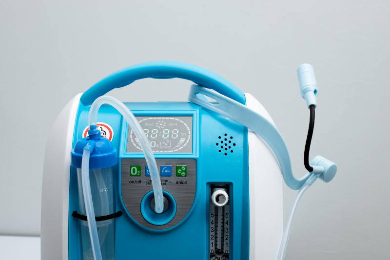
Oxygen Concentrator
Image 7 is an example of an O2 concentrator. The size and configuration vary. Oxygen concentrators convert room air into pure oxygen. People who are O2 dependent use them to allow mobility (Hardavella et al., 2019).
Delivery Systems
Several systems deliver O2 and are used to meet the doctor's prescription. In an emergency, 2 L/min per nasal cannula is the normal starting dose. The O2 level can also be set to match the level used at home. During a cardiac or pulmonary arrest, set the O2 at maximum delivery for administration with an Ambu bag.
A nasal cannula is a tubing with two prongs that go into a person's nose. It is attached to green O2 tubing to an O2 delivery device. A nasal cannula delivers low-dose O2 mixed with room air. The nasal cannula can deliver 2-6 L/min (Hardavella et al., 2019).
Nasal Cannula and Tubing
Image 8 shows a nasal cannula and tubing.
Nasal Cannula Position
Image 9 shows a properly positioned nasal cannula. The prongs go in the nares. The tubing goes over the ears and under the chin. A sliding tube can be adjusted to keep the cannula tighter. The cannula can be taken over the ears to the back of the head, but that position tends to move the prongs out of the nose as they move around in bed. If the cannula is too tight, it causes sores on the nose and ears.
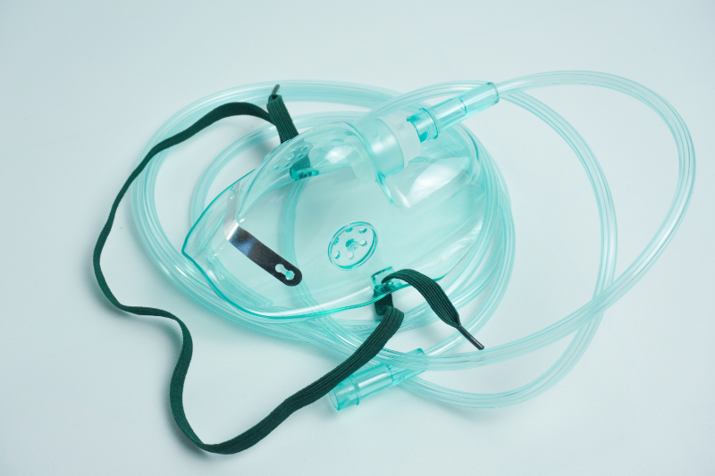
Simple face masks are used to deliver O2 at 8-10 L/min.
Simple Oxygen Mask
Image 10 is a picture of a simple oxygen mask. The metal piece can be adjusted to conform to the shape of the nose, and an elastic strap around the head keeps the mask in place. O2 is attached straight to the mask. Ports on the side of the mask allow carbon dioxide to escape and room air to mix with the O2.
Reservoir masks have a soft plastic bag attached at the end. The O2 tubing flows into the bag. The mask has valves to prevent room air mixture and increase O2 concentration. When the person inhales, they breathe in from the reservoir bag. Exhaled air escapes through vents in the side of the mask.
The two types of reservoir masks are partial rebreather and non-rebreather. A partial rebreather mask delivers 6-10 L/min. A partial rebreather mask conserves one-third of exhaled carbon dioxide in the reservoir as a respiratory stimulant.
A non-rebreather mask delivers 10-15 L/min and does not retain any exhaled carbon dioxide. Reservoir bags look the same—the difference lies in the function of the valves (Hardavella et al., 2019).
Reservoir Bag
Image 11 is of a reservoir bag.
An Ambu bag is an emergency resuscitation mask that delivers 100% O2.
Ambu Bag
Image 12 is of an Ambu bag in use. The O2 tubing connects at the end of the bag.
There are two types of artificial airways, endotracheal tubes (ET) and tracheostomy tubes. These delivery devices are inserted directly into the lungs by qualified staff. You may see people with a long-term tracheostomy tube. With a tracheostomy, O2 is delivered straight to the tube. Image 13 is of an endotracheal tube, and image 14 is of a tracheostomy tube.
The most common symptoms in persons with a pulmonary embolism (PE) are:
Dyspnea at rest or with exertion (this is the most common symptom)
The onset of dyspnea is frequently (but not always) rapid, usually within seconds or minutes.
Dyspnea may be less frequent in older persons with no previous cardiopulmonary disease.
Dyspnea is more likely present in persons with PE in the main or lobar vessels.
Pleuritic pain (seen in 66% of cases)
Approximately 10% of persons present with the symptoms of an infarcted lung, usually due to smaller, more peripheral emboli.
Pleuritic pain is typical in this population due to inflammation of the pleura.
Cough (seen in 37% of cases)
Orthopnea (seen in 28% of cases)
Calf or thigh pain or swelling (seen in 44% of cases)
Wheezing (seen in 21% of cases)
Hemoptysis (seen in 13% of cases)
Hemorrhage from the infarcted lung is also thought to be responsible for hemoptysis (Morrone & Morrone, 2018).
Risk factors can be classified as follows:
Acquired
Provoking (e.g., recent surgery, trauma, immobilization, initiation of hormone therapy, active cancer)
Non-provoking (e.g., obesity, heavy cigarette smoking)
Inherited (i.e., genetic)
Around 30 genetic risk factors have been identified, including factor V Leiden and the prothrombin gene mutation, or G20210-A (Morrone & Morrone, 2018)
Most emboli are thought to arise from lower extremity proximal veins (iliac, femoral, and popliteal), and more than 50% of persons with proximal deep vein thrombosis (DVT) have concurrent PE at presentation.
A calf vein DVT rarely embolizes to the lung, and two-thirds of calf vein thrombi resolve spontaneously after detection. However, if untreated, one-third of calf vein DVTs extend into the proximal veins, where they have greater potential to embolize. PE can also arise from DVTs in non-lower-extremity veins, including renal and upper-extremity veins, although embolization from these veins is less common (Waheed et al., 2023).
Asthma can be life-threatening. An attack may be precipitated by exertion, anxiety, infection, or exposure to an allergen. In obtaining a medical history, get some idea of the severity of attacks, precipitating factors, medication effectiveness, hospital admissions due to asthma, and the use of systemic steroids. People with asthma usually have their usual inhaler(s) with them. Asthma signs and symptoms include the following:
Shortness of breath
Tachypnea or more than 25 breaths per minute
Expiratory wheezing
Use of accessory muscles for respiration
Tachycardia (Mayo Clinic, 2022)
Life-threatening asthma attack symptoms include the following:
Cyanosis
Slow respiratory rate of fewer than eight breaths per minute
Bradycardia
Decreased level of consciousness or confusion (Mayo Clinic, 2022).
Most asthma attacks respond to an inhaler. Call EMS if there is no rapid response, or the attack becomes severe. High-flow O2 should be given.
Anxiety is the common precipitating factor in hyperventilation. Breathing too quickly depletes the carbon dioxide that is necessary for normal ventilation. Hyperventilation leads to respiratory alkalosis. Symptoms of hyperventilation include the following:
Dizziness or lightheadedness
Shortness of breath
Belching, bloating, dry mouth
Weakness, confusion
Sleep disturbances
Numbness and tingling in the arms or around the mouth
Muscle spasms in hands and feet, chest pain, and palpitations
Loss of consciousness (Johns Hopkins Medicine, n.d.)
Be calm and reassure the person. Have the person rebreathe their own exhaled air to increase the carbon dioxide level. A paper bag or the person's cupped hands can be used.
Damage from a burn continues after the source of the burn is removed. The dominant priority is to stop the burning process. If a person is on fire, smother the fire with a large cover or have them roll on the ground to extinguish any flames. Running will increase the fire. The size and severity of the burn determine the level of care needed. Optimum first aid for burns and scalds is cool, running tap water for about 20 minutes; this action is effective up to three hours after the burn. Cool only the burned area, keeping the rest of the body warm. Do not apply toothpaste, butter, oil, eggs, turmeric, or ice to burn wounds. None of these are beneficial to wound healing. Oil-based substances can trap heat in the tissues, causing further damage. Ice can cause vasoconstriction and cold injuries, further damaging the skin (Stiles, 2018).
In cases of chemical burns, i.e., drain cleaner, bleach, battery fluid, etc., the continued contact of the agent with the person's skin may not be apparent. The process may require repeated testing of the person's skin, specific chemical neutralization, and extensive lavage of the affected areas.
If the clinical history or the physical examination suggests that upper airway burns or inhalation injury may be present, early intubation and mechanical ventilation are indicated. Persons with extensive burns require large volumes of intravenous (IV) crystalloid resuscitation fluids. While this resuscitation can be delayed briefly when lifesaving interventions are being performed, early commencement is beneficial. Treatment of burns is determined by the degree of burn and the percentage of body surface burned.
Burns are classified as first, second, third, or fourth-degree, depending on how deep and severe the damage penetrates the skin's surface.
First-degree (superficial) burns affect only the outer layer of skin, the epidermis. The burn site is red, painful, dry, and has no blisters.
Second-degree (partial thickness) burns involve the epidermis and part of the lower layer of skin, the dermis. The burn site looks red and blistered and may be swollen and painful.
Third-degree (full-thickness) burns destroy the epidermis and dermis. They may go into the innermost layer of skin, the subcutaneous tissue. The burn site may look white or blackened and charred.
Fourth-degree burns go through layers of the skin, underlying tissue, and deeper tissue, possibly involving muscle and bone. There is no pain in the area since the nerve endings are destroyed (Perez et al., 2020).
Although sometimes considered burn injuries, high-voltage electrical injuries (e.g., lightning strikes, power lines) present different problems. The tissue injury from electrical injuries may not be apparent on physical examination. Massive myonecrosis, soft tissue, and bone damage may be concealed beneath normal-appearing skin between the entrance and exit wounds. Continuous cardiac monitoring is needed because of the risks of direct myocardial injury and hyperkalemia arising from myonecrosis.
The classic description of chest pain is most commonly seen in the male, middle-aged population. Females, diabetics, and older adults often describe different or atypical sensations that may be cardiac.
Chest Pain Signs and Symptoms
Retrosternal chest discomfort, not frank pain (i.e., pressure, heaviness, squeezing, burning, choking)
Positive Levine sign (presence of fist clenched over the sternum when describing the discomfort)
Diaphoresis with cold, clammy skin
Indigestion, nausea, or vomiting
Shortness of breath (dyspnea)
Lightheadedness
Anxiety with increased heart rate
Pain localized primarily in the epigastrium, back, neck, jaw, or shoulders
Report of recent exertion, eating, exposure to cold, or emotional stress
Action to take with chest pain until EMS can respond
Get help
Have someone gather available resources (AED, defibrillator, crash cart)
Monitoring vital signs
Start O2 if available
(Mayo Clinic, 2021)
Chest pain may be described as pressure, heaviness, squeezing, burning, or choking. The pattern and location of the discomfort are important.
Chronic cardiac chest pain is called angina. Angina may be a recurrent problem for some and is treated with nitroglycerin. People with chronic angina may not want EMS or emergency department care as they think nitroglycerin and rest will allow for recovery.
For cold injuries, the dominant priority is slowly rewarming, particularly in the case of systemic hypothermia, but it is equally applicable to cold injuries to the extremities (e.g., frostbite). While mild hypothermia is managed as described under the environment in the primary survey, severe cold injuries should be treated with immersion in water warmed to 40°C (Jin et al., 2021).
Type 1 diabetes, due to autoimmune β-cell destruction, usually leads to insulin deficiency. It often occurs in childhood or adolescence and requires diet control and insulin injections. Type 2 diabetes is due to a progressive loss of β-cell insulin secretion; this type usually results from insulin resistance and usually occurs in adults. It is possible to control it by diet and oral medication.
The acute complications of diabetes are hypoglycemia, diabetic ketoacidosis (DKA), and hyperglycemic hyperosmolar syndrome (HHS). The latter two are acute but develop over a more extended period and are characterized by extremely high serum glucose. DKA and HHS are primarily caused by poor compliance with the diabetic medication regimen, acute infection, or illness. These complications' signs and symptoms are non-specific but include the following:
Mental status changes
Polydipsia
Polyuria
Weakness
These are serious, life-threatening complications.
Hypoglycemia is much more common than DKA or HHS. A severe hypoglycemic event can cause death, and hypoglycemic events increase mortality risk. Hypoglycemia is most common with type 1 diabetes. The primary causes of hypoglycemia in a person with diabetes are too much insulin and a lack of sugar. Typical signs and symptoms of hypoglycemia include (but are not limited to) agitation, confusion, dizziness, drowsiness, irritability, palpitations, sweating, and weakness (Nakhleh & Shehadeh, 2021).
Hypoglycemia can occur without signs and symptoms. Some people can have significant hypoglycemia, but the body's response mechanisms fail to provide warning signs and symptoms, a condition called hypoglycemia unawareness. Hypoglycemia unawareness is not unusual, and it is very common in type 1 diabetics, affecting as many as 35% of persons with the disease. Treatment recommendations for hypoglycemia include (Nakhleh & Shehadeh, 2021):
Symptomatic hypoglycemia: Ingest carbohydrates - 15-20 grams of glucose is preferred, and repeat the blood glucose measurement in 15 minutes.
If the blood glucose is still low, ingest more glucose until the glucose level is normal, and then have a meal or a snack.
Fats can prolong the glycemic response, and proteins can increase the insulin response without increasing the glucose level, so carbohydrates are preferred for treating hypoglycemia.
Glucagon should be prescribed for anyone who has level 2 hypoglycemia.
Side effects are the secondary and undesired effects of a drug. These are expected reactions that occur with drug administration; every person receiving that drug is at risk of experiencing side effects. Examples of side effects include drowsiness with diphenhydramine administration and nausea with the administration of chemotherapeutic drugs.
Allergic reactions are aberrant immune responses to an antigen or an allergen. An antigen is a toxin or substance that incites an allergic reaction in the body, especially one that leads to antibody production. An allergen is defined as a substance that provokes an allergic reaction.
The term allergy implies an immediate hypersensitivity reaction mediated by immunoglobulin E (IgE) antibodies. However, many allergic reactions are not IgE-mediated. The subsequent reaction produces a wide range of clinical symptoms, from itching to anaphylaxis. Anaphylaxis is the most severe form of an allergic reaction, and it can occur suddenly and may, eventually, lead to death. The clinical symptoms of an allergic reaction to a drug may vary widely. A generalized allergic reaction may appear similar to serum sickness or immune-complex reactions (Hostoffer & Joseph, 2022).
Serum sickness and serum sickness-like reactions occur approximately 7-10 days after the primary exposure to an allergen, in this case, the drug, and are characterized by urticaria (hives), polyarthralgia (joint pain involving multiple joints), fever, and lymphadenopathy (swollen lymph nodes). Occasionally the persons may have systemic venalities' (inflammation of the veins during intravenous drug administration), which may progress into a full-blown systemic vasculitis. Serum sickness and serum sickness-like reactions can also occur after secondary exposure to heterologous proteins (classic serum sickness) or after secondary exposure to non-protein drugs such as sulfa or penicillin-based antibiotics (Rixe & Tavarez, 2022).
Epinephrine is the medication used to treat severe allergic reactions and anaphylaxis. It narrows blood vessels and opens airways in the lungs. These effects can reverse low blood pressure, wheezing, itching, hives, and swelling. Epinephrine is available in an EpiPen auto-injection. Highly sensitive people may carry an EpiPen. If an EpiPen is used, an EMS response is needed because the effects wear off in 10-20 minutes (Thornton, 2020).
Drug poisoning is the adverse effects experienced by exposure to chemicals, drugs, or otherwise innocuous substances. The reaction to a given toxin depends on the toxin dose. It varies in how each individual responds to the toxin, even with the same dose. The factors that affect an individual's reaction to a drug include; acquired tolerance, genetic predisposition, and other drugs in the system affecting enzymatic activation or inhibition. Depending on the route of exposure, poisoning may be local or systemic.
In most cases, the person may be confused, unconscious, or unwilling to cooperate. Any suspicious findings in the history should be further investigated. Suspicious findings include sudden unexplained illness in previously healthy persons, a history of psychiatric illness, and the onset of symptoms after ingesting foods or drinks, including alcohol or medications.
All healthcare facilities are required to have detailed information about the chemicals that are at the worksite. The information is in a standard format called a Safety Data Sheet (SDS). Help for suspected poisoning is available at POISONCONTROL.
The poison control phone number 1-800-222-1222 (U.S.) will route the call to your area's poison center.
Pam saw a group of people crowded around a person with jerking movements of the limbs. Pam approached the group and said, "I am a physical therapist. Can I help?" The friends said yes.
Meanwhile, the person became stiff, lost consciousness, and became cyanotic. Pam checked the person for a clear airway, pulse, and respirations. The airway was clear, the pulse was rapid, and the respirations were slow. Pam asked if this person had seizures. The response was that the person had seizures but had been controlled. She asked the people if they were relatives, and they said yes. Pam asked if they wanted EMS called, and they said yes. She pointed at a young man and told him to call 911. Pam directed other people to move the chairs and ice chests away from the person. She got help rolling the person flat on her back and asked anyone who knew CPR to kneel on the other side of the person in case cardiopulmonary resuscitation (CPR) was needed. By this time, the person was flaccid, not cyanotic, and still unconscious. Pam stayed with the person monitoring the pulse and respirations until the EMS staff arrived.
Pam took control of the situation and identified specific people for specific tasks. Pam's actions avoided delays in waiting for the group to decide to act. She introduced herself and asked permission to help. Pam could have obtained a more thorough medical history. In the flurry, Pam did not ask someone to time the seizures, which was needed. She correctly positioned the person and monitored the airway, breathing, and circulation.
This course discussed the outpatient healthcare provider's responsibility to act in an emergency, including obtaining consent. An initial visual assessment was described. Specific emergencies and the appropriate responses were discussed. All healthcare professionals need to be prepared to respond in an emergency.
Select one of the following methods to complete this course.
Take TestPass an exam testing your knowledge of the course material.
CEUFast, Inc. is committed to furthering diversity, equity, and inclusion (DEI). While reflecting on this course content, CEUFast, Inc. would like you to consider your individual perspective and question your own biases. Remember, implicit bias is a form of bias that impacts our practice as healthcare professionals. Implicit bias occurs when we have automatic prejudices, judgments, and/or a general attitude towards a person or a group of people based on associated stereotypes we have formed over time. These automatic thoughts occur without our conscious knowledge and without our intentional desire to discriminate. The concern with implicit bias is that this can impact our actions and decisions with our workplace leadership, colleagues, and even our patients. While it is our universal goal to treat everyone equally, our implicit biases can influence our interactions, assessments, communication, prioritization, and decision-making concerning patients, which can ultimately adversely impact health outcomes. It is important to keep this in mind in order to intentionally work to self-identify our own risk areas where our implicit biases might influence our behaviors. Together, we can cease perpetuating stereotypes and remind each other to remain mindful to help avoid reacting according to biases that are contrary to our conscious beliefs and values.
Bobko, J. P., Badin, D. J., Danishgar, L., Bayhan, K., Thompson, K. J., Harris, W. J., Baldridge, R. T., & Fortuna, G. R. (2020). How to Stop the Bleed: First Care Provider Model for Developing Public Trauma Response Beyond Basic Hemorrhage Control. Western Journal of Emergency Medicine: Integrating Emergency Care with Population Health, 21(2), 365–373.
Boddu, S. H. S., & Kumari, S. (2020). A Short Review on the Intranasal Delivery of Diazepam for Treating Acute Repetitive Seizures. Pharmaceutics, 12(12), 1167. Visit Source.
Brouhard, R. (2020). How to Recognize a Medical Emergency. Verywell Health. Visit Source.
Center for Disease Control and Prevention (CDC). (n.d.). Picture of America Poisoning Fact Sheet. Center for Disease Control and Prevention (CDC). Visit Source.
Center for Disease Control and Prevention (CDC). (2019). Psychololgy of a Crisis. Center for Disease Control and Prevention (CDC). Visit Source.
Davis, J. (2004). 5 emergencies: Do you know what to do? WebMD. Visit Source.
Erich, J. (2020). Epilepsy for EMS. EMS World. Visit Source.
Hardavella, G., Karampinis, I., Frille, A., Sreter, K., & Rousalova, I. (2019). Oxygen devices and delivery systems. Breathe (Sheffield, England), 15(3), e108–e116. Visit Source.
Hostoffer, R.W., & Joseph, N. (2022). Immunoglobulin E. In: StatPearls [Internet]. Treasure Island (FL): StatPearls Publishing. Visit Source.
Jin, H. X., Teng, Y., Dai, J., Zhao, X. D., & Members of the Emergency Medicine Committee of the People’s Liberation Army (2021). Expert consensus on the prevention, diagnosis and treatment of cold injury in China, 2020. Military Medical Research, 8(1), 6. Visit Source.
Kadam, R.A. (2017). Informed consent process: A step further towards making it meaningful!. Perspectives in clinical research, 8(3), 107–112.
Mayo Clinic. (2021). Chest pain—Symptoms and causes. Mayo Clinic. Visit Source.
Mayo Clinic. (2022). Asthma - Symptoms and Causes. Mayo Clinic. Visit Source.
Morrone, D., & Morrone, V. (2018). Acute Pulmonary Embolism: Focus on the Clinical Picture. Korean circulation journal, 48(5), 365–381. Visit Source.
Nakhleh, A., & Shehadeh, N. (2021). Hypoglycemia in diabetes: An update on pathophysiology, treatment, and prevention. World journal of diabetes, 12(12), 2036–2049. Visit Source.
National Institute of Neurological Disorders and Stroke (NINDS). (2019). NINDS Know Stroke Campaign—Needtoknow. National Institute of Neurological Disorders and Stroke (NINDS). Visit Source.
Perez, E., Foley, M., & Karlin, R. (2020). Classification of Burns—Health Encyclopedia—University of Rochester Medical Center. University of Rochester Medica Center, Health Encyclopedia. Visit Source.
Stiles, K. (2018). Emergency management of burns: Part 2. Emergency Nurse, 26(2), 36–42. Visit Source.
Thim, T., Krarup, N. H., Grove, E. L., Rohde, C. V., & Løfgren, B. (2012). Initial assessment and treatment with the Airway, Breathing, Circulation, Disability, Exposure (ABCDE) approach. International journal of general medicine, 5, 117–121. Visit Source.
Thornton, P. (2020). EpiPen: Instructions, Side Effects & Warnings. Drugs.Com. Visit Source.
Listen from your pocket or in your car, Apple CarPlay and Android Auto compatible.
Sync Between Devices
Start on your desktop, pickup on your mobile. Never lose your place.
Reinforce Your Learning
Switch between reading and listening to your course or read while you listen!
Enhance Your Experience
Interactive course outline, robust play controls, multiple voices and different playback speed options.
Listen Anywhere
Listen from your pocket or in your car, Apple CarPlay and Android Auto compatible.
Subscribe for Access
Sync Between Devices
Start on your desktop, pickup on your mobile. Never lose your place.
Reinforce Your Learning
Switch between reading and listening to your course or read while you listen!
Enhance Your Experience
Interactive course outline, robust play controls, multiple voices and different playback speed options.
×
Away for now!
We're sorry! All our support agents are unavailable to chat at the moment.
Need Immediate Assistance? Please visit our FAQ section which provides answers to many common inquiries.
Get in Touch Directly via Email If your query is urgent or you'd prefer to reach us directly, we invite you to submit a support ticket through our Contact Us page. Our dedicated support team will review your inquiry and get back to you as soon as possible.
Support Hours Our chat support is typically available Monday to Friday, 9:00 AM - 6:00 PM EST. For support outside of these hours, please use our email contact
Are you sure?
You should NOT select "Remember me for 30 days" if you are on a public or shared computer.