≥ 92% of participants will know how to correlate abnormalities in the CBC with the clinical scenario and be able to identify appropriate next steps, including further testing and diagnostics, and when to refer to a specialist.
CEUFast, Inc. is accredited as a provider of nursing continuing professional development by the American Nurses Credentialing Center's Commission on Accreditation. ANCC Provider number #P0274.
≥ 92% of participants will know how to correlate abnormalities in the CBC with the clinical scenario and be able to identify appropriate next steps, including further testing and diagnostics, and when to refer to a specialist.
Upon completion of this course, the participant will be able to do the following:
The complete blood count (CBC) is vital to a thorough health assessment. It includes information on many body systems and disease states.
This module will review each component of the CBC. Abnormal CBC results and associated differential diagnoses will be discussed. Pertinent history and physical exam components will be addressed. Additional testing that should be considered, as well as when to refer patients and to what specialists, will also be reviewed.
The CBC is a laboratory test done on peripheral blood. Its benefits include being easy to obtain, readily available, cost-effective, and having a quick turnaround time. It is also an excellent screening test and can be reassuring when the results are within the normal range (Chen et al., 2024).
Its limitation is that it is often only the first step in identifying pathology. Most abnormalities require additional testing before a diagnosis is made.
The CBC is often ordered serially, from once a year to once a day to numerous times a day. It can provide both diagnostic and prognostic information on the progression of a disease state and response to therapy over time.
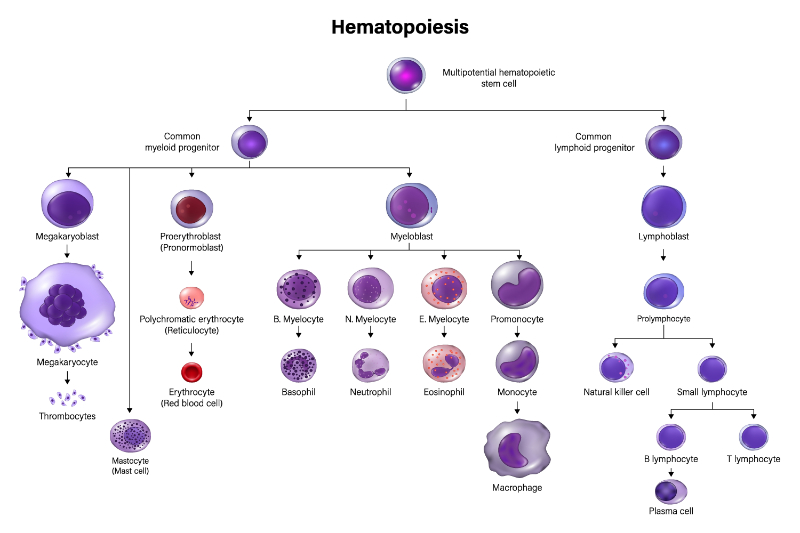
CBC results reflect information on the three primary blood cells formed in the bone marrow. To best understand the intricacies of the CBC, it is essential to review the hematopoietic stem cell division chart. This chart reviews the maturation of all blood cells within the bone marrow from which they are derived.
Blood cell growth begins with a pluripotent stem cell. Stem cells can grow and mature into the three main blood cells seen on the CBC: WBCs, RBCs, and platelets. The first division of the stem cell begins with a cell differentiating into either a myeloid or lymphoid cell.
Myeloid cells mature in stages to ultimately become platelets, erythrocytes, or a variety of WBCs. Lymphoid cells mature into B and T cell lymphocytes. Lymphocytes are WBCs that take a different maturation pattern than myeloid-derived WBCs and play a different role in our immunity. Understanding this difference can help to differentiate potential etiologies when the WBC count is abnormal.
Nowadays, most CBCs are completed using modern electronic instruments. These instruments separate and count each cell type. These counters can provide information on the average cell volume and size but cannot comment on the cell's morphology.
If abnormalities exist, a peripheral blood smear may also be necessary to examine the morphology of the abnormal cell line. Clinicians can request it once the CBC results have been reviewed, or the lab may do it automatically, depending on the individual laboratory policy and procedures guidelines. Some examples of clinical indications to request a peripheral blood smear include unexplained cytopenia, suspicion of leukemias or myeloproliferative disorders, symptoms of hemolysis, and severe sepsis.
Throughout this course, abnormal values will be considered high (H) or low (L). Specific normal values will not be listed as these vary based on each laboratory.
WBCs, or leukocytes, are recorded in a CBC. They are primarily formed in the bone marrow.
WBCs mature into five different types of cells. These are known as the differential. A standard differential is divided up as follows (Curry, 2019):
This array of WBCs protects the body against infection in various ways. Neutrophils are best at killing bacteria, fungi, and foreign debris. Lymphocytes produce antibodies to act on specific antigens entering the body. They are well known for fighting viral infections. Monocytes are part of the clean-up crew and help rid the body of damaged cells. Allergic reactions and parasite invasions activate eosinophils. Basophils are also activated in allergic responses.
The CBC usually lists differential results as both a percentage and an absolute. Occasionally, the results only report the percentage of each differential line. The percentage of abnormal differential cells is generally helpful if the WBC count is within the normal range. If the WBC is high or low, the percentage is less reliable, and it becomes vital to look at the absolute count of each differential cell line.
Let us take a deeper look at each of the differential components.
Neutrophils, known as polymorphonuclear neutrophils (PMN), are the first and primary defense against bacterial and fungal infections. A neutrophil has an approximate lifespan of ten days in the bone marrow, 2-3 days in the tissues, and less than 24 hours in circulation (Kobayashi et al., 2023).
When assessing the neutrophil count, it is important to notice the absolute neutrophil count (ANC), not just the percentage. The ANC is more representative of the patient's actual capacity to fight infections. When the ANC is low, the patient is neutropenic.
A hyperstimulated bone marrow can produce excessive amounts of neutrophils. Some of the early or immature forms of the neutrophils will spill into circulation. These premature neutrophils are called bands and will mature into neutrophils in the peripheral blood. These bands may be seen in acute severe bacterial infections. Not every lab reports bands, but it should be noted if they do.
ANC is calculated using the following equation (Clinical Advisor, 2010):
Example:
Neutropenia
Absolute neutropenia puts patients at risk for frequent and recurring infections. It is often referred to as:
An ANC of less than 0.5 mm3, along with infectious symptoms or fever, is considered emergent and must be further evaluated immediately. Broad-spectrum antibiotics, usually administered intravenously, are urgently indicated with a fever as an infectious workup is pursued (Agnello et al., 2021).
Neutropenia can be either congenital or acquired. Congenital neutropenia is usually discovered in childhood and is often associated with frequent infections and other clinical findings such as premature grey hair or skin and bone disorders.
Duffy-null associated neutrophil count (DANC) is also commonly seen. DANC was previously labeled benign ethnic neutropenia, but as it is not a pathologic finding, it has been renamed. Patients of African and other ethnic groups can present with a lower-than-normal neutrophil count with no increased risk of infection. Neutropenia may also be seen in patients from ethnic groups that are not usually associated with DANC. In these cases, it is often called familial neutropenia. Familial neutropenia is a standard variant of the low end of the baseline, typically mild, with no history of unusual or recurrent infections (Berliner, 2024).
Acquired neutropenia is seen in response to pathology. With autoimmune neutropenia, an increase in neutrophil destruction occurs. In this case, the bone marrow produces the correct amount of neutrophils; however, they are destroyed once they leave the bone marrow.
Bone marrow disorders, including myelodysplasia, leukemias, lymphomas, and aplastic anemia, can also present with neutropenia. In these situations, the bone marrow is not able to produce mature neutrophils. Other inflammatory rheumatic disorders may contribute to neutropenia, most commonly rheumatoid arthritis and lupus.
Finally, some nutritional deficiencies may present with neutropenia. Vitamin B12, copper, and folate deficiency could be responsible for newly acquired neutropenia (Berliner, 2024).
Neutrophilia
Neutrophilia is a high neutrophil count, at least two standard deviations above each lab's mean. Keep in mind that normal mean neutrophil counts do vary by age. In general, neutrophilia is either caused by an increased production of neutrophils in the bone marrow or a demargination of the cells from the walls of the bloodstream. This is a process where the cells that generally adhere to the endothelial lining of the blood vessel are released into the bloodstream, thus increasing the circulating neutrophils (Coates, 2024).
An increase in neutrophil production may be malignant or reactive. Malignant processes include myeloproliferative neoplasms, most commonly polycythemia vera or chronic myeloid leukemia.
Some genetic disorders also may cause neutrophilia, including leukocyte adhesion deficiency and Down syndrome.
Neutrophilia can be seen as a secondary symptom in certain conditions, including pregnancy, eclampsia, postpartum, and hypercortisolism. Other potential causes of neutrophilia include asplenism, cigarette smoking, and obesity (Coates, 2024).
Lymphopenia
Lymphopenia occurs when the lymphocyte count is low. The most common etiologies for this include sepsis, leukemia, and side effects of chemotherapy, immunotherapy, and radiation therapy. Immunodeficiency disorders and autoimmune disorders often have an associated lymphopenia (Cleveland Clinic, 2023). Lifestyle behaviors such as poor diet and heavy alcohol intake can contribute to lymphopenia.
Lymphocytosis
Lymphocytosis is an elevation of normal lymphocyte counts. It is often seen as a reactive process secondary to acute or chronic infections, stress, and asplenism. Both bacterial and viral infections can mount a lymphocytic reaction. Primary bone marrow disorders, such as various leukemias, non-Hodgkin's lymphoma, and Sézary syndrome, can also manifest with lymphocytosis (Hamad & Mangla, 2023).
Monocytopenia
Conversely, leukemia may also cause a low monocyte count. More commonly, this occurs in response to cancer treatment, including chemotherapy, radiation, and infections (Espinoza & Emmady, 2023).
Monocytosis
An elevated monocyte count is seen in response to infection, especially viral infections and inflammation. It is also commonly found in autoimmune disorders and trauma. Hematologic disorders, including myelodysplasia and leukemia, can also present with an elevated monocyte count (Espinoza & Emmady, 2023).
Basopenia
A low basophil count is rare, possibly because the absolute number of basophils is already so low (less than 1%). It is known to decrease with the use of glucocorticoids due to increased apoptosis. It is also seen in chronic urticaria, allergic reactions, hypothyroidism, and systemic lupus. This is likely in response to basophilic migration out of circulation while infiltrating tissues (Shah et al., 2021).
Basophilia
Eosinopenia
Infection and Cushing's syndrome may cause low eosinophils. However, a low value alone is less likely to be clinically significant. It is difficult to get a sense of a meaningful low result (Cleveland Clinic, 2022a).
Eosinophilia
As with all WBCs, eosinophils can be elevated with active infections, especially parasitic infections. Eosinophilia can also occur with autoimmune disorders, such as inflammatory bowel disease, vasculitis, and sarcoidosis. Allergic responses like seasonal allergies and asthma may also be the root cause of an elevated eosinophil count. Eosinophilic leukemia can present with an elevated eosinophil count (Cleveland Clinic, 2022a).
If leukocytosis is present, it is vital to assess the differential to consider the likely diagnosis and next steps in testing. If there are a number of immature hematopoietic cells, flow cytometry for leukemia or lymphoma can be considered. If the basophils are elevated, especially with an enlarged spleen, molecular testing for polymerase chain reaction (PCR) for the breakpoint cluster region-Abelson leukemia (BCR-ABL) gene is indicated to rule out chronic myelogenous leukemia. If lymphocytes are elevated, consider flow cytometry for chronic lymphoid leukemia.
If infection is suspected, viral serology, chest X-ray, urine culture, wound culture, and blood culture should be considered. A bone marrow biopsy is often the gold standard for diagnosing leukemias, lymphomas, and myeloproliferative disorders; however, this is often best left for the specialty team to order.
Patients with abnormal WBCs, whether high or low, may present with various complaints, including symptoms of infection, fevers and chills, fatigue, pain, shortness of breath, weight loss, and a change in appetite. Rashes or skin abscesses, headaches, and mouth sores are also reasons patients may seek medical care. Patients may also have no new concerns or complaints, as an abnormal WBC can be an incidental finding.
The physical exam should focus on potential sites of infection and any inflammatory changes. Palpable adenopathy should be assessed. An abdominal exam, including palpating for hepatosplenomegaly, is also indicated. Any recent unanticipated weight loss can also be a clue to a more serious bone marrow malignancy.
It is often difficult to understand when a referral is necessary for abnormal WBC counts and differentials. Any time a hematologic malignancy is suspected, a hematology referral is warranted. Urgent referrals are necessary when the patient has severe absolute neutropenia. If a patient also has an active infection or fever, this then becomes emergent. The patient should be referred to the nearest emergency department for expedited ongoing evaluation and treatment. Emergent referral is also indicated if acute leukemia is suspected with high or low WBC coupled with immature cells such as blasts or promyelocytes in the differential. If an emergency referral is not indicated, repeating the CBC to monitor for trends is vital to help understand the etiology of the abnormality, and is appreciated by hematologic specialists.
A 56-year-old woman presents to her primary care provider's office complaining of a lump under her right arm. She noticed it about two weeks ago. It did not change in size during that time. She has no pain or discomfort. She has no limitations in her mobility. She denies any injury to the area. She has been in her usual state of health with no recent infections. No fevers or chills are noted. Her weight is stable, and her energy has been good. She denies any night sweats. She works full-time as a book editor at home and has had no problems completing her work. The rest of her review of systems is negative.
Her past medical history includes hypothyroid and generalized anxiety disorder. Her medications include Synthroid 0.88 micrograms (mcg) daily and Celexa 20 milligrams (mg) daily.
On exam, a 2-centimeter (cm) x 2 cm mass is noted in her right axilla. It is mobile and non-tender. No associated erythema. She has no cervical, inguinal, or epitrochlear adenopathy. Her spleen is palpable just below her lower costal margin. No other abnormalities are noted.
Intervention: To further assess her mass and splenomegaly, a CBC is done. Her CBC results are as follows:
WBC Count
Differential Count (%)
Hemoglobin (HGB) - 12.5 g/dl
Hematocrit (HCT) - 26
MCV - 90
Platelets - 140K
Her lymphocyte count is very high.
As she has been clinically well with no fatigue, pain, or infections, a reactive response is unlikely, and a primary bone marrow problem is considered.
The patient has been clinically well. She has a repeat CBC in 1-2 weeks. The results are basically unchanged, with an abnormal WBC and an elevated lymphocyte count. Her physical exam is also unchanged, with mild splenomegaly and no changes in her adenopathy.
This patient has a persistent abnormal CBC. The total WBC is high, but the majority of those cells are lymphocytes. This, coupled with mild adenopathy and splenomegaly, leads one to consider a hematologic malignancy such as chronic lymphocytic lymphoma. At this point, she is referred to hematology to work up her lymphocytosis and adenopathy further.
As the patient has no B symptoms and is without neutropenia, anemia, or low platelets, there is no urgency to the referral. Her adenopathy has not been progressing. A regular hematology referral can be placed without urgency.
In this case, the serial CBC is very helpful in ruling out a reactive or transient cause of the lymphocytosis. Despite the high WBC, her general well-being supports a more conservative diagnostic approach.
The RBC count, hematocrit, and hemoglobin are all closely related and provide information on the erythrocytes in slightly different ways. They are generally assessed together to help inform the clinician of any potential pathology.
The RBC count is the number of erythrocytes circulating in 1 mm3 of venous blood. Each RBC contains molecules of hemoglobin, whose function is to transport and exchange oxygen to tissues.
The hematocrit is an indirect measurement of the circulating RBC number and volume in the bloodstream. The hematocrit is a percentage of total blood volume and, as such, closely mimics the RBC and hemoglobin results. The usual ratio of hematocrit to hemoglobin is 3:1. For example, if the hemoglobin is 12 grams (g)/deciliter (dl), the hematocrit will be 36%. This ratio assumes all RBCs are of normal size with normal amounts of hemoglobin.
As the hematocrit is a percentage of circulating blood, three variables can affect the results (Mondal, H., & Zubair, 2024).
A change in any of the above can affect the results. Dehydration will decrease the volume, which will increase the percentage of RBCs. The opposite is true of overhydration or hemodilution. If a patient is microcytic or has very small RBCs, the percentage will be less than someone who has macrocytosis or large RBCs, even though the actual production and number of RBCs remain the same.
The hematocrit is most useful when both the size of the RBCs and the patient's hydration status are normal.
The hemoglobin result is a more direct measure of the RBC count.
The normal values of RBCs, hematocrit, and hemoglobin differ by gender, with males having a higher baseline than women. The mean count also tends to decrease with age. Each specific lab has different normal values based on a bell curve. It is important to always refer to each specific lab's reported normal results.
Elevated RBCs, hematocrit, and hemoglobin
Increases in the RBCs, hematocrit, and hemoglobin can occur for a variety of reasons. These results can be elevated as primary pathology or secondary in response to other ailments.
Primary pathology includes proliferative bone marrow disorders, most commonly polycythemia vera, where the bone marrow inappropriately produces too many erythrocytes. This is often seen in combination with other elevated cell lines, including WBCs and platelets.
Secondary causes include dehydration and any disorder that causes chronic hypoxia, such as chronic obstructive pulmonary disease (COPD), heart disease, and sleep apnea. Even heavy smoking can cause a rise in erythrocytes. This occurs as the body recognizes oxygen deprivation and attempts to improve the situation by increasing the cells that deliver oxygen to the tissues.
These results can also be elevated secondary to medications.
Decreased RBCs, hematocrit, and hemoglobin
When the hematocrit and hemoglobin are low, the patient is anemic. This common and complex diagnosis requires additional information to ascertain the root cause of the anemia and its subsequent treatment.
Anemia Categories
Differential diagnosis for low hemoglobin and hematocrit generally fall into three categories: a decrease in the production of erythrocytes, an increase in the destruction of erythrocytes, and blood loss (Turner et al., 2023).
A decrease in production often includes nutritional deficiencies such as iron, B12, and folate deficiency.
Renal disease is another common etiology for low production of erythrocytes. Erythropoietin is a hormone made in the kidney which drives the bone marrow to produce RBCs. When a kidney is not functioning well, as often seen with chronic diseases such as diabetes mellitus, less erythropoietin is released, and the RBC production decreases, leading to anemia.
An increase in the destruction of RBCs once they are formed occurs in hemolytic anemias and with mechanical shearing. An example of this includes prosthetic valve replacements, which can cause mechanical shearing of the cells as they pass through the valve (Turner et al., 2023).
To best evaluate for a specific anemia diagnosis, the RBC indices must next be evaluated.
The RBC indices include the MCV, the mean corpuscular hemoglobin (MCH), and the mean corpuscular hemoglobin concentration (MCHC). These results provide information on the size, weight, and hemoglobin concentration when attempting to classify anemias.
Mean corpuscular volume
The MCV is the average size or volume of a single RBC. It is derived by dividing the hematocrit by the RBC count.
MCV = Hematocrit (%) x 10
RBC (million/mm3)
When the MCV is elevated, the patient is macrocytic; when low, the patient is microcytic. This is usually the first determination used when assessing a patient's type of anemia (El Brihi & Pathak, 2024).
Differential Diagnosis
Microcytosis, a low MCV, is associated with the most common anemia, iron deficiency, but also with anemia of chronic disease and thalassemia.
Mean corpuscular hemoglobin
The MCH reflects the average weight of hemoglobin within an RBC. It is calculated by dividing the hemoglobin by the RBCs.
MCH = Hemoglobin (g/dl) x10 / RBC (million/mm3)
Large macrocytic cells contain more hemoglobin, and conversely, small cells have less. The MCH results closely mimic the MCV results and provide minimal additional information. The differential diagnosis for a low MCH is the same as for microcytic anemia, and an elevated MCH with anemia differential diagnosis is the same as for macrocytic anemia.
Mean corpuscular hemoglobin concentration
The MCHC measures the concentration within a single RBC. It is derived using the following formula (Ware, 2020):
MCHC = hemoglobin (g/dl) x100
Hematocrit (%)
We report a low MCHC as being hypochromic and normal as normochromic. An RBC can not actually be hyperchromic as it can only fit a maximum amount of hemoglobin in each cell; however, false positives reported as an elevated MCHC exist.
If the hemoglobin surpasses the maximum volume, changes in the cell membrane ensue, causing alterations in the cell shape. A common example of this is spherocytosis. This can be seen in a blood smear report but may be reported as an elevated MCHC. Intravascular hemolysis and cold agglutinin disease can also report a falsely elevated MCHC.
Low MCHC can be seen in iron deficiency and thalassemia.
Red Cell Distribution Width
We understand that an RBC formed when a patient has a nutritional deficiency may be abnormally big or small (MedlinePlus, 2024). We also understand that the lifespan of an RBC is approximately 120 days. If a patient has had a nutritional deficiency for more than three months, all the cells should be approximately the same size, whether they are big or small. Once treatment begins, the formation of new cells should include cells at a standard size. When a CBC is done, we would then see a variety of sizes, some of the older cells, which are still big or small, and some normal-sized cells, causing a variation in the RDW.
An increase in RDW can be seen in early nutritional deficits, including iron, B12, and folate deficiencies. It can also increase in response to therapy as new cells developed are in the normal range. Diseases that cause fragmentation of RBCs, such as sickle cell disease and some hemolytic anemias, will also have an elevated RDW. Post-hemorrhagic anemia will also often have an increased RDW due to the bone marrow releasing immature cells, which are larger.
Associated Testing for Anemia
While not included in the CBC, the reticulocyte count is a useful tool when assessing anemia. If a patient is anemic, a reticulocyte count adds valuable information on both the classification and the response to therapy. A reticulocyte is an immature RBC developed in the bone marrow. Reticulocytes live for a few days in the bone marrow and a few days in the bloodstream before maturing into adult erythrocytes (Mast et al., 2008).
An increase in the reticulocyte count suggests normal bone marrow function in response to anemia and the treatment of anemia. For example, post-operative hemoglobin levels for many surgeries are below baseline. At the same time, in an otherwise healthy patient with no baseline anemia or nutritional deficit, the bone marrow will recognize the anemia and begin to produce more RBCs. This is seen with an increase in the reticulocyte count; at the same time, if an anemic patient has a nutritional deficiency or is not secreting enough erythropoietin, the bone marrow will not be able to produce the RBCs, and the reticulocyte count will be low or normal. In a patient with anemia, one would expect to see an elevated reticulocyte count. The reticulocyte count is reported in both percentage and absolute numbers. As always, the absolute result is more meaningful if the hemoglobin is outside of the normal range.
Let us take this one step further.
Many lab analyzers now also include the reticulated hemoglobin in the reticulocyte count. The reticulated hemoglobin is a measure of the amount of available iron within the cell. This makes it an excellent marker for iron-deficient anemia. A low reticulated hemoglobin has been shown to be both sensitive and specific when used to assess for iron-deficient anemia.
The reticulocyte count, including reticulated hemoglobin, should be considered an ancillary study when evaluating anemia. It is also beneficial when evaluating responses to treatment for iron deficiency anemia.
Iron Studies
Haptoglobin, lactic dehydrogenase, and direct antiglobulin test
If a destructive process is being considered, haptoglobin should be added to the lab panel as this reduces intravascular hemolysis. The reticulocyte count also is usually quite high when hemolysis is the culprit. Other labs to help identify hemolysis include lactic dehydrogenase (LDH) and direct antiglobulin test (DAT).
Erythropoietin Level
Erythropoietin levels should also be evaluated in anyone with anemia and chronic renal insufficiency. When this is low, coupled with anemia, exogenous erythropoietin can be very helpful.
Patients are often asymptomatic if erythrocytes are only mildly elevated or low. Patients with elevated counts may present with headache, fatigue, and pruritus, especially after a hot shower. Patients with more severe erythrocytosis may have symptoms of hyperviscosity, including chest pains, shortness of breath, numbness, blurred vision, abdominal pain, and early satiety (especially when combined with splenomegaly), and unusual bleeding such as frequent nosebleeds.
As anemia limits a patient's oxygen-carrying capacity, patients often present with cardiopulmonary symptoms. Fatigue is the most common complaint. Chest pains, palpitations, and respiratory distress require immediate intervention. Patients also complain of cold extremities, headaches, dizziness, and lightheadedness.
Patients with anemia may also present with symptoms of bleeding, either acute or chronic. Patients with B12 deficiency may experience peripheral neuropathy.
Patients with iron deficiency anemia often complain of difficulty concentrating. This has been shown to equate to poor school performance in school-age children with iron deficiency anemia (Yeboah et al., 2024).
When assessing patients with abnormalities in hematocrit, hemoglobin, and red cell indices, it is important to do a thorough history. As a common cause is blood loss, clinicians should evaluate for any recent significant bleeding, such as surgeries, trauma, rectal bleeding, or menorrhagia. Minimal microscopic chronic bleeding can also play a role. This can often present with gastrointestinal symptoms such as gastroesophageal reflux disease (GERD), known ulcers, and bleeding hemorrhoids. Medications that increase the risk of bleeding should also be considered, including aspirin, non-steroidal anti-inflammatory drugs (NSAIDs), and anticoagulants.
Understanding potential nutritional deficiencies is necessary. Inquire about diet and any restrictions and food availability. It is also important to ascertain if this is the first time someone has presented with anemia or if they have had it in the past.
It is important to monitor trends in anemia. Assessing previous CBC results to monitor for changes over time is extremely helpful. If patients do not have previous CBC available, inquire if they have ever donated blood. Patients who have met the requirements for blood donation are much less likely to have a genetically acquired cause of anemia.
A complete physical exam is warranted, as there are many potential root causes of anemia. Patients with anemia often present with pallor; however, this is usually not severe until the hemoglobin drops below 8 g/dl. Skin can also become jaundiced if the patient has hemolytic anemia. Tachycardia and hypotension may be seen, as well as orthostatic hypotension. It is important to evaluate for splenomegaly as this can quickly direct further workup if present. Potential sites of bleeding should be evaluated, including a rectal exam and stool for guaiac, as well as a gynecology exam as necessary.
The majority of anemias can be worked up and treated in primary care offices. However, severe anemia causes symptoms such as chest pains and shortness of breath, and those requiring transfusions or immediate intervention should be referred to a specialist. Transfusion parameters are specific to each medical center and blood bank.
Anemia caused by bleeding is best referred to specialists of the bleeding site.
A referral is also indicated if hemolysis is suspected or if there is no response to initial therapy, regardless of the etiology.
A significant risk of polycythemia vera is thrombosis. Patients with a hematocrit above 45% from a primary condition, such as polycythemia vera, can be at risk for thrombotic events (Tremblay et al., 2025). This is a cause for concern and a trigger for a hematology referral.
A 21-year-old female college student presents to the office with fatigue and trouble concentrating. She has been a vegan for the past year. She has no significant past medical history. She does not take any medications. She denies any pain but gets short of breath in her hot yoga class. She has had some dizziness on and off. She has not had any falls. She denies any recent infections and no weight loss. She has her menses once a month, for 5-6 days, and describes her flow as heavy. She denies any other blood loss, no blood or black, tarry stools, and no indigestion or reflux symptoms. She denies any pica symptoms. The remainder of her review of systems is negative.
Her physical exam is negative.
A CBC is drawn to evaluate her fatigue further.
Results:
This patient has a strong history of potential for iron deficiency anemia. She has not been eating meat for the past year, and she has heavy menses.
Her ferritin is 10 mg/l, and her reticulated hemoglobin is low. This confirms her diagnosis of iron deficiency anemia.
She is treated with an oral iron supplement and educated on how to add high-iron foods to her diet. It is also recommended that she consult with gynecology to consider interventions to minimize her heavy menstrual flow.
She returns to the clinic in one month for a repeat CBC, ferritin, and reticulocyte count. Her hemoglobin has improved to 12 g/dl with an MCV of 85 mm3. Her ferritin is up to 50 mg/l, and her symptoms have resolved.
Platelets are the smallest of all the cells on the CBC. They are formed in the bone marrow, deriving from megakaryocytes. The function of the platelet is to maintain vascular integrity. They basically clump, or aggregate, together to prevent bleeding and form clots in the vasculature. The majority of platelets live in the bloodstream, with a small percentage, less than 25%, living in the liver and spleen. The average lifespan of a platelet is ten days (Williams & Sergent, 2022).
Thrombocytopenia
The platelet count represents the number of platelets per cubic millimeter of blood.
Increased destruction of platelets once they leave the bone marrow can happen in a number of different ways. Immune dysfunction is one common etiology. Antibodies see platelet antigens as foreign, and the platelets are destroyed. This is often idiopathic (idiopathic thrombocytopenia [ITP]) but can also occur with post-transfusion reactions, viral infections, lymphoproliferative disorders, or drug-induced thrombocytopenia.
Common medications that may cause thrombocytopenia include chemotherapy and immunotherapy, quinine, ranitidine, Bactrim, rifampin, and vancomycin. If medication is the culprit, the platelet count usually goes back to normal once the medication is stopped. Heparin-induced thrombocytopenia (HIT) is also an immunologic response.
The shearing of platelets can occur as a mechanism of increased destruction. The most common cause of this is prosthetic heart valves.
Platelet counts can also be low due to splenic sequestration. If a patient has an enlarged spleen, that may be the culprit. Enlarged spleens often have increased platelet sequestration, which results in less circulating platelets.
Hemorrhage is another cause of thrombocytopenia - both from the loss of blood and from platelets being consumed while attempting to stop the bleeding. Consumption of platelets causing a drop in platelet count can also be seen with disseminated intravascular coagulation (DIC) (Sallman et al., 2021).
Thrombocytosis
An elevated platelet count is equally concerning. A rise in the platelet count can put the patient at risk for a thrombotic event. Depending on the etiology of the elevated count, a patient may also be at risk for bleeding.
An elevated platelet count can be seen in a number of benign and malignant hematologic conditions, including leukemias, lymphomas, and even certain myelodysplastic syndromes.
Myeloproliferative disorders, including essential thrombocytosis and polycythemia vera, also present with a high platelet count.
A mild increase in platelets is often seen with iron-deficient anemia, thyroid disorders, and rheumatoid arthritis.
Platelets can also have a reactive thrombocytosis response to stress, injury, inflammation, hemorrhage, and trauma. This is usually a transient increase, quickly resolving once the acute event has resolved (Teffari, 2024).
Patients with high and low platelets may present with abnormal bleeding and bruising. If the platelet count is extremely high, thrombotic events may occur. Headache, weakness, fatigue, and pruritus, especially after contact with hot water, are all symptoms of elevated platelet count. Patients may also have vision changes and difficulty concentrating.
Basic history questions should focus on any unusual bleeding or bruising. Any recent infections and new medications should also be noted. Surgical history, especially splenectomy, should be reviewed. Evaluate for any signs or symptoms of thrombotic events, such as deep vein thrombosis, transient ischemia, and cerebral vascular accidents.
A full neurological exam is indicated to help evaluate for potential neurologic bleeding or thrombosis. Evaluating the skin for any signs and symptoms of bleeding, including petechiae and ecchymosis, is required. An abdominal exam to evaluate for any hepatosplenomegaly is vital, as is palpating for any adenopathy.
Patients with persistent mild elevation or decrease in platelet count should be referred to hematology for ongoing evaluation.
A 70-year-old man presents to an urgent care walk-in clinic with complaints of spontaneous bruising and mouth blisters. He started noticing bruises about 3-4 days ago, and this morning, he woke up with blisters in his mouth. He is otherwise in his usual state of health and feels quite well. His past medical history includes hypertension, coronary heart disease, hypercholesterolemia, and osteoarthritis. His medications list includes lisinopril, metoprolol, aspirin, atorvastatin, and ibuprofen.
He denies any pain. No recent injury or unusual activities. His review of systems is negative other than his usual hip and knee pains, which feel better mid-morning after he takes his ibuprofen. His physical exam reveals multiple ecchymotic areas from 2-4 cm in diameter to his left extremity, right chest wall, and left inner thigh. Petechiae is noted bilaterally on his lower extremities. He also has a number of 3-4 mm purple blisters bilaterally on his mucus membranes.
A stat CBC is drawn.
This CBC shows a critically low platelet count and anemia. This, coupled with his physical findings, leads one to consider an acute thrombocytopenia with associated bleeding.
This patient requires urgent transport to an emergency room for further evaluation and immediate treatment.
Although he feels quite well, immediate evaluation and treatment by a hematologist is warranted. The risk of significant bleeding is very high. This is most consistent with acute idiopathic thrombocytopenic purpura but requires further evaluation and treatment by a specialty team.
We have reviewed each possible result on the CBC, what the high and low numbers may indicate, and what the clinical presentation and symptoms may be. Now, let us discuss how to assess the results together and what they may indicate when more than one abnormality is present.
Pancytopenia
Pancytopenia occurs when all three cell lines—WBCs, RBCs, and platelets—are low. This occurs as either a production problem in the bone marrow or a destruction problem after the cells are made.
Primary bone marrow disorders such as aplastic anemia, leukemias, lymphomas, myelodysplastic syndromes (MDS), and myelofibrosis can all be associated with pancytopenia. Metastatic bone disease from a primary solid cancer can also infiltrate the bone marrow and cause pancytopenia.
Infectious diseases, including human immunodeficiency virus (HIV), Hepatitis C, parvovirus, Epstein-Barr virus (EBV), and tuberculosis, may also present with pancytopenia. Autoimmune disorders and many of their immunotherapy treatments may also cause low blood counts. Chemotherapy also classically causes transient pancytopenia.
Patients with splenic sequestration and cirrhotic liver diseases also often have associated pancytopenia (Chiravuri & De Jesus, 2023).
Polycythemia
Patients can also present with all cell lines elevated. This usually is a result of an overactive or hyperproliferative bone marrow activity. Polycythemia vera, the most common myeloproliferative neoplasm, is often thought to be an elevation of the RBCs alone. However, it is actually an increase in all three cell lines.
We now understand that most myeloproliferative neoplasms are caused by genetic mutations. The most common mutations include changes in the JAK2, CALR, and MPL1 genes. These can be tested on peripheral blood and are standard when patients have multiple, persistently elevated counts (Pillai et al., 2023).
Mixed Results
At times, patients may present with mixed high and low CBC results. It is important to analyze each abnormality alone and in combination with the other results in order to narrow the differential diagnosis. The abnormal results may sometimes be related; for example, someone with severe iron deficiency will often present with a mild thrombocytosis, or a patient with elevated WBC, lymphocytes, and low hemoglobin and platelets, which could be consistent with a leukemia diagnosis.
Patients, especially those with multiple comorbidities, may have more than one thing going on. For example, an older man with diabetes, hypertension, and heart disease on numerous medications who presents with upper respiratory infection symptoms may have mild neutrophilia and mild anemia. It is plausible to explain the neutrophilia secondary to the infection and the anemia secondary to chronic disease. This also helps to illustrate why baseline and serial CBCs can be very helpful in patients with comorbidities and those on multiple medications.
A thorough history and physical is necessary for any patient presenting with mixed results. Differential diagnosis can be two or more separate issues or all related, as in the case of bone marrow-based etiology.
Referral points remain the same for single abnormalities except for the case of severe pancytopenia, which would require emergency care. Pancytosis should also be considered for an urgent referral if any of the high values are critical in nature.
A 72-year-old female presents to the primary care office for her routine visit to follow up on her hypertension and type 2 diabetes mellitus and subsequent chronic renal insufficiency secondary to her diabetic nephropathy. She has had well-controlled blood pressure. However, her blood sugars have been slightly elevated for the past year. Her medications include metoprolol, losartan, metformin, and aspirin. She also takes Naprosyn when needed for joint aches and supplements with calcium and vitamin D.
She is in her usual state of health with the exception of feeling more tired. She notices it has been more difficult to get her chores done, and she needs a break when she is doing more active chores like making the bed and vacuuming.
To further evaluate, a CBC is done.
The differential diagnosis that is most concerning for this patient includes:
The absolute neutrophil count is calculated to be 1.08, which we understand is mild neutropenia. The hemoglobin is also low, with an elevated MCV. This patient has macrocytic anemia. In addition, the platelet count is low. It is noted that all three major cell lines are decreased.
The last CBC this patient had drawn was months ago, which showed mild normocytic anemia with a hemoglobin of 11.0 and MCV of 95. All other counts were in the normal range but on the low end of normal.
While this patient has known renal insufficiency, which may explain anemia, the elevated MCV, together with low ANC and platelets, are more concerning for a primary bone marrow disorder, because of this pancytopenia, which is a significant change from six months ago. A hematology referral is placed.
As the patient awaits the referral appointment, the patient is educated on the importance of infection prevention, such as handwashing and avoiding sick contacts. They are aware that should they develop a febrile illness, they are to contact healthcare providers immediately. The concern is that the ANC continues to decline, and the patient develops a more significant neutropenia, which would put them at increased risk for infection. Urgent or emergent treatment is indicated if they develop an infection with a low ANC.
The CBC is a common and valuable tool in all areas of health care. It provides information on numerous body systems and can be helpful when looked at serially. It is helpful for clinicians to understand the potential various etiologies of common abnormal results in order to best diagnose and treat the patient. Putting together the patient's history and physical exam findings with the CBC results can be very powerful when trying to ascertain the specific diagnosis. Follow-up laboratory work based on abnormal findings is often required. This requires a narrow list of differential diagnoses. Once diagnosed and treated, the CBC is also used to monitor response to therapy. At times, significant and persistent abnormalities will require subspecialty consultation with hematology.
CEUFast, Inc. is committed to furthering diversity, equity, and inclusion (DEI). While reflecting on this course content, CEUFast, Inc. would like you to consider your individual perspective and question your own biases. Remember, implicit bias is a form of bias that impacts our practice as healthcare professionals. Implicit bias occurs when we have automatic prejudices, judgments, and/or a general attitude towards a person or a group of people based on associated stereotypes we have formed over time. These automatic thoughts occur without our conscious knowledge and without our intentional desire to discriminate. The concern with implicit bias is that this can impact our actions and decisions with our workplace leadership, colleagues, and even our patients. While it is our universal goal to treat everyone equally, our implicit biases can influence our interactions, assessments, communication, prioritization, and decision-making concerning patients, which can ultimately adversely impact health outcomes. It is important to keep this in mind in order to intentionally work to self-identify our own risk areas where our implicit biases might influence our behaviors. Together, we can cease perpetuating stereotypes and remind each other to remain mindful to help avoid reacting according to biases that are contrary to our conscious beliefs and values.