Sign Up
You are not currently logged in. Please log in to CEUfast to enable the course progress and auto resume features.

View Full Course Library

Kawasaki Disease

This Course Has Expired

Sorry, but this course is no longer active. We are keeping the course material here for research puposes. View our full course library
REQUEST THIS COURSE BE RENEWED
Author:    Pamela Downey (MSN, ARNP)

Introduction

Kawasaki disease (KD), previously called mucocutaneous lymph node syndrome, is one of the most common vasculitides of childhood, particularly in East Asia. KD rarely occurs in adults. It is typically a self-limited condition, with fever and manifestations of acute inflammation lasting for an average of 12 days if not treated (Sundel, 2020a). The underlying etiology is unknown.

KD can cause a variety of cardiovascular complications, particularly coronary artery (CA) aneurysms. These, in turn, can lead to coronary occlusion and cardiac ischemia and result in significant morbidity or even mortality. The frequency of CA aneurysm development and associated morbidity and mortality has dramatically decreased due to treatment with intravenous immune globulin (IVIG). This therapy effectively prevents CA abnormalities, but the benefits in children who have already developed CA aneurysms are more equivocal (Sundel, 2020d). Other cardiovascular complications include cardiomyopathy with depressed myocardial contractility and heart failure, myocardial infarction (MI), arrhythmias, and peripheral arterial occlusion. These complications may cause significant morbidity and mortality, particularly in children who are inadequately treated (Sundel, 2020c). Thus, early diagnosis and timely treatment are critical to achieving the optimal clinical outcome.

Children suspected of having KD who do not fulfill diagnostic criteria (i.e., have fever ≥five days but less than four signs of mucocutaneous inflammation) may have incomplete or atypical KD. "Incomplete KD" is the preferred term since these patients do not appear to differ from those with classic KD in any way except that they lack a sufficient number of criteria to fulfill the epidemiologic case definition (Sundel, 2020b). Children with incomplete KD are also at risk for cardiovascular sequelae.

Epidemiology

KD is second only to immunoglobulin A (IgA) vasculitis (Henoch-Schönlein purpura) as the most common vasculitis of childhood.

Geographic Variation (Sundel, 2020c)

The incidence of KD is greatest in children who live in East Asia or are of Asian ancestry living in other parts of the world. The incidence in underdeveloped countries is largely unknown, and ascertainment is likely to be incomplete. KD is particularly difficult to diagnose in areas where measles is still prevalent because the presentation is similar. Many nations worldwide have demonstrated an increase in the number of children diagnosed with KD since the early to mid-2000s. However, it is not clear whether this represents an actual increase in the incidence of the disease, increased awareness of the condition, or a greater tendency to classify children with incomplete clinical features as having KD.

The following studies illustrate the geographic and ethnic variation in the incidence of KD:

  • Japan:
    • KD is a reportable disease in Japan, and almost 250,000 cases of KD have been registered there since its initial description by Tomisaku Kawasaki in 1967.
    • The incidence among children younger than five years of age was approximately 215 per 100,000 per year in 2007 and 2008, with the incidence in boys aged zero to four years reaching as high as 240 per 100,000. The highest incidence was among children aged 6 to 11 months. Thus, approximately 1 in 100 Japanese children develop KD by five, making KD a common illness in Japan.
    • Although epidemics of KD were observed in Japan in 1979, 1982, and 1986, there has not been an epidemic since that time. However, incidence rates now exceed the rates observed during the three previous epidemics.
    • In Japan, KD is most prevalent in the winter, with a smaller peak evident in summer.
  • Taiwan and China:
    • Data from the Taiwan National Health Insurance Database indicate that the incidence of KD among children younger than five years was 69 per 100,000 per year between 2003 and 2006.
    • A hospital-based survey of KD in all 45 hospitals in Beijing reported an increase in children younger than five years of age from 41 per 100,000 in 2000 to 51 per 100,000 in 2004.
  • United States (U.S.):
    • Studies of hospital discharge records by the U.S. Centers for Disease Control (CDC) estimated an overall annual incidence of 20 per 100,000 children younger than five years of age in the U.S.
    • The annual incidence was highest among Asians and Pacific Islanders (30 per 100,000), intermediate among non-Hispanic African Americans (17 per 100,000) and Hispanics (16 per 100,000), and lowest among Caucasians (12 per 100,000).
    • A winter-spring predominance of cases is characteristic, and the peak incidence of illness is less than one year of age.
    • In contrast to Japan, surveillance in the U.S. is passive, and many cases may be missed.
  • England:
    • In an analysis of national admissions data in England, the annual incidence of KD for children under five years of age averaged 8 per 100,000 from 1998 to 2003.
    • The incidences were higher for children of Chinese ethnicity and those who lived in the greatest urban density areas and with the greatest degree of poverty.
  • Israel:
    • A retrospective study using the Israel National Hospital Discharge Register indicated an increase in KD incidence in Israel from 5 per 100,000 children under five years of age from 1996 to 1999 to 7 per 100,000 in 2000 to 2004.
    • For male infants under one year of age, the KD incidence rate doubled between 1996 to 1998 and 1999 to 2009.

Other Risk Factors (Sundel, 2020c)

  • Boys are affected as much as 50% more commonly than girls.
  • Eighty to 90% of cases occur in children younger than five years of age. However, KD is relatively uncommon among children younger than six months (approximately 10% of KD hospitalizations in the U.S.).
  • Occurrence beyond late childhood is rare, although older children can develop KD and may experience delays in diagnosis and higher coronary artery disease (CAD).
  • Fewer than 100 cases of classic KD in adults had been reported in the literature at the time of a 2010 case series and review.

The epidemiology of the illness is illustrated by analyzing data from the Pediatric Health Information System (PHIS) that identified 4,811 patients treated in 27 hospitals in the U.S. between 2001 and 2006.

  • The median age at first admission was 3.4 years (range one month to 21.3 years), and 60% of the patients were between one and four years of age.
  • Sixty percent of the patients were male.
  • Patients of Asian ancestry were overrepresented in the KD group compared with the overall patient group within the PHIS data set.

In Japan, there is a reported 10-fold increased risk of KD for children with an affected sibling and a twofold increased risk for those with a previously affected parent. In North America, there are case reports of families with multiple affected members, but data are insufficient to determine whether there is an increased familial risk for developing KD.

Incomplete KD (Sundel 2020b)

The incidence of incomplete KD is unknown. In a retrospective report of 242 Japanese children with KD treated at a single center over nine years, 10% of patients were diagnosed with incomplete KD. The incidence appears to be greater in infants younger than six months of age. This incidence was illustrated in a retrospective review of 44 children with KD. Five of 11 infants (45%) had incomplete KD compared with 4 of 33 (12%) older children. KD rarely occurs in adults and presents in an incomplete form more often than in children.

Possible Etiologic Factors(Sundel, 2020c)

The etiology of KD remains unknown. A variety of theories have been proposed based upon pathologic, epidemiologic, and demographic data. Infection by one or more agents that usually cause an asymptomatic or nonvasculitic condition in most children but results in KD in genetically predisposed individuals fits the epidemiologic data well. Various case series over many years have reported localized outbreaks of KD, each associated with a different bacterial or viral pathogen such as parvovirus B19, Propionibacterium, human bocavirus, and numerous others.

The major debate concerns whether KD is caused by a single, previously unidentified agent or an immunologic response to various triggers. This point remains unresolved, although the possibility of a unique KD infection seems less likely with each negative serologic, genetic, and immunologic study or case series.

Immunologic Response (Sundel, 2020c)

KD is a systemic, inflammatory illness that particularly affects medium-sized arteries, especially the CAs. Pathologic studies indicate that multiple organs and tissues are involved, but long-term sequelae appear only in the arteries. Blood vessel damage appears to result from inflammatory cell infiltration into vascular tissues. The stimulus for this infiltration is unknown, but it is most profound in the CAs. It can involve the destruction of luminal endothelial cells, elastic lamina, and medial smooth muscle cells in severe cases. The destruction of elastin and collagen fibers and the loss of the arterial wall's structural integrity led to dilatation and aneurysm formation. Inflammatory cells infiltrating the CAs may include neutrophils, T cells (particularly CD8 T cells), eosinophils, plasma cells (particularly IgA producing), and macrophages. Macrophages are not prominent participants in any other type of vasculitis.

A neutrophilic infiltrate is observed in the arterial wall in KD fatalities that occur within the first two weeks after fever onset and may represent an innate immune response. A study of gene expression patterns in acute KD peripheral blood using DNA microarrays reflects the predominance of neutrophils early in the course of KD. Expression of genes associated with neutrophils and inflammatory processes, including adrenomedullin, grancalcin, and granulin, was high during the acute phase of the illness. These transcripts' levels tended to decrease over time, while gene expression in CD8 T cells and natural killer (NK) cells increased as the illness evolved. CD8 T cells were prevalent in the arterial wall in fatalities that occurred after two weeks in another study, consistent with developing an acquired immune response. Gene expression in the peripheral blood from individual patients tended to be more consistent than were transcript levels in different subjects on the same day of illness. This suggests the possibility that DNA polymorphisms affected individuals' gene expression.

Plasma cells producing oligoclonal IgA antibodies are found in the arteries and respiratory tract of children with KD. A synthetic monoclonal antibody derived from prevalent IgA sequences in the KD arterial wall identified intracytoplasmic inclusion bodies consistent with aggregates of proteins and nucleic acids in 85% of KD cases but not in infant control tissues. Immune complexes are sometimes detected in the peripheral blood in KD, but they are not observed to form deposits in affected tissues. They do not appear to correlate with the development of CAD.

Infectious Etiology (Sundel, 2020c)

Many epidemiologic data suggest that KD is caused or triggered by a transmissible agent or agents. Support for this theory is derived from the following similarities between KD and other pediatric infectious conditions:

  • KD is characterized by a febrile exanthem with lymphadenitis and mucositis.
    • These are features similar to contagious diseases, such as adenovirus infection, measles, and scarlet fever.
  • There is a seasonal increase in the winter and summer.
  • The disease often occurs in epidemics, and a geographic wave-like spread of illness during epidemics has been noted.
  • Boys are more susceptible than girls.
    • In general, the "set-point" of the immune system varies between sexes, leading to an overall higher incidence and morbidity due to infections in males (e.g., meningitis, Campylobacter enteritis, etc.) and a higher incidence of autoimmune diseases in females.
  • Siblings of children with KD in Japan are at increased risk for developing the disease, which usually occurs within one week of onset of the index case.
  • The disease is common among children younger than five years but rare in those younger than six months. The rarity in infants may be explained by transferring passive immunity to the relatively common infectious trigger(s) by transplacentally acquired maternal antibodies.
  • There is spatial and temporal clustering of cases.

The cause(s) of KD remain(s) unknown despite clinical and epidemiologic data suggesting a relationship to infections.

  • A retrospective review of 129 consecutive patients diagnosed during 24 months from 1997 to 1998 at a single tertiary-care hospital found that 33% of children with typical KD had at least one confirmed infection at KD diagnosis. However, a wide variety of different bacterial and viral infections were identified. Similarly, screening patients with "universal" prokaryotic and eukaryotic primers has not identified a single infectious agent as the cause. Thus, the significance of findings, such as aggregates of particles that share morphologic features with several RNA virus families in a small number of patients with KD, remains unclear.

Multisystem Inflammatory Syndrome in Children (MIS-C) (Sundel, 2020c)

Coronavirus disease 2019 (COVID-19) is associated with hyperinflammatory syndromes.

  • Children may develop organ failure involving the gastrointestinal tract, heart, central nervous system (CNS), kidneys, and other systems.
    • The severe inflammation, cytopenias, coagulopathy, and hyperferritinemia are similar to macrophage activation syndrome (MAS) or toxic shock in some children.
  • Other children develop mucocutaneous symptoms like those in KD.
    • In a percentage of such cases, CA dilatation, and even giant coronary aneurysms, may develop.

Genetic Factors (Sundel, 2020c)

Genetic factors appear to contribute to this disorder's pathogenesis, as suggested by the increased frequency of the disease in Asian and Asian-American populations and family members of an index case.

Variants or polymorphisms of the following genes are associated with increased susceptibility to KD:

  • Inositol 1,4,5-trisphosphate 3-kinase C (ITPKC) gene on chromosome 19q13.2.
    • ITPKC acts as a negative regulator of T cell activation, including transcription of interleukin 2 (IL-2). The single nucleotide polymorphism (SNP) associated with KD susceptibility results in a weaker inhibitory effect upon T cell activation. Patients with this SNP may have a more vigorous T cell response during inflammatory diseases, such as KD, compared with those without this allelic change. However, this polymorphism is not common enough even in the Japanese population to explain the vast majority of KD cases.
  • Angiopoietin 1 (ANGPT1) and vascular endothelial growth factor A (VEGFA) genes.
    • Expression of angiopoietin one is upregulated, and VEGF is downregulated in patients with acute versus convalescent KD, suggesting disruption of vascular homeostasis.
  • The genes encoding the chemokine receptor CCR5 and its major ligand CCL3L1.
  • The adenosine triphosphate (ATP) binding cassette, subfamily C, member 4 (ABCC4) gene.
    • ABCC4 is a cyclic nucleotide transporter involved in the migration of dendritic cells and cellular efflux of prostaglandin

Genome-wide association studies have revealed other potential susceptibility loci, including a functional polymorphism in the immunoglobulin G receptor gene (FCGR2A). ITPKC is associated with an increased risk of CA aneurysms in addition to being a putative susceptibility factor.

Environmental Factors (Sundel, 2020c)

Environmental factors have also been proposed as the triggers of KD. The overlap between the cutaneous and acral manifestations of KD to acrodynia, a disease caused by mercury toxicity, led to proposals that mercury might play a causative role in KD. Similarly, the frequent association between KD and atopy, particularly atopic dermatitis, has inspired a common immune susceptibility search. This has led to various hypotheses involving dust mites, rug shampoo, and pollen release in the etiology of KD. However, once again, additional data accrued over time have not provided support to these theories. Analysis of seasonal variations in epidemics of KD suggested that cases are linked to largescale wind currents from Central Asia, potentially carrying an airborne antigenic trigger in the troposphere.

Despite these and numerous other theories' plausibility, it is difficult to understand how a single environmental factor could account for a ubiquitous disease such as KD that occurs at all times of the year and in virtually every country on Earth. Thus, like other pediatric vasculitides such as IgA vasculitis (Henoch-Schönlein purpura), KD may occur in genetically susceptible individuals following exposure to any of a variety of infectious or environmental triggers.

Clinical Manifestations (Sundel, 2020a)

The clinical features of KD reflect widespread inflammation of primarily medium-sized muscular arteries. Diagnosis is based upon evidence of systemic inflammation (e.g., fever) in association with signs of mucocutaneous inflammation. The characteristic bilateral nonexudative conjunctivitis, erythema of the lips and oral mucosa, rash, extremity changes, and cervical lymphadenopathy typically develop after a brief nonspecific prodrome of respiratory or gastrointestinal symptoms. These characteristic clinical signs are the basis for the diagnostic criteria for KD. (Table 1).

Table 1 Diagnostic Criteria for Kawasaki Disease
The diagnosis of KD requires fever lasting at least five days without any other explanation combined with at least 4 of the five following criteria. A significant proportion of children with KD have a concurrent infection. Consequently, ascribing the fever to such an infection or KD requires clinical judgment.
  • Bilateral bulbar conjunctival injection
  • Oral mucous membrane changes, including injected or fissured lips, injected pharynx, or “strawberry tongue”
  • Peripheral extremity changes, including erythema of palms or soles, edema of hands or feet (acute phase), and periungual desquamation (convalescent phase)
  • Polymorphous rash
  • Cervical lymphadenopathy (at least one lymph node >1.5 cm in diameter)
If ≥4 of the above criteria are present, a diagnosis of KD can be made on day 4 of illness

Variations in age have the greatest impact on a patient's likelihood of developing mucocutaneous manifestations of KD. For examples:

  • Oral mucous membrane findings are seen in approximately 90% of cases of KD
  • Polymorphous rash findings are seen in approximately 70% to 90% of cases of KD
  • Extremity changes are seen in approximately 50% to 85% of cases of KD
  • Ocular changes are seen in >75% of cases of KD
  • Cervical lymphadenopathy is seen in approximately 25% to 70% of cases of KD

These findings are often not present simultaneously, and there is no typical order of appearance. For examples:

  • Some patients have only developed a fever and cervical lymphadenopathy by admission (so-called KD with isolated cervical lymphadenopathy, KDiL).
    • In one case series, these patients tended to be older and have a more severe course, with increased CA disease risk and lack of response to IVIG.
      • Consequently, repeated histories and physical examinations are important for making a timely diagnosis of KD in children who fail to meet diagnostic criteria and appropriate consideration of alternative diagnoses.

Fever

An elevated body temperature is the most consistent manifestation of KD. Fever is minimally responsive to antipyretic agents, and it typically remains above 38.5ºC (101.3ºF) during most of the illness. Alternately, fever may be intermittent and may be missed by parents who use tympanic, temporal, axillary, or similar temperature measurement methods that are less reliable than oral or rectal methods. Thus, the diagnosis should be considered in all children with prolonged, unexplained fever ≥five days but should still be considered in seemingly afebrile children who have other findings consistent with KD.

Conjunctivitis

Bilateral nonexudative conjunctivitis is present in more than 90% of patients. A predominantly bulbar injection typically begins within days of the onset of fever, and the eyes often have brilliant erythema, which characteristically spares the limbus. (Image 1)

Image 1: Conjunctivitis in KD

Conjunctivitis in KD

Children also are frequently photophobic. Also, anterior uveitis may develop in up to 70% of children with ocular findings. Therefore, a slit-lamp examination may be helpful in ambiguous cases. The presence of uveitis provides further evidence for the diagnosis of KD since it is more commonly seen in KD than in other diseases with similar presentations.

Mucositis

Mucositis often becomes evident as KD progresses. Cracked, red lips (Image 2), and a "strawberry tongue" (Image 3) are characteristic.

Image 2: Cracked, Red Lips Seen in KD

Cracked, Red Lips Seen in Kawasaki Disease

Chapped Lips.jpg Attibute: By Lesion - Own work, CC BY-SA 3.0

Image 3: Strawberry TongueStrawberry Tongue

“Strawberry tongue” results from sloughing of filiform papillae and denuding of the inflamed glossal tissue. The bumps on the "strawberry" are the remaining fungiform papillae. These manifestations of oral mucositis may occur singly, in a very mild form, or not at all. Discrete oral lesions, such as vesicles or ulcers, and tonsillar exudate suggest a disease process other than KD.

Rash

The cutaneous manifestations of KD are polymorphous. The rash usually begins during the first few days of illness, typically as perineal erythema and desquamation, followed by macular, morbilliform, or targetoid skin lesions of the trunk and extremities. Vesicular or bullous lesions generally are not observed, but KD may trigger a psoriasiform eruption in children not previously recognized to have psoriasis. Patients may also have redness or crust formation at the site of Bacille Calmette-Guérin (BCG) inoculation. This finding is more useful for increasing the suspicion for KD in countries where the BCG vaccine is routinely given.

Extremity Changes

Changes of the extremities are generally the last manifestation to appear. Children develop an indurated edema of the dorsum of their hands and feet (Image 4) and diffuse erythema of their palms and soles.

Image 4: Indurated Edema the Hands as seen in KD (Acute Phase)

Indurated Edema of the Dorsum of the Hands as seen in Kawasaki Disease

Attribution: Kawasaki_symptoms.jpg: Dong Soo Kimderivative work: Natr, CC BY 2.0, via Wikimedia Commons

The erythema overlying the metacarpophalangeal and proximal interphalangeal joints indicates arthritis of the small joints of the hand.

The convalescent phase of KD is often characterized by sheet-like desquamation that begins in the periungual region of the hands and feet (Image 5A and B) and linear nail creases (Beau's lines). The prevalence of periungual desquamation in patients with KD has been reported to vary from 68% to 98%.

Image 5: Characteristic Periungual Desquamation of the Hands and Feet seen in KD

Characteristic Periungual Desquamation of the Hands and Feet

By: Dong Soo Kimderivative work: Natr (talk) - Kawasaki_symptoms.jpg, CC BY 2.0,

Skin peeling usually begins under the nails during the second week of illness. Peeling of large sheets of skin progresses proximally over the next several days.

Lymphadenopathy

Cervical lymphadenopathy is the least consistent feature of KD, absent in as many as one-half to three-quarters of children with the disease, especially those under one year of age.

  • When present, lymphadenopathy tends to involve the anterior cervical nodes overlying the sternocleidomastoid muscles primarily.
  • Often, only a single, large node is palpable, although ultrasound imaging of the neck typically reveals numerous discrete nodes arranged like a bunch of grapes.
  • Diffuse lymphadenopathy or other signs of reticuloendothelial involvement (e.g., splenomegaly) should prompt a search for alternative diagnoses.

Cardiovascular Findings

Cardiovascular findings are not part of the diagnostic criteria of KD, but they support the diagnosis since most conditions that mimic KD do not involve the heart.

  • Cardiac manifestations during the first week to 10 days of illness may include tachycardia out of proportion to the degree of fever and gallop sounds.
    • These physical exam findings are the result of lymphocytic myocarditis that is ubiquitous in children with KD.
    • Additionally, heart sounds may be muffled due to a pericardial effusion, which is detected in approximately 30% of children with KD.
      • Pericardial effusions are usually small.
      • Significant fluid collections and tamponade are rare.
    • With improved echocardiographic techniques and a better understanding of age and sex norms for CA diameters, approximately 30% of patients with KD are found to have CA dilatation at diagnosis.
      • Frank aneurysms are usually not seen until after day 10 of illness.
      • Severely ill patients, particularly young infants, may develop fusiform aneurysms of other nonvisceral medium-sized arteries, most characteristically involving the brachial arteries.
        • These are easily palpable or visible in the axillae, although they may be mistaken for enlarged lymph nodes.
        • Additionally, young infants may have cold, pale, or cyanotic digits of the hands and feet due to reduced perfusion. Gangrene may, in rare cases, cause loss of fingers or toes during this acute period.

Arthritis

Arthritis is not included in the diagnostic criteria but has been reported in 7.5% to 25% of KD patients.

  • The prevalence of arthritis was 7.5% in a retrospective Canadian study of 414 consecutive patients diagnosed with KD.
    • The large joints (i.e., knee, ankle, and hip) were primarily involved.
    • Oligoarticular involvement (arthritis of four or fewer joints) occurred in 16 patients, and polyarticular involvement (arthritis of five or more joints) in 15 patients.

With only very rare exceptions, the arthritis is self-limited and nondeforming.

Patients with arthritis were more likely to have increased levels of inflammatory markers (C-reactive protein [CRP] or erythrocyte sedimentation rate [ESR]) and neutrophils. Otherwise, there were no differences in clinical features, response to therapy, or clinical outcomes between patients with or without arthritis.

Other Findings

The following nonspecific symptoms commonly occur during the prodrome of the illness, 7 to 10 days before the typical mucocutaneous features develop:

  • Diarrhea, vomiting, or abdominal pain – 61%
  • Irritability – 50% (older children with KD more commonly present with lethargy than irritability)
  • Vomiting alone – 44%
  • Decreased oral intake – 37%
  • Cough or rhinorrhea – 35%
  • Joint pain – 15%

Patients with gastrointestinal involvement often have pseudo-obstruction on radiologic studies. The presentation of gastrointestinal symptoms before typical KD features may delay the diagnosis and lead to unnecessary invasive procedures, including surgery.

Infants

Infants are at increased risk of CA aneurysms, possibly because of delay in treatment due to their lack of complete diagnostic criteria.

  • In a retrospective Taiwanese study of 120 patients with KD that included 20 infants ≤six months of age, the infants were more likely to present with incomplete KD than patients older than six months of age (35% versus 12%), have CA involvement (65% versus 19%), and receive late immune globulin therapy.

Even in infants diagnosed and treated before the 10th day of illness, CA abnormalities are significantly higher than in older patients.

  • In a retrospective study of 720 children with KD, 88 of whom were <6 months of age, a larger proportion of infants <6 months old had a dilated or aneurysmal CA on the initial echocardiogram with those ≥six months old (43.4% versus 19.5%).
    • Of infants <6 months old who had a normal echocardiogram at diagnosis, 18.6% developed a dilated or aneurysmal CA on a subsequent echocardiogram within eight weeks of diagnosis.

Adults

Approximately one-fourth of adult KD cases have occurred in patients with human immunodeficiency virus (HIV) infection.

  • One review found that cervical lymphadenopathy, hepatitis, and arthralgia were all more common in adults with KD than in children, and meningitis, thrombocytosis, and CA aneurysms were less common.
  • A subsequent review of 43 adult patients with KD in France, which excluded cases associated with HIV, found more sobering outcomes.
    • The median time to diagnosis was 13 days, and morbidity was correspondingly high:
      • 26% of patients demonstrated CA vasculitis
      • 19% had CA aneurysms
      • 9% had a MI
    • Patients diagnosed before day 10, however, had outcomes similar to those seen in children.
      • In a separate case report, splenomegaly and elevated serum ferritin levels were reported in one adult patient, most likely due to macrophage activation syndrome (MAS) complicating KD.

Clinical Presentation of Complete Versus Incomplete KD (Sundel, 2020b)

Signs and symptoms appear to parallel those in children who fulfill diagnostic criteria for complete KD (see Table 1 above) when a clinical judgment of reliable observers is used to define incomplete KD.

  • Fever for five or more days is the one common finding.
    • However, fever may be absent or missed in some infants.
    • Patients with incomplete KD have less than four signs of mucocutaneous inflammation.
    • Some infants present only with fever and no other clinical features of KD.

One report studied 242 patients hospitalized for KD in Japan for nine years and found that 25 (10%) ultimately failed to meet diagnostic criteria. Three criteria were met in 17 of the 25 patients (68%), and 7 (28%) met only two criteria. Only one patient ultimately developed transient dilatation of a CA. In this review, a comparison of physical findings among patients with complete and incomplete KD revealed the following:

  • Cervical lymphadenopathy was the cardinal manifestation most often absent in children with either complete or incomplete KD.
    • Adenopathy was missing in up to 90% of children with incomplete KD versus 40% to 50% of those who met the complete KD criteria.
  • Rash was not present in 50% of children with incomplete KD than 7% to 10% of children with complete KD.
  • Peripheral extremity changes were absent in approximately 40% of incomplete KD.
    • By comparison, only 15% of those with complete KD failed to develop palmar erythema, dorsal edema, or periungual desquamation.
  • Mucous membrane changes were most characteristic of complete KD and were present in more than 90% of children with incomplete or incomplete KD.

Laboratory Findings (Sundel, 2020a)

No laboratory studies are included among the diagnostic criteria for complete KD. However, certain findings may support the diagnosis of KD, particularly in incomplete cases.

  • Systemic inflammation is characteristic of KD. Typical manifestations include:
    • Elevation of acute-phase reactants (e.g., CRP or ESR)
      • CRP elevations resolve well before the ESR does
        • However, patients with more severe disease can have persistently high levels of CRP for weeks
        • Control of inflammation by IVIG accelerates the decrease in CRP, making this a more useful marker of disease activity in a treated child
      • Treatment with IVIG usually raises the ESR, so this lab test should not be measured after a child receives IVIG
    • Ferritin is another acute-phase reactant that is elevated in inflammatory conditions such as KD, usually less than five times the upper limit of normal
      • Much higher values, typically >5,000 ng/mL, are seen in MAS
        • This serious but rare complication of KD is associated with increased resistance to IVIG treatment and an increased risk of CA abnormalities. Elevations of that magnitude are essentially diagnostic of MAS in the setting of KD
    • Thrombocytosis that generally develops after the seventh day of illness
      • Platelet counts generally rise by the second week of illness and may reach 1,000,000/mm3 (reactive thrombocytosis) in the most severe cases
      • In some studies, the degree of thrombocytosis correlates with the risk of CA changes in KD
      • On the other hand, rare children with KD develop thrombocytopenia due to a consumptive coagulopathy
        • These patients are at significantly increased risk of morbidity and mortality, particularly the development of CA abnormalities
    • Leukocytosis
      • Elevated WBC (leukocyte) count
    • Lymphocyte numbers typically drop during the acute phase of KD, then rise dramatically during convalescence
      • Early in the disease course, a complete blood count with a lymphocytic rather than neutrophilic preponderance suggests a viral illness
    • A left-shift (increased immature neutrophils) in the white blood cell (WBC) count
    • Thrombocytopenia, high triglycerides, low sodium, elevated liver function tests (LFTs), and monocytes/macrophages in the cerebral spinal fluid (CSF) can all be signs of subclinical MAS and may warrant further diagnostic testing
  • Children with KD often present with normocytic, normochromic anemia
    • Hemoglobin concentrations more than two standard deviations below the mean for age are noted in one-half of patients within the first two weeks of illness
  • Urinary microscopy commonly reveals WBCs
    • Pyuria is usually of urethral origin and may be missed on urinalyses obtained by bladder tap or catheterization
    • The WBCs are not polymorphonuclear leukocytes and therefore are not detected by dipstick tests for leukocyte esterase
    • Thus, children with suspected KD should have a clean, voided, or bagged urine specimen collected for microscopic examination to detect this characteristic feature
  • In one retrospective series of 259 patients, 45% had at least one abnormal LFT
    • In a case-control series, approximately 30% of 280 patients with KD had a mild-to-moderate elevation of transaminases (e.g., serum alanine aminotransferase >50 units/L)
      • The reason for this transaminitis is unclear
      • Additionally, a minority of children develop obstructive jaundice from hydrops of the gallbladder
  • CSF may display a mononuclear pleocytosis without hypoglycorrhachia (decreased CSF glucose) or elevation of CSF protein
    • In a retrospective review, 46 of 520 children with KD underwent lumbar puncture
      • In this subset of patients, 39% had elevated CSF WBC counts
        • The median count was 22.5 cells/mm3 with 6% neutrophils and 92% mononuclear cells, although cell counts as high as 320/mm3 with up to 79% neutrophils were reported
  • Similarly, arthrocentesis of inflamed joints in KD typically demonstrates a pleocytosis, with 125,000 to 300,000 WBCs/mm3, primarily neutrophils
  • Children with KD develop significant perturbations in serum lipid profiles, including elevated triglycerides and low-density lipoproteins, and depressed high-density lipoproteins, as is often observed in various infectious and inflammatory conditions
    • A normal return occurs within weeks or months following IVIG therapy, though abnormalities may persist for years in children not treated with IVIG
  • Hyponatremia (serum sodium <135 mEq/L) may be seen and is associated with an increased risk of CA aneurysms

Diagnosis (Sundel, 2020a)

Diagnosis of KD according to the criteria established by Tomisaku Kawasaki in 1967 requires the presence of fever lasting ≥five days, combined with at least four of the five following physical findings, without an alternative explanation:

  • Bilateral bulbar conjunctival injection (Image 1 above)
  • Oral mucous membrane changes, including injected or fissured lips (Image 2 above), injected pharynx, or “strawberry tongue” (Image 3 above)
  • Peripheral extremity changes, including erythema of palms or soles, edema of hands or feet (acute phase) (Image 4 above), or periungual desquamation (convalescent phase) (Image 5 above)
  • Polymorphous rash
  • Cervical lymphadenopathy (at least one lymph node >1.5 cm in diameter)

Approximately 40% of children with KD have a concurrent infection. Ascribing the fever to such an infection or KD requires clinical judgment.

Redness or crust formation at the Bacille Calmette-Guérin (BCG) inoculation site is also suggested as a useful sign in several diagnostic guidelines.

  • In one series of 15,524 patients with KD and a history of BCG vaccination, 50% had this finding compared with none of the 53 children admitted with respiratory syncytial virus or rotavirus infection who served as the control group.

Rash and conjunctival injection are seen with many illnesses. Still, other KD features, such as red, cracked lips and redness and swelling of the hands and feet, are unusual in the illnesses in the differential diagnosis and should increase the suspicion for KD.

Laboratory Evaluation (Sundel, 2020a)

As with all clinical criteria, these are imperfect guidelines with less than 100% sensitivity and specificity. Also, Dr. Kawasaki published his guidelines before cardiac involvement was recognized in this disease, so they were never intended to identify children at risk for developing CA abnormalities. Thus, it is not surprising that at least 10% of children who develop CA aneurysms never meet complete KD criteria. Incomplete KD should be suspected in patients less than six months of age with unexplained fever ≥seven days, even if they have no clinical findings of KD. In patients of any age with unexplained fever ≥, five days and only two or three clinical criteria. An algorithmic approach can help identify such cases and significantly decrease the number of children who develop CA abnormalities despite not meeting the disease criteria.

Laboratory Evaluation (Sundel, 2020a)

As noted above, Dr. Kawasaki identified the first 50 cases of "mucocutaneous lymph node syndrome" based on clinical findings rather than laboratory or imaging studies. Thus, no laboratory values are included in the classical diagnostic criteria, but they nonetheless may support a diagnosis of KD in ambiguous cases. Some laboratory tests are explicitly included in the algorithm for the diagnosis of incomplete KD.

The following blood tests are typically obtained on children in whom a diagnosis of KD is being considered:

  • Complete blood counts (CBC) with differential white blood cell (WBC) counts, i.e., CBC with diff
  • CRP and ESR
  • LFTs including aspartate transaminase (AST), alanine transaminase (ALT), and albumin
  • Urinalysis

Elevated WBC and platelet counts, transaminases, acute-phase reactants, and anemia and pyuria suggest KD.

Also, when specific mimics of KD are strongly suspected, studies that are more specific for these alternative diagnoses may help confirm the diagnosis. These can include:

  • Blood cultures
  • Rapid viral testing (e.g., adenovirus)
  • Serologic testing for leptospirosis and other bacterial infections

Severe acute respiratory syndrome coronavirus 2 (SARS-CoV-2) is associated with an emerging syndrome (MIS-C) comprised of persistent fever, systemic inflammation, and multiorgan failure mimicking children with some features of KD. Polymerase chain reaction (PCR) testing for SARS-CoV-2 should be performed in children who present with MIS-C features, particularly if there is evidence of coronavirus disease 19 (COVID-19) exposure.

Echocardiography (Sundel, 2020a)

Echocardiography should be performed in all patients with KD as soon as the diagnosis is suspected of establishing a reference point for longitudinal follow-up and treatment efficacy. Additionally, initial CA diameter is a factor in identifying patients at high risk of developing a CA aneurysm and therefore warranting augmentation of initial IVIG therapy. Finally, CA diameters are useful for identifying patients treated with IVIG despite failing to meet classical diagnostic criteria for KD.

Differential Diagnosis (Sundel, 2020a)

KD is most commonly confused with infectious exanthems of childhood. Early in the course, KD is often mistaken for more routine childhood illnesses, such as viral gastroenteritis, viral upper respiratory tract infection, or pneumonia, depending upon the other presenting symptoms, such as vomiting or cough. Concurrent viral infections are common, and, therefore, the presence of respiratory symptoms or positive respiratory viral PCR testing does not exclude the diagnosis of KD. Meningitis is sometimes suspected due to irritability.

Infectious diseases and other mimics of KD may have the following clinical features not commonly found in KD:

  • Bullous or vesicular rash (e.g., Stevens-Johnson syndrome [SJS])
  • Discrete intraoral lesions (e.g., Koplik spots in measles)
  • Exudative conjunctivitis (e.g., adenovirus)
  • Exudative pharyngitis (e.g., streptococcal pharyngitis)
  • Generalized lymphadenopathy (e.g., Epstein-Barr virus [EBV] infection)

The presence of any of these findings or the absence of fever should suggest a diagnosis other than KD. Of note, concurrent infections (both viral and bacterial) are common in patients with KD, found in up to 33% of children in one study. In this retrospective analysis of 129 consecutive children seen with KD in Toronto, infection at the time of diagnosis did not affect response to therapy or outcome. In any event, the diagnosis of an infectious condition does not preclude a concurrent diagnosis of KD.

The differential diagnosis of KD includes:

  • Measles, echovirus, adenovirus, and EBV
    • These viral illnesses may share many of the signs of mucocutaneous inflammation, but they typically have less evidence of systemic inflammation and generally lack the extremity changes seen in KD
    • Additionally, EBV typically causes a polyclonal gammopathy, while serum immunoglobulin G (IgG) in KD is low or low normal
  • Toxin-mediated illnesses, especially group A streptococcal infections (e.g., scarlet fever and toxic shock syndrome)
    • These usually lack the ocular and articular involvement typical of KD, though patients with staphylococcal toxic shock syndrome occasionally have conjunctival erythema
    • Patients with toxic shock often have generalized edema
      • The edema is rarely confined to the hands and feet, as it is in children with KD
    • Patients with scarlet fever may have periungual desquamation
  • Rocky Mountain spotted fever and leptospirosis
    • Headache and gastrointestinal complaints typically are prominent features of these infections
  • Drug reactions such as SJS or serum sickness
    • These may mimic KD but with subtle differences in the ocular and mucosal manifestations, such as keratitis in SJS rather than uveitis in KD
    • Further, laboratory markers of inflammation are generally normal or only mildly elevated
  • Systemic juvenile idiopathic arthritis (JIA)
    • Children with this condition generally lack the conjunctival and oral findings of KD. Lymphadenopathy also is generalized, and it may be accompanied by splenomegaly, unlike in KD. While systemic JIA is not generally associated with cardiac involvement other than pericarditis, several case reports have documented moderate coronary involvement (Z-score <3.0) in children with systemic JIA

Delayed Diagnosis (Sundel, 2020a)

Treatment with IVIG within the first ten days of illness reduces the prevalence of CA aneurysms fivefold compared with children not treated with IVIG. Consequently, it is desirable to diagnose KD as soon as possible after the onset of symptoms to initiate treatment and reduce CA lesions' risk. However, timely identification is challenging because the diagnosis is based on nonspecific clinical signs, and there is no definitive diagnostic test. Thus, the clinicians in a medical facility with the most experience taking care of patients with KD should be consulted as early as possible to evaluate suspected KD. These clinicians may include pediatric rheumatologists, infectious disease specialists, cardiologists, or hospitalists, depending upon the institution.

In a retrospective study of 562 patients diagnosed with KD at eight North American centers, 92 cases (16%) were diagnosed after the first ten days of illness (i.e., late diagnosis). Predictors of a delay in diagnosis of KD included:

  • Age below six months
  • Clinical presentation of incomplete KD
  • Greater distance from a tertiary center
  • Variability between clinical centers

In contrast, socioeconomic status was not associated with a delay in diagnosis.

These findings suggest that practice variation in confirming a KD diagnosis may contribute to a delayed diagnosis. This study's results underscore the need for a high KD suspicion index, especially in young infants and patients who present with incomplete KD, to identify and treat patients promptly.

Evaluation of Incomplete KD (Sundel, 2020b)

Complete KD diagnosis is problematic because the correct diagnosis rests upon clinical judgment and supportive laboratory findings but remains uncertain unless the child develops CA abnormalities. However, the goal of adding this category of patients was to help providers identify children at risk of developing CA abnormalities, which would benefit from treatment for KD regardless of whether they meet diagnostic criteria. As such, the American Heart Association (AHA) and the American Academy of Pediatrics (AAP) have created an algorithm for the evaluation of incomplete KD that uses laboratory studies and echocardiography to aid in the diagnosis of patients whose clinical presentation is consistent with KD but who do not meet diagnostic criteria. Patients who do not initially meet diagnostic criteria for complete or incomplete KD should be reevaluated if they have a persistent fever. At any point in the evaluation, consultation with an expert is recommended if assistance is required or the diagnosis is in question.

Incomplete KD should be suspected and laboratory evaluation performed in patients less than six months of age with unexplained fever ≥seven days, even if they have no clinical findings of KD. In patients of any age with unexplained fever ≥, five days and only two or three clinical criteria. Other illnesses associated with fever, rash, and similar laboratory abnormalities must be carefully excluded before the label of incomplete KD is applied. Children with various inflammatory and infectious conditions, including adenovirus, atypical measles, sarcoid, polyarteritis nodosa, and systemic JIA, have been mislabeled as having incomplete KD before establishing the correct diagnosis. The main diagnoses to exclude in adults are drug hypersensitivity reactions and toxic shock syndrome.

Laboratory Tests/Diagnostic Tests (Sundel, 2020b)

The AHA/AAP-recommended laboratory evaluation includes the following tests:

  • Acute-phase reactants (CRP or ESR)
  • CBC with differential
  • Serum alanine aminotransferase level
  • Serum albumin
  • U/A, preferably clean catch. Avoid bladder tap

Laboratory findings suggestive of KD include the following:

  • Elevated acute-phase reactants (CRP ≥3 mg/dL [≥30 mg/L] or ESR ≥40 mm/hour)
  • Non-neutrophilic (sterile) pyuria due to urethritis in KD (≥10 WBCs/high-power field)
  • Normocytic, normochromic anemia for age
  • Platelet cell count ≥450,000/microL after seven days of illness
  • Serum alanine aminotransferase level >50 units/L
  • Serum albumin ≤three g/dL
  • WBC count ≥15,000/microL

Echocardiography (Sundel, 2020b)

Although not included in the diagnostic criteria for KD, the presence of cardiac abnormalities detected by echocardiography provides support in ambiguous (incomplete) cases of KD. Echocardiographic abnormalities suggestive of KD include:

  • CA aneurysms (unusual before ten days of disease)
  • Decreased left ventricular contractility
  • Findings consistent with coronary arteritis (e.g., CA Z-score >2.5 and lack of tapering of the CAs)
  • Mild valvular regurgitation (primarily mitral valve)
  • Pericardial effusion

None of these findings are pathognomonic for KD, but significantly dilated CAs are unusual in other conditions. Mild CA dilatation is sometimes observed in children with other febrile illnesses.

  • A prospective study of echocardiograms in febrile children found that conditions other than KD may be associated with some degree of CA dilatation.
  • In one study, only children with KD had CA Z-scores >2.5, suggesting that this cutoff is a reliable marker of KD.

The recommendation for echocardiography depends upon the clinical course and initial laboratory findings. Echocardiography is helpful when KD is suspected in a child who does not meet classical criteria for KD but who has the following features:

  • Elevation of CRP or ESR and less than three supplemental abnormal laboratory findings
    • KD is less likely if the echocardiogram is normal, particularly if the fever abates and the CA Z-score is <2.0
    • Repeat echocardiography and consultation with a clinician who has expertise in KD are recommended if fever persists
  • Periungual desquamation after the resolution of the fever in someone not meeting epidemiologic criteria for KD (see Picture 5 above)

Criteria for Treatment (Sundel, 2020b)

The AHA and the AAP criteria to diagnose incomplete KD and initiate treatment include clinical features compatible with KD. Such children commonly have a fever for at least five days, two or three additional clinical criteria, and elevated CRP or ESR. IVIG therapy should not be withheld when KD is the most likely diagnosis, even without any additional disease manifestations.

Features increasing the likelihood that such a child has incomplete KD that would benefit from IVIG therapy include any of the following:

  • Abnormal echocardiography
  • Fever for seven or more days, even without supportive clinical or laboratory findings, in a child under six months of age who is felt to be more likely to have KD than any alternative diagnosis
  • Three or more abnormal supplemental laboratory findings

This determination is best made by a clinician experienced in diagnosing KD to avoid misdiagnosis and unnecessary treatment. The above recommendations are based upon clinical criteria and reflect the consensus of experts in the field, although they are not based upon firm data and have not been validated. Ultimately, they reflect a bias in favor of treatment in uncertain cases that have developed due to the availability of effective and safe therapy. As such, children with a variety of other febrile conditions may end up receiving IVIG treatment using these guidelines. However, the presence of a concurrent infection does not exclude the diagnosis of KD since up to 33% of children with KD have a concurrent infection.

Even though the diagnosis is uncertain, treating children at risk should result in fewer children with incomplete KD, subsequently developing CA aneurysms. This was demonstrated in a study that evaluated the performance of the 2004 AHA/AAP criteria in 195 children at four centers, 58 of whom developed CA aneurysms despite failing to meet criteria for KD.

  • Fifty-three of these children had incomplete KD and would have received IVIG at presentation according to the 2004 AHA/AAP guidelines.
  • Two of the remaining five patients would have received IVIG after further monitoring.
  • Overall, the application of the 2004 AHA/AAP guidelines would have led to IVIG treatment of 97% of the children who developed aneurysms.
  • The corresponding number of children without KD who would have received unnecessary IVIG treatment was not calculated.
  • In any event, as long as the disadvantages of undertreating appear to outweigh the disadvantages of overtreating, some children who have a condition that mimics KD will receive unnecessary IVIG. Thus, continued consideration of alternative diagnoses in "atypical cases" is essential, particularly in children who fail to respond or respond only incompletely to IVIG.

Prognosis (Sundel, 2020b)

Earlier reports suggested a poor prognosis for infants and children with incomplete KD.

  • One review cited 41% mortality, although only children with CA aneurysms were included in this series.
  • However, subsequent studies have indicated that children with incomplete KD are comparable with those with complete KD.

Nonetheless, children with incomplete KD features are more likely to be diagnosed later in the illness, and prolonged fever is the strongest predictor of CA aneurysm formation. Thus, children will be best served if KD is considered in any case of unexplained fever lasting five or more days. The AHA's goal and the AAP guidelines cited above are to precisely remove this obstacle to early diagnosis and treatment.

Identification of Patients at High Risk for IVIG Resistance (Sundel, 2020d)

Once the KD diagnosis has been made, the next step is to determine the risk of IVIG resistance since it is associated with a higher risk of CA abnormalities. Patients who do not respond to IVIG treatment may benefit from more aggressive initial therapy for KD.

The first retrospective study to look at risk factors for refractory KD used the database of the 2003 to 2004 nationwide survey of KD in Japan. Of the 15,940 patients with KD, 20% did not respond to initial IVIG therapy. These nonresponders had a dramatically higher risk for CA aneurysms and giant CA aneurysms compared with responders. One of the risk factors identified was initial treatment at or before the fifth day of illness. However, a review of data from 1997 to 2004 nationwide KD surveys suggests that earlier initial treatment might have reflected more clear-cut and, therefore, more severe disease at presentation. Thus, the high incidence of retreatment among children initially treated before the sixth day of illness could support the impression that severe vasculitis requires more aggressive therapy.

No single risk factor identifies most children likely to have an incomplete response to IVIG's initial dose. Scoring models based upon combinations of risk factors have been developed to predict initial response to IVIG therapy. However, most such attempts have been inadequate to be clinically used, with sensitivities ranging from 86% to 33% and specificities from 87% to 62%. Also, risk factors are not necessarily transferable between populations. For example, the Kobayashi score effectively-identified 95% of Japanese children in Japan likely to respond incompletely to IVIG's initial dose. However, when used in the U.S., 64% of patients who were resistant to IVIG were not flagged as high risk by the Kobayashi score. Three other Japanese risk scores sensitive and specific for identifying children in Japan at high risk of being resistant to the first IVIG dose are similarly insensitive in American patients with KD.

Japanese Children (Sundel, 2020d)

Risk stratification has proven useful in identifying Japanese children treated in Japan for KD who are at high risk of IVIG resistance. Further studies are needed to validate criteria for identifying high-risk patients in other populations prospectively. The Japanese studies have led to the augmentation of initial therapy with glucocorticoids or other agents in certain patients at high risk for IVIG resistance (refractory KD). Extrapolating the data from Japanese children in Japan to other populations, including children of Japanese ethnicity in other countries, is increasingly common because of the strength of the benefit in Japan.

The risk of IVIG resistance in Japanese children with KD can be ascertained using the Kobayashi score or similar validated scores. These scores can be used to determine which patients might benefit from augmented initial therapy. The data are strongest for administering glucocorticoids to initial IVIG treatment in children at high risk of failing to respond to IVIG alone.

  • For example, the RAISE (Randomized controlled trial to Assess Immunoglobulin plus Steroid Efficacy for Kawasaki disease) trial examined the addition of glucocorticoids to initial IVIG in children with a Kobayashi score ≥ 5.
    • In this randomized trial, 125 patients received IVIG and aspirin plus prednisolone, while 123 received IVIG and aspirin alone.
    • The incidence of CA abnormalities was significantly lower in the group that received prednisolone (4 patients [3%] versus 28 patients [23%]).
    • Serious adverse events were similar between both groups.

The Kobayashi score includes the following risk factors:

  • Age ≤12 months (1 point)
  • AST ≥100 int. units/L (2 points)
  • CRP ≥10 mg/dL (≥100 mg/L) (1 point)
  • Early diagnosis, with initial treatment at or before the fourth day of illness (2 points)
  • Neutrophils ≥80% of the WBC count differential (2 points)
  • Platelet count ≤300,000/mm3 (1 point)
  • Sodium ≤133 mmol/L (2 points)

Additional risk factors for nonresponsiveness to initial IVIG therapy identified in retrospective studies include:

  • Elevated ALT
  • Low serum albumin
  • Male sex
  • Recurrent episodes of KD

Non-Japanese Children (Sundel, 2020d)

Non-Japanese children are also at increased risk of failing to respond to initial therapy with IVIG alone when they fulfill risk scores (such as the Kobayashi score). Still, these scores have low sensitivity and poor negative predictive value outside of Japan. In non-Japanese populations, different risk factors can identify children in whom significant CA dilatation is likely to occur despite treatment with IVIG. Extrapolating from the RAISE trial, some centers elect to treat such children with adjunct glucocorticoids because they believe that the potential benefits outweigh potential risks. However, it is important to stress that limited data are available, demonstrating that such an extrapolation is valid.

One risk model appears to successfully predict risk factors for developing CA aneurysms despite initial treatment with IVIG in children in the U.S. with KD. The development database retrospectively analyzed 903 patients with KD from Boston and San Diego. The validation cohort consisted of 185 well-characterized subjects in a Pediatric Heart Network clinical trial at eight North American centers. The final score used several of the same risk factors central to the Japanese models, with the unique addition of enlarged CAs at presentation (Z-score >2). Predictors of CA aneurysms at two to eight weeks were:

  • Baseline Z-score of left anterior descending or right CA ≥2.0
  • Age <6 months
  • Any Asian race
  • CRP >13 mg/dL

Each variable is given 1 point, except 2 points for baseline Z-score of left anterior descending or right CA >2.0. Patients are grouped into low (0 to 1 point), moderate (2 points), or high (3 to 5 points) risk. The odds of developing aneurysms (Z-score >2.5) two to eight weeks after the onset of KD was 40-fold greater in high- than low-risk groups in the validation cohort.

  • Children with enlarged CAs at presentation, before IVIG treatment
    • A retrospective study of 500 children with CA aneurysms found that CA Z-scores at presentation were highly predictive of outcomes
      • The suggested cutoff for initial Z-scores ranges from 2.0 to 3.0, depending upon the desired balance between minimizing unnecessary use of glucocorticoids versus minimizing the development of CA aneurysms in the greatest number of children
    • A subsequent retrospective study of 121 children with CA aneurysms (Z-score ≥2 to <10) at the time of diagnosis who were treated with glucocorticoids plus IVIG (n = 30), infliximab plus IVIG (n = 58), or IVIG alone (n = 33) found that children treated with glucocorticoids were 31% less likely to have a progression of CA aneurysm size compared with IVIG alone
      • Additionally, no children treated with glucocorticoids needed additional treatment due to persistent fever compared with 21% treated with IVIG alone
    • Another retrospective study of 157 European children of mixed ethnic backgrounds with KD found that the presence of any echocardiographic abnormality on the initial echocardiogram (CA dilatation, CA aneurysm, pericardial effusion, perivascular brightness of the CAs, left ventricular dysfunction) was strongly associated with IVIG resistance and development of CA lesions within the first six weeks. These findings are relatively subjective and difficult to quantify. Thus, further studies are needed before other echocardiogram abnormalities are included as a criterion for the addition of glucocorticoids
  • Young infants with KD
    • Children under six months of age have up to a 35% risk of developing CA aneurysms despite timely treatment with IVIG, and children from 6 to 12 months of age are also at higher risk of failing to respond to IVIG
    • Young age is included in many risk scores, including the Kobayashi score, and is the most sensitive marker of increased CA risk. Accordingly, it is used by some centers as an independent indication for augmentation of initial therapy with glucocorticoids
  • Children with KD associated with shock
    • Though the literature consists mostly of case reports and case series, children with KD who present with shock are at higher risk of failing to respond to IVIG
    • Studies have not been performed to determine whether adjuvant glucocorticoids improve CA outcomes. Still, they may have other benefits in nonseptic shock, such as decreasing capillary leak and other shock manifestations
  • Children with KD presenting with MAS
    • From 1% to 5% of patients with KD develop MAS, with cytokine storm, cytopenias, hepatic dysfunction, and thrombosis risk
    • These patients have a higher rate of failing to respond to IVIG and an increased mortality rate
    • Outcomes are poor in children with KD and MAS, whose diagnosis and treatment are delayed
    • Thus, augmentation of initial IVIG is an attractive option even though it is unclear whether glucocorticoids alone affect outcomes

Initial Therapy (Sundel, 2020d)

The treatment of patients who are diagnosed with KD is consistent with the AHA and AAP guidelines. The recommended initial therapy includes:

  • IVIG (2 g/kg) was administered as a single infusion over 8 to 12 hours.
    • The risk of IVIG resistance should be determined before initiating therapy.
    • Patients who are at high risk for IVIG resistance should be additionally treated with systemic glucocorticoids.
  • Aspirin (30 to 50 mg/kg daily divided into four doses).
    • Aspirin may be contraindicated.

Children who meet partial criteria for incomplete KD are treated no differently from children who fulfill diagnostic criteria for KD.

A review of almost 16,000 cases enrolled in the 17th Japanese nationwide survey of KD found that 16.1% of children with CA abnormalities had incomplete KD, meeting fewer than five diagnostic criteria. A retrospective review evaluated 195 patients with KD who developed CA aneurysms at four centers in the U.S. from 1981 to 2006, 53 (27%) of whom were not treated for KD because they did not fulfill diagnostic criteria. Application of the 2004 AHA/AAP guidelines4 would have resulted in IVIG therapy administration to all but three of these children (98%).

Patients are usually observed for 24 hours (minimum 12 hours) following completion of initial IVIG therapy to confirm fever resolution. Recommendations concerning the timing of follow-up of children discharged after responding to IVIG depend upon whether CAs are normal or dilated and the patient's risk factors for ultimately failing to respond to IVIG. Refractory KD is defined as persistent or recurrent fever of any magnitude between 36 hours to approximately two weeks after the start of treatment.

Initial Therapy (Sundel, 2020d)

Theoretically, it should be possible to stratify therapy for KD according to disease severity defined by the likelihood of developing CA aneurysms. However, no criteria have been developed that can reliably distinguish children who are not at risk of developing the severe disease at the time of initial presentation. Thus, all children diagnosed with complete or incomplete KD are treated with IVIG and also aspirin, unless contraindicated, at the time of diagnosis.

  • IVIG
    • Since the first report of IVIG therapy in patients with KD in 1983, randomized, controlled studies and meta-analyses have confirmed that IVIG started within ten days of fever onset reduces the risk of CA aneurysms from approximately 25% to <5%.
    • IVIG has additional beneficial effects in KD, such as the rapid resolution of the almost universal lymphocytic myocarditis seen in the disease.
    • The recommended IVIG dose for treating patients who are newly diagnosed with KD is based upon the superiority of a single 2 g/kg dose in a randomized control trial compared with five daily doses of 400 mg/kg/day.
    • Treatment is most effective if given within the first 7 to 10 days of the illness. However, after ten days in patients with persistent fever, ongoing signs of systemic inflammation or CA abnormalities are still given.
    • IVIG is given as a single infusion over 8 to 12 hours.
    • The mechanism of the beneficial effect of IVIG remains unknown.
      • IVIG appears to have a generalized anti-inflammatory effect, reducing fever and acute-phase reactants such as CRP and fibrinogen.
      • However, the ESR may rise further after IVIG because positively charged proteins such as immunoglobulins increase the sedimentation of red blood cells, regardless of the degree of inflammation.
      • Possible mechanisms by which IVIG improves KD outcomes include modulating cytokine levels and production, augmenting T cell suppressor activity, downregulating antibody synthesis, and providing anti-idiotypic antibodies.
    • Cost-benefit analysis reveals that the IVIG treatment of KD is one of the most cost-effective medical therapies available, leading to tremendous short- and long-term savings.
    • Efficacy and dose:
      • IVIG with aspirin is effective in decreasing the risk of CA aneurysms.
      • There is a dose-response effect of IVIG (the highest dose studied is 2 g/kg), but several meta-analyses have shown that CA outcomes are not affected by the addition of aspirin.
      • Also, treatment with IVIG usually results in faster resolution of fever and more rapid normalization of acute-phase reactants, serum lipoprotein profiles, and myocardial contractility.
      • A meta-analysis reported a decreasing risk of CA aneurysms with increasing doses of IVIG. The duration of fever also decreased with increasing IVIG dosing. The relative risks (RRs) of developing CA aneurysms 30 days after receiving varying doses of IVIG plus aspirin, compared with aspirin alone, were:
        • IVIG dose of 1 g/kg, RR 0.81
        • IVIG dose of 1.2 g/kg, RR 0.51
        • IVIG dose of 1.6 g/kg, RR 0.35
      • IVIG's dose-response effect implies that additional modifications of this treatment regimen might lead to further improvement in outcome.
        • However, no data document the effects of treating patients with doses greater than 2 g/kg.
        • Further dose increases are limited by the cost and availability of IVIG and potential side effects.
        • There is also a concern for the large volume that is administered to patients who may not be able to tolerate the fluid load, although, in clinical practice, this has not been a significant problem in patients with KD.
        • Another concern is the potential for additional benefit with CA complications' prevalence rate already reduced fivefold with IVIG standard therapy. Nevertheless, the dose-response to IVIG provides the theoretical basis for the practice of IVIG retreatment of patients who have a persistent or recrudescent fever after initial IVIG therapy.
      • There are no randomized, controlled studies comparing IVIG therapy alone with combined IVIG and aspirin therapy.
        • In one retrospective report from Taiwan, IVIG 2 g/kg was initially administered to patients without concomitant aspirin treatment.
          • Low-dose aspirin (as an antiplatelet agent) was subsequently prescribed following resolution of fever.
          • In 128 of 162 patients (80%), fever resolved within 24 hours of completion of IVIG therapy.
          • At the time of diagnosis, 10% of patients had CA aneurysms. Subsequent CA aneurysms formed in 3% of patients whose fever normalized within 24 hours of completing IVIG therapy. These results are comparable with those seen in studies of children treated initially with both IVIG and aspirin.
    • Administration:
      • IVIG is most effective when administered in a single infusion given over 8 to 12 hours. The following studies illustrate this:
        • A randomized trial of 549 patients with KD demonstrated that a single dose of IVIG (2 g/kg), compared with a four-day treatment regimen (400 mg/kg for four consecutive days), led to a more rapid resolution of fever, normalization of laboratory evidence of acute inflammation, and lower risk of CA abnormalities.
        • In the previously mentioned meta-analysis, two studies demonstrated that a single infusion of IVIG (2 g/kg), compared with a regimen of 400 mg/kg per day for five days, was more effective at reducing the frequency of CA aneurysms. Additionally, the duration of fever and the length of hospital stay also were decreased.
    • Timing of therapy:
      • IVIG therapy's effectiveness is best established for patients treated within the first 7 to 10 days of illness.
      • There are few data on the efficacy of IVIG therapy administered more than ten days after the onset of KD in preventing CA aneurysms.
      • The AHA and AAP guidelines recommend that IVIG be administered to children with KD within the first ten days of illness and, if possible, within the first seven days of illness because the rate at which children with KD develop aneurysms increases significantly after the ninth day of illness.
      • Nonetheless, IVIG should still be administered to patients with KD. They present after the 10th day of illness if they have a persistent fever without another explanation, aneurysms, or evidence of ongoing systemic inflammation (e.g., elevated CRP or ESR).
      • Two retrospective studies reported a lower incidence of cardiac sequelae and a shorter duration of clinical symptoms (e.g., fever) for patients treated before day 5 or 6 of illness.
      • On the other hand, in the nationwide surveys of KD in Japan, there was no difference in the incidence of CA aneurysms between 4,731 patients treated early (≤ day 4 of illness) and 4,020 patients treated between days 5 and 9.
        • However, patients treated early were more likely to require retreatment with IVIG.
      • In one report of 16 children with CA aneurysms, patients treated after a mean of 17 days of illness appeared to benefit with improvement in echocardiographic abnormalities already present at the time of treatment.
      • A case-control study of 150 children treated with IVIG 10 to 20 days (cases) or 4 to 8 days (controls) after the onset of illness found that the rate of CA lesions (CALs); dilatation or aneurysm) during the convalescent period was significantly higher in cases (27%) versus controls (1%).
        • However, after ten days, IVIG treatment's effectiveness was unclear because approximately one-half of patients in the late group had already developed CALs before receiving IVIG.
        • In fact, among patients without CALs before treatment with IVIG, the percentage developing de novo CALs after treatment was 8% in both the early- and late-treated groups.
    • Type of IVIG:
      • IVIG is a biologic product pooled from donor plasma that undergoes many manufacturing procedures, including different sterilization methods.
      • These processes may result in variable efficacy in treating patients with KD among the different brands of IVIG.
      • However, data are not sufficient to identify a brand of IVIG that is most efficacious in the treatment of KD.
    • Adverse effects:
      • Despite its advantages, IVIG is an expensive and potentially toxic intervention.
      • Adverse effects include:
        • Aseptic meningitis
        • Hemolytic anemia
        • Transmission of bloodborne pathogens
      • Significant hemolysis requiring transfusion can occur within 5 to 10 days of infusion due to isoagglutinins in the IVIG products.
        • The risk of hemolysis is dose-related and particularly increased in patients who receive more than one IVIG dose.
        • Children with non-type O blood are also at increased risk of developing significant hemolysis following IVIG therapy.
      • The greatest long-term concern with any blood product is the potential transmission of bloodborne pathogens.
        • For example, more than 100 cases of hepatitis C occurred in recipients of a single brand of IVIG in 1994 (none were in children with KD).
        • Since that time, manufacturers have introduced a variety of elaborate sterilization procedures, including lyophilization, pasteurization, and addition of solvent detergents.
          • These are generally effective in rendering the product free of at least lipid-soluble viruses so that transmission of hepatitis C is no longer a risk. However, other pathogens, such as parvovirus, might escape neutralization by these procedures.
          • Other IVIG purification stages may inactivate detergent-resistant viruses, and confirmed reports of transmission of viruses are rare with available preparations of IVIG.
          • Clinicians should remain cognizant of such risks, but significant toxicity is rare, and the benefits outweigh the risks in children with confirmed KD.
  • Aspirin
    • Aspirin was one of KD's first treatments because of its anti-inflammatory effects (e.g., reduction in fever duration) and antiplatelet effects.
    • However, it is unclear if the addition of aspirin provides greater anti-inflammatory effects than does IVIG alone.
    • Several meta-analyses have shown that aspirin does not affect aneurysm formation.
    • The question of whether the benefits of aspirin warrant its continued use in KD is unlikely to be answered since all prospective studies that have demonstrated the effectiveness of IVIG in treating KD also employed moderately to high doses of aspirin.
    • The dose of aspirin used to achieve an anti-inflammatory effect during the acute phase of the illness is relatively high, with a recommended range of 30 to 100 mg/kg per day in four divided doses.
    • The lower end of the dose range, 30 to 50/kg per day (maximum four g/day) recommended by the AHA and AAP guidelines, is advocated because higher doses have an increased potential for adverse effects without confirmed benefits.
      • Aspirin should be held for any contraindication, particularly infection with, or exposure to, varicella or influenza.
      • Once the fever has been absent for 48 hours, patients are generally switched to a low dose of aspirin, 3 to 5 mg/kg per day, for its antiplatelet effect.
        • This low-dose aspirin regimen is continued until laboratory markers of ongoing inflammation (e.g., platelet count and ESR) return to normal unless CA abnormalities are detected by echocardiography.
      • CRP usually normalizes within one to two weeks of IVIG treatment. However, normalization of ESR typically takes an additional one to two months.
      • Thus, aspirin therapy generally is complete within two months of the onset of disease in children with no CA abnormalities.
      • Alternative regimens, such as treatment with high-dose aspirin until the 14th day after fever onset or low-dose aspirin (ASA; 3 to 5 mg/kg) from the start of therapy, is used by other practitioners.
      • Other anti-inflammatory agents, such as ibuprofen, may be used if the patient has prolonged arthritis and is not taking high-dose aspirin.
    • Efficacy and dose:
      • There are no randomized, controlled studies comparing only high-dose aspirin (>80 mg/kg per day) with moderate-dose aspirin (≤50 mg/kg per day) in resolving the signs and symptoms of inflammation in KD.
      • A review of six randomized, controlled studies, which evaluated varying doses of IVIG and aspirin and aspirin alone using blinded echocardiographic assessment to detect CA aneurysms, demonstrated no difference in the prevalence of CA aneurysms between patients receiving moderate-dose aspirin (30 to 50 mg/kg per day) and those receiving high-dose aspirin (80 to 120 mg/kg).
      • The prevalence of CA aneurysms at respective subacute (30 days) and convalescent (>60 days) time points based upon IVIG dose was as follows:
        • Aspirin alone, 26% and 18%
        • IVIG dose of <1 g/kg and aspirin, 18% and 14%
        • IVIG dose of 1 to 1.2 g/kg and aspirin, 16% and 10%
        • IVIG dose of 1.6 g/kg and aspirin, 9% and 6%
        • IVIG dose of 2 g/kg and aspirin, 4% and 4%
      • The risk of CA aneurysms was only dependent upon the IVIG dose.
        • For each IVIG dose, CA aneurysms' risk was the same in each of the aspirin subgroups when evaluated at 30 days (subacute) and 60 days (convalescent) after diagnosis.
          • This suggests that aspirin at any dose does not affect the incidence of CA changes.
      • Several retrospective studies have examined different aspirin doses, with no differences seen in the rate of CA abnormalities.
        • In a retrospective study from Taiwan, children with KD received either >30 mg/kg/day of aspirin until the fever resolved and then 3 to 5 mg/kg/day of aspirin (n = 305) or low-dose aspirin (3 to 5 mg/kg/day) from the onset of initial treatment (n = 546).
          • There were no significant differences between the groups about IVIG resistance rate, CA lesion formation, or hospitalization duration. The group on initial moderate-to-high-dose aspirin had significantly lower hemoglobin levels and higher CRP levels and hepcidin than the low-dose aspirin group.
          • In another retrospective study, children with KD at two Canadian centers received either low-dose aspirin (3 to 5 mg/kg) from diagnosis or initial high-dose aspirin (80 to 100 mg/kg) used for a mean of 4.5 days.
            • The odds of IVIG resistance were greater in the low-dose aspirin group in a multivariable logistic regression analysis. A second IVIG dose was given in 28/122 patients (23%) in the low-dose group compared with 11/127 (7.3%) in the high-dose group.
            • There was no significant difference in the CA aneurysms rate in the subacute phase (1.7%) in the low-dose group compared with 4.8% in the high-dose group.
          • A retrospective, nonrandomized cohort study avoided some of the risks of other retrospective studies by comparing outcomes at three institutions that routinely used high-dose aspirin (80 mg/kg/day) to outcomes at three centers that used low-dose aspirin (3 to 5 mg/kg/day), rather than different doses used at the same institution (potential bias by indication).
            • Overall, 1,213 subjects were included, 848 receiving high-dose, and 365 low-dose aspirin.
            • There were no differences in the risk of CA abnormalities in the low-dose versus high-dose groups (22.2% versus 20.5%).
    • Adverse effects:
      • The risks of aspirin therapy are low and appear to be similar to those reported in other settings, including:
        • Chemical hepatitis with elevated transaminases
        • Transient hearing loss
        • Dose-related hemolytic anemia
        • Reye syndrome (rare)
      • Theoretically, these risks may be increased in patients with KD.
        • Aspirin-binding studies have suggested that the hypoalbuminemia of children with KD predisposes them to toxic levels of free salicylate despite measured (total) salicylate levels within the therapeutic range.
        • Additionally, several Reye syndrome cases in varicella or influenza infection settings have been documented in patients on high-dose aspirin therapy for KD.
        • The recommended therapeutic regimens are designed to maintain any potential beneficial effects of aspirin in KD while minimizing the risk of toxicity by lowering the dose to antiplatelet levels as soon as inflammation improves. The result is that serious aspirin side effects are rare among children treated for KD.

Additional Therapy for Patients at High Risk for IVIG Resistance (Sundel, 2020d)

Patients at high risk for IVIG resistance are at increased risk for CA abnormalities and are usually treated with additional therapy.

  • Glucocorticoids
    • Glucocorticoids have been shown to decrease the rate of CA abnormalities in Japanese patients with KD at high risk for IVIG resistance. Still, findings regarding glucocorticoids' benefits vary in other patients with KD and with different glucocorticoid regimens.
    • It is suggested to add glucocorticoids to the initial treatment regimen for KD in Japanese patients with a Kobayashi score ≥5 (or other positive, validated, high-risk score).
    • The addition of glucocorticoids is suggested in non-Japanese children who meet one or more of the high-risk criteria for IVIG resistance outlined above, such as age <6 months or CA Z-score ≥3.0.
    • The protocol in Japan, where children are typically hospitalized longer than in the U.S., is to give children 2 mg/kg/day of intravenous prednisolone for five days, then switch to oral dosing starting with 2 mg/kg/day for five days, then 1 mg/kg/day for five days, and then finally 0.5 mg/kg/day for five days or until afebrile.
      • A randomized, open-label, blinded-endpoints trial of 248 Japanese patients with severe KD at high risk for IVIG resistance found that CA abnormalities were significantly decreased in those treated according to the "RAISE" protocol with prednisolone in addition to IVIG and aspirin compared with IVIG and aspirin alone (3% versus 23%).
        • The IVIG dose was 2 g/kg given over 24 hours.
        • Aspirin was dosed at 30 mg/kg/day until the patient was afebrile and then decreased to a dose of 3 to 5 mg/kg/day.
    • In some U.S. institutions, patients receiving adjuvant glucocorticoids are given a total of 15 days of prednisone, five days each on 2 mg/kg, 1 mg/kg, and 0.5 mg/kg.
      • Most patients defervesce quickly on intravenous glucocorticoids, and if their echocardiograms are stable, they are switched to oral glucocorticoids before five days have passed.
      • As long as fever or coronary vasculitis is not fully controlled; however, intravenous therapy is continued for at least five days to ensure that glucocorticoids' absorption is optimized.
      • Patients are switched from intravenous to oral glucocorticoids 12 to 24 hours before expected discharge to allow time for monitoring the patient's condition on the oral regimen.
      • CRP is measured two to three times per week until the concentration is ≤five mg/L.
      • Patients who fail glucocorticoid therapy should be reassessed to confirm that they have received the proper diagnosis.
  • Overall Efficacy
    • A systematic review and meta-analysis that examined treatment with glucocorticoids plus IVIG compared with IVIG alone in both randomized and observational studies in Japanese and non-Japanese populations found a significantly lower rate of CA abnormalities in the combination therapy group.
      • This effect was more pronounced if glucocorticoids were given as part of initial therapy or if patients were predicted to be at high risk for IVIG resistance at baseline based upon criteria similar to those listed above.
      • Effects were less pronounced in patients who received glucocorticoids as rescue therapy two or more days after initial IVIG therapy.
      • Similar results about the efficacy of initial treatment with glucocorticoids were found in another systematic review and meta-analysis.
      • Therapeutic recommendations based upon these analyses are limited by the fact that different studies have used varying formulations (e.g., oral prednisolone, intravenous prednisolone, intravenous methylprednisolone [IVMP], pulsed-dose IVMP), durations (e.g., single-dose, five days, 15 days), and doses (2 to 30 mg/kg/day) of glucocorticoids.
  • Adverse Effects
    • Many studies support the utility of glucocorticoid treatment in children at high risk of failing IVIG therapy alone, but clinicians should be aware of the potential toxicity of glucocorticoids.
    • Though not well reported in the literature, rare patients with KD develop extremely high WBC counts (>40,000/mm3), acute hepatomegaly and transaminitis, and even gastrointestinal hemorrhage due to the combination of aspirin and glucocorticoids.
    • Bradycardia is the most common cardiovascular abnormality seen in patients with rheumatologic or immunologic disease treated with systemic glucocorticoids.
      • In a retrospective Japanese cohort study of 176 patients with severe KD, the incidence of bradycardia (heart rate less than the first percentile for normal children) in patients who received initial therapy with IVIG plus prednisolone from February 2012 onward was significantly higher compared with those treated with IVIG alone before this date (79% versus 7%).
        • Slowing the glucocorticoid infusion rate effectively alleviated the bradycardia, and bradycardia was associated with initial therapy responsiveness.
  • Cyclosporine
    • Cyclosporine, a calcineurin inhibitor, has shown some efficacy for refractory KD and initial therapy of KD in patients at high risk for IVIG resistance.
      • A multicenter, randomized, open-label, blinded endpoints trial of 175 children in Japan at high risk for IVIG resistance based upon a Kobayashi score ≥5 found a lower incidence of CA abnormalities at any evaluation point between treatment day three and week 12 in the group treated with cyclosporine (5 mg/kg/day for five days) as part of the initial treatment regimen compared with placebo (14% versus 31%, respectively.
        • The incidence of adverse events was similar between the two groups.
        • This study had several limitations, including a lack of blinding.
        • Further studies are needed before cyclosporine can be recommended as an additional therapy for KD at high risk for IVIG resistance.

Therapies Not Recommended for Initial Treatment (Sundel, 2020d)

  • Tumor necrosis factor inhibition (TNF)
    • Elevated levels of TNF alpha are identified in some patients with KD.
    • Anti-TNF-alpha agents, such as etanercept or infliximab, have been studied as adjuvant therapy for primary disease and as monotherapy for refractory KD.
    • A randomized trial of 201 patients at eight centers in North America that compared initial therapy with IVIG plus etanercept versus IVIG plus placebo showed no significant overall difference in treatment resistance.
    • One trial randomly assigned 196 children with KD to a single intravenous dose of infliximab 5 mg/kg or placebo in addition to standard initial therapy.
      • No difference was seen between the two groups in the treatment resistance rate, defined as a temperature of 38°C or higher at 36 hours to seven days after completion of the IVIG infusion (11% for both).
      • The median number of days with fever was shorter in the infliximab group than placebo (one versus two days), and IVIG reaction rates and some inflammation markers were lower in the treatment group. Still, no effect on the incidence of CA abnormalities was noted.
    • A trial comparing infliximab versus IVIG for salvage therapy of IVIG-resistant KD is enrolling patients at 30 sites in North America. However, smaller studies do not show clear benefits on CA outcomes from the addition of infliximab as augmented initial or salvage therapy.
    • Accordingly, while awaiting further data, the routine use of TNF-alpha inhibitors in KD is not endorsed because of the data supporting glucocorticoids' benefits in IVIG-resistant KD.
  • Other therapies
    • Ulinastatin, a medication available in only certain countries, is a urinary trypsin inhibitor with anti-inflammatory effects.
      • It may prevent tissue and organ damage, a particularly neutrophil-mediated injury suspected of playing a role in refractory KD.
      • Ulinastatin was less effective as initial monotherapy than IVIG in a small, randomized trial in Japan.
      • A retrospective study suggests that it may benefit when used with IVIG and aspirin for initial therapy, but further studies are needed before ulinastatin can be recommended to treat KD.

Refractory KD (Sundel, 2020d)

Fever persists or returns in 10% to 15% of patients with KD who are initially treated with IVIG and aspirin. Persistent fever of any magnitude usually indicates ongoing vasculitis, although other causes of fever should be excluded. Barring extenuating circumstances, children are not usually retreated until at least 36 hours after starting their initial IVIG infusion. A fever before this time may represent a reaction to the medication or a slow response to therapy. In children with severe CA involvement or other signs of active vasculitis and inflammation, rescue therapy need not be postponed for more than 18 to 24 hours after the first dose of IVIG.

It is extremely important not to dismiss mild temperature elevations in children with KD because persistent or recrudescent fever is the single strongest risk factor for developing CA aneurysms. Additional therapy is indicated in any patient with KD who does not respond fully to initial therapy. Indeed, this is the reason that researchers are focusing on augmented initial therapy for patients at high risk of failing to respond to the initial dose of IVIG.

Resistance to Initial Course of IVIG (Sundel, 2020e)

Persistent or recurrent fever of any magnitude between 36 hours to approximately two weeks after the start of treatment in patients with KD is generally assumed to be the result of failure to abort the disease process. Patients who have persistent or recurrent fever more than 24 hours after completing the initial treatment should also be assessed for intercurrent infection. The diagnosis of KD should be re-evaluated. However, these patients should be retreated for presumed recrudescence of KD unless there is clear evidence of another explanation for fever since numerous studies have confirmed an association between prolonged fever and the development of CA abnormalities. Fever within 36 hours of the start of intravenous IVIG therapy does not warrant retreatment because it may represent a reaction to the medication or a slow response to therapy.

The clinician needs to remain alert to mild temperature elevations in children treated for KD. In one study of 378 patients, those who remained febrile had an almost ninefold increased risk of developing CA abnormalities than those who responded to initial IVIG (12.2% versus 1.4%). Accurate core temperature measurements are important for KD patients' optimal management because of the strong association between elevated temperatures and CA abnormalities. Temperatures should be taken orally or rectally. Inaccurate temperature measurement may explain the rare reports of CA abnormalities after children became afebrile based upon axillary temperature measurement.5 Thus, the most prudent approach to a child recovering from KD is regular follow-up during the convalescent period regardless of whether there are any signs of smoldering vasculitis.

Incidence (Sundel, 2020e)

Fever persists or returns within 48 hours in approximately 10% to 15% of patients with KD who are initially treated with IVIG and aspirin. For unclear reasons, however, this fraction can vary significantly. In 2006, the incidence of refractory KD in Boston was 8%. In contrast, the likelihood of having KD refractory to initial IVIG therapy in San Diego increased to 38% in 2006 from a range of 10% to 20% between 1998 and 2005. This increase was not associated with any changes in the formulations of IVIG used. The same IVIG brands and lots administered in San Diego to treat KD patients were also used in Boston.

Risk Factors (Sundel, 2020e)

Just as IVIG's KD mechanism is not understood, some patients with KD fail initial IVIG therapy is also not clear. Retrospective studies have identified potential factors that predict which patients will require further therapy for refractory disease. The presence of one or more of these risk factors for treatment failure should alert clinicians to an increased likelihood that the patient may not respond adequately to the initial IVIG therapy. Scoring models based upon these risk factors have been developed to predict the initial response to IVIG therapy. Still, their sensitivity (range from 86% to 33%) and specificity (range from 87 to 62) are not adequate for identifying all children who would potentially benefit from augmented initial therapy. Also, risk scores useful for identifying children in Japan who will have IVIG resistance are less effective in predicting which children are at risk for refractory KD in other populations. Nonetheless, many centers add glucocorticoids or other agents to initial IVIG for children at high risk of failing to respond to initial IVIG, even as ongoing studies seek to improve the sensitivity and specificity of algorithms for identifying such high-risk patients. Studies are ongoing to determine whether patients with these risk factors may benefit from more aggressive initial therapy.

Other Causes of Fever (Sundel, 2020e)

Fever that occurs during or within 36 hours after the IVIG infusion may be due to a medication's reaction. Intercurrent infection or MAS (a form of hemophagocytic lymphohistiocytosis) is another potential cause of recurrent or prolonged fever in KD patients. Alternative diagnoses, such as systemic JIA or chronic vasculitis (e.g., polyarteritis nodosa), should be considered when KD findings are prolonged beyond three to four weeks. In general, a child diagnosed with KD who has persistent or recurrent fever plus the reappearance of one or more of the presenting signs of mucocutaneous inflammation 36 hours after the start of the IVIG infusion is likely to have IVIG resistance. However, an absence of signs of KD other than fever, or the appearance of manifestations of infection, MAS, or other conditions, should lead to evaluation for other causes of fever.

Retreatment with IVIG (Sundel, 2020e)

Children who are febrile more than 24 to 36 hours after completing the initial intravenous IVIG infusion require additional therapy unless an alternative explanation for persistent or recurrent fever is unambiguously demonstrated. Children are generally re-treated with persistent fever after an initial IVIG dose with a single infusion of IVIG (2 g/kg) for a total cumulative IVIG dose of 4 g/kg because of the apparent dose-response effect of IVIG and its general safety.

There are no prospective trials evaluating approaches to salvage therapy in children who fail initial treatment for KD. Observational studies suggest that most patients with persistent fever after initial IVIG therapy will resolve their fever after retreatment with IVIG. It also appears that retreatment with IVIG (resulting in a higher total cumulative dose of IVIG) decreases CA aneurysms' risk. However, some clinicians prefer to use alternative salvage agents such as infliximab, even though data demonstrating beneficial effects on CAs are minimal.

IVIG carries a dose-related risk of causing hemolytic anemia, with up to 15% of children receiving 4 g/kg requiring transfusions. The hemolysis is due to antibodies against major blood types in IVIG. Children with type AB blood are at the highest risk, while those with type O do not develop hemolysis. Thus, in some children with non-type O blood, glucocorticoids may be used instead of a second IVIG dose for resistant KD. Cumulative doses of IVIG above 4 g/kg are typically not beneficial in KD, so this amount is usually not exceeded during the acute phase of KD. Overall, at least 5% of children remain febrile despite multiple doses of IVIG.

Therapeutic Options in Patients Who Fail IVIG Retreatment (Sundel, 2020e)

Even multiple doses of intravenous IVIG may not completely control recalcitrant cases of KD. These patients are at high risk of developing CA abnormalities and long-term sequelae of the disease. The greatest risk factor is prolonged fever. Ideally, such patients should be identified prospectively before CA abnormalities develop, allowing supplemental therapy to prevent or ameliorate vascular damage. One systematic review and meta-analysis of 16 controlled studies involving 2,746 patients treated with IVIG plus glucocorticoids versus IVIG alone concluded that glucocorticoid's efficacy in providing CA abnormality protection was inversely related to the duration of illness before glucocorticoids were administered. Evidence is strong that glucocorticoids' addition to initial IVIG treatment decreases the likelihood that high-risk patients will develop CA aneurysms. However, analysis of rescue therapy with glucocorticoids does not provide statistically significant evidence of a benefit on CA outcomes. Unfortunately, risk scores are insufficiently sensitive or specific to identify high-risk patients reliably. Instead, many patients wind up failing to respond to IVIG fully and then are candidates for salvage therapy. The ultimate goal, both with initial IVIG treatment and salvage therapy with glucocorticoids or other agents, is to prevent or minimize dilatation of the CAs. Still, no salvage agent unequivocally has been shown to achieve this goal in children who have failed to respond to IVIG.

Therapeutic options for rescue therapy are based upon agents that have demonstrated effectiveness in other vasculitides, including glucocorticoids, TNF inhibitors, other immunosuppressive agents, and plasmapheresis. These rescue therapies have not been compared in controlled studies, and all have been used in small numbers of children who are refractory to IVIG. While many second-line agents appear to control fever in children with KD, none have demonstrated a protective effect on CAs in prospective clinical trials. Also, fever in KD is self-limiting, and apparent response to therapy reported in uncontrolled studies may represent abatement of the disease process that is not directly related to the therapy administered. Thus, while a wide variety of salvage agents are reported to be safe and effective in small, uncontrolled studies, their therapeutic utility remains unproven. Clinicians are thus left with numerous imperfect options and inadequate data to make a fully informed choice when their patient remains febrile and is at high risk of developing CA abnormalities.

Glucocorticoid Therapy (Sundel, 2020e)

A regimen of methylprednisolone (30 mg/kg per day) for up to three consecutive days in patients who demonstrate ongoing signs suggestive of active vasculitis (e.g., persistent or recurrent fever) despite two doses of IVIG (total 4 g/kg) or whose risk of hemolysis with a second dose of IVIG warrants use of an alternate agent may be employed. Methylprednisolone is administered over two hours once daily until a full response (i.e., resolution of fever and clinical manifestations of mucocutaneous inflammation) is obtained or the patient has received three doses of methylprednisolone. At that point, children who continue to have evidence of active vasculitis should be treated with one of the less-studied salvage therapies (e.g., an anti-TNF agent or plasmapheresis).

As a result of experience with glucocorticoids for treating a broad range of vasculitides, glucocorticoids were the first agents used as salvage therapy for children with KD who failed to respond to two courses IVIG. Several case reports, series, and uncontrolled clinical trials have reported the benefits of a short course of systemic glucocorticoids in patients with KD resistant to IVIG therapy. Most found that many patients had a good clinical response, with a persistent resolution of fevers. One retrospective study found daily prednisolone rescue therapy for more than 15 days was effective in preventing CA abnormalities. However, not all found a difference in the incidence of CA aneurysms in those treated with systemic glucocorticoids versus an additional dose of IVIG. One systematic review and meta-analysis concluded that salvage therapy with glucocorticoids might be ineffective in protecting CAs because too much time has passed, and damage has occurred. Until a randomized trial comparing the efficacy of glucocorticoids and other medications for salvage therapy provides high-quality data, clinicians will continue to have to rely on extrapolations from imperfect studies.

Various glucocorticoid regimens are used in patients who fail to respond to IVIG therapy, generally paralleling glucocorticoid regimens for augmented initial therapy of KD. In North America, the first prospective trial of augmented primary treatment used IV pulsed-dose methylprednisolone (30 mg/kg per day) for up to three consecutive days. This is the agent most often used to treat children who remain febrile after initial IVIG therapy. In Japan, children with KD are typically hospitalized longer than in the U.S., and salvage therapy is based upon the RAISE protocol, with daily doses of prednisolone 1 to 2 mg/kg/day IV or orally (PO) for at least 15 days.

The following studies are illustrative:

  • In a Japanese case series, 63 of 411 patients (11%) did not respond to an initial IVIG infusion of 2 g/kg IVIG and aspirin (30 mg/kg per day).
    • Thirty-four of 44 nonresponding patients treated with IV methylprednisolone (30 mg/kg per day for three days) had persistent resolution of their fevers.
    • The remaining 19 nonresponding patients were treated with an additional IVIG dose, and 12 patients had fever resolution.
    • All patients who failed second-line therapy were given another course of either methylprednisolone or IVIG.
      • Overall, there were eight cases of CA aneurysms.
      • One case occurred in a patient who responded to initial therapy, while the other seven cases occurred in patients who failed second-line therapy.
      • Five of the ten patients who failed to respond to methylprednisolone and two of the seven patients who failed to respond to the second dose of IVIG developed CA aneurysms.
  • In another trial, the safety and efficacy of pulsed-dose glucocorticoids versus additional IVIG doses were compared in patients with IVIG-resistant KD.
    • In this trial, 35 of 262 patients (13%) did not respond to an initial infusion of 2 g/kg of IVIG plus aspirin (30 mg/kg per day) and were retreated with an additional dose of IVIG (1 g/kg).
      • Seventeen patients did not respond to the second dose of IVIG and were randomly assigned to receive either a single additional IVIG dose (1 g/kg) or pulsed-dose glucocorticoids.
        • Those who received glucocorticoids had a shorter duration of fever and lowered overall medical costs.
        • CA aneurysms incidence was high in both groups and did not differ significantly between the two (five of seven in the group retreated with IVIG and seven of nine in the glucocorticoid group).
        • This study was limited by the small number of patients and perhaps by the suboptimal IVIG dose used for retreatment.
  • In a retrospective study, Japanese researchers reviewed records of 1,947 consecutive patients with KD between 2000 and 2011 at 13 hospitals in Japan's two prefectures.
    • Of the 1,745 eligible patients who received initial therapy with IVIG (either 2 g/kg for one day or 1 g/kg/day over two consecutive days plus aspirin 30 mg/kg/day), 359 patients had a fever for more than 24 hours after receiving IVIG or fever that recurred after an afebrile period and were eligible for analysis of first-line rescue therapy.
    • Three groups were identified:
      • 140 patients were treated with a second dose of IVIG (1 or 2 g/kg).
      • 80 patients were treated with IV prednisolone, 2 mg/kg/day in three divided doses until CRP normalized (<0.5 mg/dL [<5 mg/L]), after which the dose was switched to enteral administration and tapered over 15 days.
      • 154 patients received both the IVIG and prednisolone regimens. If the fever recurred, the duration or dose of prednisolone could be increased.
      • The results showed:
        • Failure to respond to rescue therapy was less common in the IVIG plus prednisolone group (11.9%) than in the IVIG-alone group (28.7%) or the prednisolone-alone group (30.6%).
        • Similarly, CA abnormalities were less prevalent in the IVIG plus prednisolone group (6.6%) than in the IVIG-alone group (15.4%) or the prednisolone-alone group (16.7%). However, these differences did not reach statistical significance.

Other Therapies (Sundel, 2020e)

The increased risk of CA aneurysms in children who fail IVIG and glucocorticoid therapy has led to a search for alternative therapy in these patients. Several agents with the potential for causing more serious toxicity than either IVIG or glucocorticoids are discussed below. These agents are reserved for children with severe refractory KD who have failed initial standard therapy and at least one repeat infusion of IVIG and a course of glucocorticoid therapy. These therapies should also be given under the supervision of a clinician with expertise in treating children with KD. The most commonly used of these agents is the TNF-alpha inhibitor infliximab.

  • TNF inhibition
    • Anti-TNF alpha agents such as infliximab and etanercept have been studied for the treatment of KD because elevated levels of TNF are a characteristic of KD.
    • TNF inhibition or blockade is a promising treatment for TNF-driven signs and symptoms in KD, based upon its relative safety, rapidity of action, and reported efficacy compared with plasmapheresis, cyclosporine, and cyclophosphamide.
    • In particular, these agents can rapidly and safely lower fever and help normalize laboratory markers of inflammation in IVIG-resistant cases of KD.
    • As with glucocorticoids, however, TNF inhibition has not convincingly been demonstrated to affect the development of CA abnormalities.
    • The potential adverse events associated with the use of these agents include infection, and malignancy.
      • However, in general, few serious adverse effects have been reported in association with immunosuppressive agents in KD.
      • Presumably, KD is a self-limited condition, so patients do not need prolonged, repeat dosing of second-line agents.
      • Unlike long-term use in chronic diseases, children have brief exposure to the medications and therefore have less profound suppression of cytokines, lymphocytes, and neutrophils.
    • Infliximab
      • Infliximab is a chimeric monoclonal antibody that binds to TNF-alpha.
      • Studies suggest that infliximab is an effective agent for treating fever and laboratory abnormalities in refractory KD.
      • Its focused but potentially prolonged immunosuppressive effects can lead to significant complications in some patients, particularly children with infections mimicking or coincident with KD.
      • Studies:
        • In a small, randomized, multicenter trial, patients who failed an initial IVIG dose were randomly selected to receive either an IV dose of infliximab (5 mg/kg) or a second IVIG dose (2 g/kg).
          • Fever resolved within 24 hours in 11 of 12 patients treated with infliximab and 8 of 12 who received a second IVIG dose.
          • Two of the four patients who failed to respond to the second dose of IVIG responded with fever resolution after crossing over to infliximab. The one patient who initially failed to respond to infliximab responded to a second IVIG infusion.
          • The progression of echocardiographic changes in CAs was similar between the two groups.
          • There were no severe adverse events directly attributed to treatment in either group.
            • Transient hepatomegaly (but with normal serum ALT) was observed in five patients from the infliximab group and one from the IVIG group.
            • Three patients treated with infliximab developed low-titer antibodies to infliximab.
            • Findings were similar in a subsequent larger retrospective review.
          • In a prospective study, 76 children with KD refractory to two IVIG therapy courses were treated with a single dose of IV infliximab (5 mg/kg).
            • Fever resolved in 92% of patients, mostly within 48 hours.
            • Improvements in a rash and conjunctival injection were also seen in many patients.
            • Five patients had CA dilatation, and one had a CA aneurysm before treatment with infliximab.
            • One month after disease onset, 12 patients had CA dilatation, and three had CA aneurysms, but with longer-term follow-up, all cases of CA aneurysms and all but three cases of CA dilatation resolved.
            • Transient drug eruptions and self-limited threefold elevations of hepatic transaminases were reported in seven patients.
            • Eight patients developed synovitis, which was resolved using nonsteroidal anti-inflammatory drugs. Four developed infections after treatment with infliximab, including Pseudomonas bacteremia in one, phlebitis in one, and urinary tract infections in two.
              • All of the infections were successfully treated with antibiotics.
          • In a small observational study, 8 of 11 patients with refractory KD responded to infliximab therapy with immediate fever resolution.
            • Four patients developed CA lesions.
            • Proinflammatory cytokines (i.e., soluble TNF receptor and interleukin [IL] 6), which were initially elevated, decreased with infliximab therapy in responsive patients. Still, elevated markers of cell-mediated vasculitis (i.e., damage-associated molecular pattern molecules and vascular endothelial growth factor) were not suppressed.
            • In contrast, in patients who responded to IVIG, all cytokines decreased markedly after IVIG therapy.
            • These results suggest that infliximab effectively suppresses cytokine-mediated inflammation but may not completely block vasculitis.
    • Etanercept
      • Etanercept is a recombinant protein that binds TNF-alpha and blocks its interaction with TNF receptors.
      • It is under study as adjunctive therapy for primary KD.
      • There is one case report of its use in addition to methotrexate in a refractory case of KD.
  • IL-1 inhibition
    • KD and systemic-onset juvenile idiopathic arthritis (SoJIA) share several phenotypic and epidemiologic characteristics and evidence of elevated interleukin levels (IL) 1.
    • Additionally, IL-1-induced inflammation has been shown to play a role in acute MI and the development of CA vasculitis in the Lactobacillus casei cell-wall extract mouse model of KD.
    • This has led to several trials using both anakinra, a short-acting competitive inhibitor of IL-1 binding to the IL-1 type 1 receptor (IL-1R1), and canakinumab, a monoclonal antibody against IL-1beta.
    • Preliminary data suggest that the agents are safe in children with KD, but efficacy data are not available.
  • Plasmapheresis
    • Plasmapheresis is generally most effective in acute inflammatory conditions, whereas it lacks long-term benefit in chronic diseases.
    • Consequently, KD appears to be an ideal disorder for this intervention.
    • On the other hand, plasmapheresis is a technically complex and medically hazardous intervention, restricting its utility in a self-limited condition such as KD.
    • Because of the risks associated with plasmapheresis and the potential efficacy of alternative salvage regimens, plasmapheresis is usually reserved for children who have active vasculitis despite multiple IVIG doses, IV methylprednisolone, and a trial of infliximab.
    • Despite its limitations, rare case reports of apparent dramatic responses of KD to plasmapheresis appeared in the literature during the period in which glucocorticoids were viewed to be contraindicated.
      • In a nonrandomized study of 75 children with KD who were unresponsive to two doses of IVIG, 27 children underwent plasmapheresis, and 48 received an additional dose of IVIG.
        • A lower percentage of children in the plasmapheresis group developed CA lesions (11% versus 48% in the IVIG group).
      • In another study, six patients unresponsive to two doses of IVIG, and one dose of infliximab were treated with plasmapheresis.
        • All six patients referenced had resolution of clinical symptoms and improvement in laboratory abnormalities.
        • CA outcomes, however, were not reported.
  • Other immunosuppressive agents
    • The potential role of more potent immunosuppressive agents (e.g., cyclophosphamide or cyclosporine) in KD is not clear.
    • These medications are relatively toxic, with onsets of action in other conditions that are often delayed for days or weeks.
    • Even patients with severe KD generally have a fever for less than three weeks.
      • Thus, few patients will remain ill enough to consider these immunosuppressive drugs by the time standard therapies have been exhausted.
    • These drugs are reserved as last resort agents in patients with refractory KD because these agents' potential toxicity exceeds the benefits in most patients with KD.
    • At least one group with considerable experience in diagnosing and treating KD has reported treating severe, refractory cases with cyclophosphamide, and another group has reported success with cyclosporine. A large-scale, multicenter study of cyclosporine treatment of KD has closed enrollment, but results are not yet available.
    • Additional anti-inflammatory agents, including biologic response modifiers, have been used to treat KD.
      • Rituximab, a B cell-depleting monoclonal anti-CD20 antibody, was administered in a single case involving a child with KD refractory to IVIG and glucocorticoids.
      • Tocilizumab, a novel anti-IL-6 monoclonal antibody, has also been used in several IVIG-nonresponsive KD cases, although its effectiveness has been inconsistent.
        • In one case series, two of four patients with refractory KD treated with tocilizumab developed giant CA aneurysms despite rapid improvement in fever, other clinical symptoms, and laboratory measures.
        • This finding raises the concern that IL-6 blockers such as tocilizumab may disrupt the process of reparative inflammation, thereby increasing the risk of CA aneurysms.
  • Miscellaneous
    • Pentoxifylline is a methylxanthine compound that is used in the treatment of peripheral vascular disease.
    • Its precise mechanism of action in various conditions is not clear. Still, it inhibits erythrocyte phosphodiesterase, leading to increased cyclic adenosine monophosphate (cAMP) activity, and also inhibits TNF-alpha messenger RNA transcription.
      • A 1994 report compared patients treated initially with IVIG and aspirin alone to those in whom low-dose (10 mg/kg per day) or high-dose (20 mg/kg per day) pentoxifylline was added.
        • CA aneurysms were detected in 3 of 18 patients (17%) who received IVIG and aspirin alone, 2 of 18 patients (11%) who also received low-dose pentoxifylline and none of 22 patients who also received high-dose pentoxifylline.
        • No adverse effects were observed.
        • Of note, no further trials of pentoxifylline in KD have been reported since this study.
    • Many other agents, including antioxidants and neutrophil-elastase inhibitors, are under evaluation to treat refractory KD, but their efficacy remains unclear.
    • More precise genetic characterization of children who develop the disease and a better understanding of the factors that mediate persistent inflammation and affect outcomes may allow clinicians to tailor therapy to an individual child's needs one day.

Complications (Sundel, 2020d)

Complications in patients with KD primarily result from cardiovascular involvement and include:

  • Arrhythmias
  • CA aneurysms
  • Depressed myocardial contractility
  • Heart failure
  • MI
  • Peripheral arterial occlusion

Noncardiac complications are generally uncommon and include:

  • Acute abdominal catastrophes
  • Altered renal function
  • MAS
  • Multiple organ dysfunction syndrome
  • Sensorineural hearing loss
  • Shock

Patients with KD shock syndrome or MAS are at higher risk of failing to respond to IVIG and are often given augmented initial therapy.

Prognosis (Sundel, 2020d)

Mortality due to KD is rare among children treated with IVIG. Long-term morbidity is primarily related to the degree of CA involvement. The recurrence of KD is uncommon.

Mortality (Sundel, 2020d)

The reported mortality rate of KD is low (0.1% to 0.3%) since the advent of IVIG therapy. The rare fatal outcomes from severe cardiac involvement in KD are generally the result of either MI or arrhythmias, although aneurysm rupture can also occur. Mistaken or late diagnosis, or complete lack of IVIG treatment, is associated with potentially fatal outcomes.

In Japan, a registry of 6,576 patients with KD has been established for longitudinal evaluation of ongoing morbidity and mortality. As of 1998, standardized mortality rates based upon Japanese vital statistics data demonstrated an increased mortality rate within the first two months of the disease. After the acute phase, the mortality rate was not increased compared with the general population. A subsequent follow-up of this cohort was published in 2013. At that time, subjects were 17 to 39 years of age, representing a follow-up of 17 to 27 years. Overall age-adjusted mortality remained no higher than in the general Japanese population, and subjects with no cardiac sequelae decreased the standardized mortality rate (SMR). However, the SMR was 1.86 for those with cardiac sequelae of KD, including 7 of 14 deaths among those with sequelae of KD definitely or presumptively ascribed to late effects of KD.

Long-Term Morbidity (Sundel, 2020d)

Long-term morbidity for patients following KD depends upon the severity of CA involvement.

  • Children without cardiovascular abnormalities detected in the acute and subacute phase (up to eight weeks after onset of the disease) appear to be clinically asymptomatic 10 to 21 years later. However, the long-term effect on cardiovascular health is unknown. It is unclear whether these patients will be at increased risk for atherosclerotic heart disease as adults compared with those who never had KD.
  • CA dilatation <8 mm generally regresses over time, and most aneurysms <6 mm in diameter fully resolve by echocardiogram. Decreases in luminal diameter result from thrombi or myofibroblastic proliferation. Thrombi may calcify, though even grossly normal vessels without calcifications do not demonstrate normal vascular reactivity. Thus, children with KD who develop CA dilation following the illness should be followed indefinitely after KD, a point highlighted by a report of sudden death in a 3.5-year-old child three months after dilated CAs had regained a normal echocardiographic appearance. An autopsy revealed obliteration of the lumen of the left anterior descending CA because of fibrosis, with evidence of ongoing active inflammation in the epicardial arteries.
  • Patients with giant aneurysms (maximum diameter ≥8 mm) are at the greatest risk for MI resulting from CA occlusion. In these lesions, thrombogenesis is promoted by the combination of sluggish blood flow through the massively dilated vessel and the frequent development of stenoses at the proximal or distal ends of the aneurysms. One Korean case series suggested that mortality in such children is low (1 out of 47 patients after a mean duration of follow-up of 12.5 years), though morbidity is significant.

Recurrence (Sundel, 2020d)

There appears to be a low rate of recurrence for KD, as illustrated by data from the 13th and 14th nationwide surveys of KD in Japan. After three years of follow-up, 2% of patients were reported to have a recurrence of KD with a rate of 6.9 per 1000 person-years. The highest incidence was in children less than three years of age who had cardiac sequelae during the first episode. Recurrences most commonly occurred within the first 12 months after the initial episode of KD.

In this report, however, a recurrent episode was defined as the rehospitalization of a patient who satisfied the diagnostic criteria for KD. To determine a true recurrence rate, follow-up studies must use a more precise definition of recurrence, i.e., a separate episode fulfilling KD criteria after an earlier occurrence has fully resolved, typically at least two months later. Episodes that occur sooner may well represent recrudescent or persistent KD and not truly a recurrent disease. Nonetheless, patients with recurrent disease appear to be at increased risk for cardiac sequelae. Consequently, practitioners should adopt a more conservative approach to possible recurrences of KD, including a lower threshold for using IVIG if the diagnosis is uncertain and earlier use of salvage therapy for incomplete responses to IVIG.

Follow-Up (Sundel, 2020d)(Sundel, 2020d)

Follow-up after discharge includes monitoring for recurrence of fever and repeat echocardiograms to assess for cardiac involvement. Live-virus vaccines are postponed due to decreased immunogenicity in children who have received IVIG treatment.

Monitoring for Fever (Sundel, 2020d)

Caregivers are typically instructed to:

  • Check the child's temperature orally or rectally every six hours until 48 hours after the last fever.
    • At this point, the aspirin is decreased to 3 to 5 mg/kg once daily, and the temperature is taken daily before the aspirin dose.
    • The temperature should also be taken if the child feels warm or recurrent clinical signs of KD seem to be developing.

Such heightened vigilance should be continued until the next outpatient follow-up visit, which usually occurs 7 to 10 days after discharge. Any child who develops fever should be evaluated for recurrence of other manifestations of inflammation, interval CA dilatation on echocardiogram, and other fever causes. These patients should be retreated for presumed recrudescence of KD unless there is clear evidence of another explanation for fever.

Cardiac Evaluations (Sundel, 2020d)

After the baseline echocardiogram is obtained at diagnosis, echocardiography is usually repeated at approximately two and six weeks of illness to evaluate CA involvement. Children who remain clinically well following IVIG therapy and have a normal echocardiogram at two weeks seldom develop new abnormalities. Conversely, those with CA aneurysms, or those at higher risk for developing CA dilatation, warrant more frequent echocardiograms. Patients should also have repeated clinical evaluations during the first one to two months following KD's diagnosis to detect arrhythmias, heart failure, valvular insufficiency, or myocarditis.

The relative risk for MI-based upon CA abnormalities detected by echocardiogram can be assessed six to eight weeks after illness onset. Based on this risk, the AHA and the AAP guidelines have been developed for medical therapy, physical activity, and the schedule and content of follow-up visits. Children with CA abnormalities generally receive antithrombotic therapy with aspirin, warfarin, or other agents and regular cardiac evaluation.

Physical Activity (Sundel, 2020d)

Children generally do not feel completely well for several weeks after KD, and they, therefore, tend to limit their activity level. Restrictions are dependent upon the risk of MI. They should be imposed only in children with increased risk of thrombosis during the disease's convalescent stage, particularly those with giant CA aneurysms. The restrictions should be determined in consultation with the child's cardiologist.

Vaccinations (Sundel, 2020d)

The administration of live-virus vaccines, including measles and varicella, should be postponed for at least 11 months in children who have been treated with IVIG. Passive-acquired antibodies persist for an extended time (up to 11 months) following IVIG administration and may interfere with vaccine immunogenicity. Patients may be vaccinated during a measles outbreak or after a varicella exposure as long as the vaccine is repeated at least 11 months after administering IVIG (unless there is serologic evidence of adequate immunity). Schedules for other routine childhood vaccinations do not need to be altered.

Influenza immunization, recommended in all children over six months of age, is particularly important in those who require long-term high-dose aspirin therapy because of the possible increased risk of Reye syndrome. Additionally, we suggest giving the varicella vaccine to patients receiving long-term low-dose aspirin therapy, even though epidemiologic data only implicate high- and medium-dose aspirin in the development of Reye syndrome. As of 2017, no Reye syndrome cases had been reported in children receiving influenza or varicella vaccines while on long-term low-dose aspirin therapy.

Case Study

Scenario/Situation/Patient Description

John Samuels, a four-year-old Japanese/Caucasian male, is transported to the Emergency Room via EMS on August 3, 2020, at 0700, accompanied by his mother, Zhua Samuels, and father, William Samuels. His mother states that he spiked a temperature the previous Friday, July 31, 2020, to 102.5° Fahrenheit (oral). The child had been being given Children’s Tylenol and Children’s Motrin every four hours, but the lowest temperature ever got was 100.5°. Last Children’s Tylenol was given at 2400.

The child has been eating and drinking little since the previous Friday. He also has been sleeping a lot, and when awake, he is very fussy and irritable. He also has “bloodshot” eyes since Saturday and a rash “between his upper legs and buttocks” since Sunday. His mother states that his cheeks, hands, and feet are red.

Social History:

  • Mother: Japanese (100%)
  • Father: German/Irish

Current Medical History:

  • None

Past Medical History:

  • None

Past Surgical History:

  • T&A – age 3

Current Medications:

  • None

Interventions/Strategies

Vital signs:

  • Temperature (oral): 103.2°
  • BP right arm: 70/35
  • Pulse oximetry: 75% on room air: placed on 2 L per nasal prongs
  • Cardiac monitor shows sinus tachycardia without ectopy

IV access (left forearm): 1000 ml 0.9 NS hung at 50 ml/hr.

Blood was drawn:

  • CBC with differential
  • LFTs including AST and ALT
  • CRP
  • ESR
  • Ferritin
  • ABGs
  • Antigen test for SARS-CoV-2

Other labs sent:

  • Blood cultures
  • Urinalysis

Other imaging studies were done:

  • 12-lead ECG
  • CXR

Emergency physician obtained an initial physical examination:

  • Neurologic:
    • Alert. Lethargic
    • Appears confused upon awakening but then can follow simple commands when the question is repeated
    • Speech comprehensible
    • PERRLA, EOMs intact
    • MAEs
    • Muscle strength 5/5 bilaterally
    • No resting tremors, fasiculations, or seizure activity were noted
    • Cerebellar testing is deferred at present
  • HEENT:
    • Cheeks reddened
    • Sclera reddened bilaterally
    • No drainage from the eyes
    • Lips cracked, reddened
    • Mucous membranes dry, reddened
    • Tongue beat red
  • Integument:
    • Red rash between upper legs
    • Perianal erythema
    • Hands and feet reddened bilaterally
    • Enlarged anterior cervical lymph nodes
  • Cardiac:
    • Sinus tachycardia without ectopy
    • Heart sounds muffled
    • No murmurs, rubs
  • Respiratory:
    • Lung sounds diminished bilaterally without rales, rhonchi, or wheezes
  • GI/GU:
    • Abdomen soft, non-tender with bowel sounds in all quadrants
  • Extremities:
    • +1 pitting edema of lower legs, feet bilaterally

Echocardiogram ordered stat.

Cardiology consult ordered stat.

Infectious disease consults ordered stat.

Awaiting results of laboratory tests

Notified PICU concerning the need for bed in the isolation room or negative pressure with airborne precautions room.

Discussion of Outcomes

In the ED, John Samuels was monitored, awaiting echocardiogram results, cardiology and infectious disease recommendations, and laboratory results. Tentative diagnoses: Incomplete Kawasaki disease versus MIS-C.

Strengths and Weaknesses

John Samuels’s health history and physical examination were performed quickly with appropriate orders written. Appropriate interventions implemented.

Summary and Recommendations

KD (also called mucocutaneous lymph node syndrome) is one of the most common vasculitides of childhood. The incidence of KD is greatest in children who live in East Asia (e.g., Japan, Korea, Taiwan) or are of Asian ancestry living in other parts of the world. Other risk factors include male sex, age between six months and five years, and family history of KD. KD occurs only rarely in adults.

KD is typically a self-limited condition, with fever and manifestations of acute inflammation lasting for an average of 12 days without therapy. Still, cardiac complications can lead to significant morbidity and mortality. Timely diagnosis and initiation of appropriate therapy minimize cardiac sequelae and improve clinical outcomes.

The etiology of KD remains unknown. Inflammatory cell infiltration into KD vascular tissue leads to vascular damage, but the inflammatory infiltration stimulus has not been identified.

The similarities between KD and other pediatric infectious conditions suggest that a transmissible agent causes KD. However, no studies have convincingly identified a specific virus, bacterium, or bacterial toxin, or other pathogen-associated with KD. The etiology may be a previously unidentified infectious agent. An alternative theory to a specific inciting agent is that KD represents a final common pathway of immune-mediated vascular inflammation following various inciting infections or environmental antigens.

Genetic factors appear to contribute to this disorder's pathogenesis, as suggested by the increased frequency of the disease in Asian and Asian-American populations and family members of an index case. Several gene polymorphisms are associated with increased susceptibility to KD, and some of these variants are also associated with coronary artery lesions and aneurysm formation.

KD is characterized by systemic inflammation manifested by fever and mucocutaneous involvement, including:

  • Bilateral nonexudative conjunctivitis
  • Cervical lymphadenopathy
  • Erythema of the lips and oral mucosa
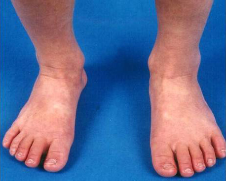
  • Extremity changes
  • Rash

These findings are the basis for the diagnostic criteria for KD. Patients who lack a sufficient number of findings to fulfill the classic criteria may have incomplete KD.

These findings are often not present at the same time. Thus, repeated histories and physical examinations are important in making a timely diagnosis of KD in children with fever and signs of mucocutaneous inflammation.

Infants and possibly adults are more likely to present with incomplete KD. Infants are at greater risk for cardiovascular sequelae, possibly due to a delay in diagnosis and intervention. Thus, infants six months of age or less with unexplained fever for at least seven days should be evaluated for KD, even if they have no clinical findings of KD.

No laboratory studies are included among the diagnostic criteria for complete KD. But certain findings characteristic of KD may support the diagnosis in ambiguous cases. However, the presence of compatible laboratory features strongly supports the diagnosis.

According to classical criteria, the diagnosis of KD requires the presence of fever ≥five days, combined with at least four of the other five signs of mucocutaneous inflammation, without any other explanation. A significant proportion of children with KD have a concurrent infection. Therefore, ascribing the fever to such an infection or KD requires clinical judgment.

Additional clinical and laboratory features are often used to guide diagnosis in children who have fewer than five KD criteria (incomplete KD). The criteria to diagnose incomplete KD and initiate treatment in a child in whom an experienced clinician believes that KD is the probable diagnosis include elevated CRP or ESR and three or more abnormal supplemental laboratory findings OR abnormal echocardiography. A child with incomplete KD whose diagnosis is delayed is more likely to develop CA abnormalities. Thus, KD should be in the differential diagnosis of any child with unexplained fever lasting five or more days.

KD is most commonly confused with infectious exanthems of childhood. The presence of clinical features not commonly found in KD, including exudative conjunctivitis, exudative pharyngitis, discrete intraoral lesions, bullous or vesicular rash, splenomegaly, or generalized lymphadenopathy, suggest another diagnosis. Nonetheless, KD is sufficiently pleomorphic that none of these findings can definitively exclude the diagnosis. Children with KD can have concurrent infections, particularly with viruses circulating in the community at their diagnosis.

Patients who fulfill the criteria for complete KD or incomplete KD require treatment because of the risk of cardiovascular complications that may result in significant morbidity and even mortality.

A single IVIG dose (2 g/kg administered over 8 to 12 hours) is recommended in patients with KD. IVIG is most effective if administered within the first ten days of illness before aneurysms typically develop. Nonetheless, IVIG should be administered even beyond this 10-day window in patients with evidence of persistent vasculitis or systemic inflammation (e.g., persistent fever, elevated acute-phase reactants).

In patients with KD, aspirin should be administered during the acute phase of the illness. The AAP and AHA have recommended a broad range of aspirin doses (30 to 100 mg/kg/day), but it is not clear that any amount of aspirin has long-term benefits. A total daily aspirin dose of 30 to 50 mg/kg/day in four divided doses (maximum dose 4 g per day) is advised. Still, it should be held for any contraindication, particularly exposure to varicella or influenza. The aspirin dose is decreased to 3 to 5 mg/kg/day 48 hours after fever resolution. Aspirin is continued until laboratory markers of ongoing inflammation (e.g., platelet count and ESR) return to normal unless CA abnormalities are detected by echocardiography, in which case aspirin therapy is continued.

For Japanese children, well-validated criteria, such as the Kobayashi criteria or similar, can be used to select patients at increased risk of IVIG resistance. Such high-risk patients are most likely to benefit from adjuvant treatment with glucocorticoids in addition to IVIG.

For non-Japanese patients, the criteria for identifying at-risk children are not well validated. In these patients, the following may be used to select those who are most likely to benefit from augmented treatment with glucocorticoids in addition to initial IVIG:

  • Age ≤12 months (and particularly age <6 months)
  • Enlarged CAs at presentation (before IVIG treatment)
  • KD associated with shock
  • KD presenting with MAS

In children with KD who are determined to be at increased risk of IVIG resistance, glucocorticoids should be added to initial IVIG therapy. A total of 15 days of prednisone/prednisolone (five days each of 2 mg/kg, 1 mg/kg, and 0.5 mg/kg), starting with an intravenous formulation and switching to oral glucocorticoids 12 to 24 hours before expected discharge, is suggested. A reasonable alternative, particularly in non-Japanese patients, is to treat with IVIG and aspirin alone since most children with KD recover with standard therapy and have a low risk of CA aneurysms or other complications.

Prognosis is based upon the severity of CA involvement as a marker of risk for MI. After the baseline echocardiogram is obtained at diagnosis, echocardiography is usually repeated at approximately two and six weeks of illness to evaluate CA involvement. Examinations and echocardiograms are repeated more frequently in children with CA abnormalities. Children also should be monitored carefully for any signs of persistent illness during the first two weeks after treatment, as those with ongoing inflammation, particularly fever of any degree, are at the highest risk of developing CA abnormalities.

The AHA and AAP have developed guidelines for subsequent therapy, physical activity, and follow-up visits (schedule and content) based on MI's relative risk.

Patients without any cardiovascular abnormalities appear to be clinically healthy at long-term follow-up (range, 10 to 21 years). However, it is unknown whether they are at increased risk for atherosclerotic heart disease.

Administration of live-virus vaccines (e.g., measles, varicella) for at least 11 months in children who have been treated with IVIG should be postponed because passively acquired antibodies can interfere with vaccine immunogenicity. One exception to this postponement is in children residing in communities experiencing an outbreak of a vaccine-preventable disease. Another exception is children on long-term aspirin therapy. Children who are ≥12 months of age and are on long-term, moderate- or high-dose aspirin therapy (30 to 100 mg/kg/day) should receive the varicella vaccine because of the increased risk of Reye syndrome. In situations where vaccination is not postponed, the vaccine should be repeated at least 11 months after administering IVIG. Influenza immunization, recommended in all children over six months of age, is also particularly important in those who require long-term high-dose aspirin therapy because of the possible increased risk of Reye syndrome.

Some patients with KD have persistent or recurrent fever despite treatment with IVIG and aspirin. Prolonged fever is a risk factor for CA sequelae in KD. In these patients, other causes of fever should be considered. Risk factors for failing to respond to a single dose of IVIG include:

  • Age <6 months
  • Elevated CRP
  • Low platelet levels
  • Low sodium
  • Male sex

Patients with refractory KD are at increased risk of developing CA aneurysms.

Additional therapy is recommended in children with KD who have persistent fever 24 hours after completing initial therapy or whose fever returns after an afebrile period (up to two weeks after starting treatment). Retreatment is suggested with a single dose of IVIG (2 g/kg). Additional dose(s) of IVIG may be administered before considering other medications in those who remain febrile. However, the benefits of a total dose of more than 4 g/kg of IVIG have not been demonstrated.

For patients who do not respond to retreatment with IVIG, treatment with glucocorticoids is suggested. Pulsed-dose methylprednisolone (30 mg/kg per day administered on one to three consecutive days) once daily until a full response (e.g., resolution of fever) is obtained or the patient has received three doses is suggested. A 15- to 30-day course of twice or thrice daily oral prednisone (total daily dose 1 to 2 mg/kg) may be equally or more effective than IV methylprednisolone in children with KD who are resistant to IVIG.

Therapeutic alternatives, which have demonstrated effectiveness for other vasculitis forms, include TNF-alpha inhibitors, other immunosuppressive agents, and plasmapheresis. However, there are limited data concerning these agents' risks and benefits for refractory KD and only a few small trials to evaluate them. The most commonly used of these agents is infliximab, a monoclonal antibody against TNF-alpha. In multiple trials, it effectively lowers fever, laboratory markers of inflammation, and signs of mucocutaneous inflammation, but there is minimal evidence that it improves CA outcomes. Infliximab (5 mg/kg) is typically given to children who have evidence of active vasculitis despite receiving two IVIG courses (total 4 mg/kg) and one to three IV doses of methylprednisolone.

Implicit Bias Statement

CEUFast, Inc. is committed to furthering diversity, equity, and inclusion (DEI). While reflecting on this course content, CEUFast, Inc. would like you to consider your individual perspective and question your own biases. Remember, implicit bias is a form of bias that impacts our practice as healthcare professionals. Implicit bias occurs when we have automatic prejudices, judgments, and/or a general attitude towards a person or a group of people based on associated stereotypes we have formed over time. These automatic thoughts occur without our conscious knowledge and without our intentional desire to discriminate. The concern with implicit bias is that this can impact our actions and decisions with our workplace leadership, colleagues, and even our patients. While it is our universal goal to treat everyone equally, our implicit biases can influence our interactions, assessments, communication, prioritization, and decision-making concerning patients, which can ultimately adversely impact health outcomes. It is important to keep this in mind in order to intentionally work to self-identify our own risk areas where our implicit biases might influence our behaviors. Together, we can cease perpetuating stereotypes and remind each other to remain mindful to help avoid reacting according to biases that are contrary to our conscious beliefs and values.

References and Resources

Sundel, R. (2020a, Sept). Kawasaki disease: clinical features and diagnosis. UpToDate. Visit Source.

Sundel, R. (2020b, Sept). Incomplete (atypical) Kawasaki disease. UpToDate. Visit Source.

Sundel, R. (2020c, Sept). Kawasaki disease: epidemiology and etiology. UpToDate.Visit Source.

Sundel, R. (2020d, Sept). Kawasaki disease: initial treatment and prognosis. UpToDate. Visit Source.

Sundel, R. (2020e, Sept). Refractory Kawasaki disease. UpToDate.Visit Source.