Sign Up
You are not currently logged in. Please log in to CEUfast to enable the course progress and auto resume features.

This Course Has Expired

Sorry, but this course is no longer active. We are keeping the course material here for research puposes. View our full course library
REQUEST THIS COURSE BE RENEWED
Author:    Jassin Jouria (MD)

Introduction

With the recent upsurge of confirmed leprosy cases in Florida, there is a heightened need to improve medical knowledge and public awareness about leprosy. This course is developed to bridge the gap across three pivotal areas – clinical studies on leprosy, public awareness of the disease, and current medical practice in leprosy management. This course, in its entirety, will discuss disease pathophysiology, classification, and therapy strategies.

Also known as Hansen's disease, leprosy has etched a long history as far back as modern civilization. The first case reports of leprosy with an express description of clinical presentation reportedly date from 600 B.C. to early 1400 B.C. in India. Of noteworthy account are the disease's chronic infectious cycle and its causative organism – Mycobacterium lepraeM. leprae. The bacilli infect the host body and spread progressively through the respiratory path, developing an affinity for the peripheral nerve cells and preferentially attacking the Schwann cells. Continued progression of cellular degradation results in collective stripping of the myelin sheaths of the cells, loss of neuronal conductance, and, subsequently, numbness.

Case Study

Ryna, a 33-year-old female with no underlying medical conditions, presented to the New York Municipal Hospital dermatological unit in October 2022. She had lived in the United States for the past 15 years and had only taken a short trip to Mexico in 2009 on a vacation visit. She had presented to the clinic with hyperpigmentation and numerous lesions on her back and the trunk region. Ryna informed the dermatologist that she first noticed the strange formation on her back about two years before the presentation. Remembering vividly, she described macular lesions with no infiltration but with poorly demarcated edges.

She had self-medicated on a topical antimicrobial agent containing hydrocortisone, clindamycin, and clotrimazole. Two weeks after consistent use of the topical agent, the formation had resolved significantly. However, the pigmentation only faded slightly. A few months later, she explained how the pigmentation had slowly resolved, leaving a dry patch on the skin. Strangely, she noticed the dry patch was poorly sensitive to touch, heat, or needle pricks. She also noticed a significantly reduced sweating response to increased room temperature during this time.

Clinical examination

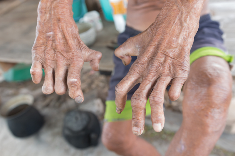
On examination, Ryna's lesions presented as multiple, asymmetrically distributed, and hypopigmented plaques with poorly defined borders. She reported little to no sensory response to sensation on her trunk, back, and left cheek. There were infiltrated plaques on her earlobes and a few on the back. On neurological examination, the sensation of pain on the tips of her fingers and toes was minimal. Peripheral nerve examinations were unremarkable, as there were no signs of nerve enlargement.

Diagnosis and therapy plan

A provisional diagnosis of leprosy was made pending the result of laboratory investigations. The dermatology teams obtained slits of skin smears of plaques from her trunk and earlobes. The acid-fast staining of these samples confirmed the presence of acid-fast bacilli in all samples. With the presenting evidence – generalized distribution of plaque on the back, trunk, cheeks, and earlobes, the presence of bacilli in skin smears, and decreased sensory function on skin patches – the dermatological team made a confirmed diagnosis of lepromatous leprosy.

Following the WHO therapy regimen, Ryna was placed on the following:

  • Tab Rifampicin 600mg plus Clofazimine 300mg once per month and
  • Tab Dapsone 100mg plus Clofazimine 50mg daily for two years.

About seven months after presentation, Ryna's lesion had significantly reduced, and sensory functions had improved. A follow-up visit plan was drafted annually for the next five years.

Overview of Leprosy as a Clinical Disease and Not a Mythological Condition

To a large extent, leprosy is largely considered one of the oldest diseases ever documented in human colonies. With actual documentation dating back to at least the 5th century B.C., descriptions of different cases of a 'skin disfiguring condition' are found in Egyptian and Persian scripts. In the Middle Ages, more descriptions of the disease emerged throughout Europe, with different submissions describing the disease as 'incurable' and 'devilish.' As common practice during these times, individuals with these diseases were isolated as outcasts in different leper colonies located many miles away from uninfected populations.

photo of skin in leprosy

Skin in Leprosy

In medieval times, despite measures to separate the infected individuals from the uninfected population, leprosy reportedly became endemic in Europe. Records exist documenting about 2,000 leper colonies in France alone by the 13th century. During the Renaissance, strict isolation of all infected individuals reportedly yielded results as records of new infections significantly reduced. In central Europe, isolation interrupted chain transmissions of the disease, and it reportedly 'vanished.'

However, the disease would resurface and persist into the 20th century in many parts of Europe. Regions particularly affected include Sicily, Scandinavia, the Iberian Peninsula, the Baltics, and the Balkans. Although there were exhaustive descriptions of the disease conditions, features, and characteristic manifestations in different groups, clinical investigations into pathophysiology and transmission were limited. The first recorded clinical interest in the diseases was published by Boeck and Danielsen  - two Norwegian scientists who had authored pathological and anatomical descriptions of the disease in many people. In 1873, Armauer Hansen eventually identified and isolated M. leprae as the causative organism in multiple disease cases (Fischer, 2017).

The discovery of M. leprae also confirmed the infectiousness of the condition, later described as "specdalsked" – a Norwegian term for leprosy. Since this discovery by Hansen, clinical studies on leprosy have focused largely on understanding the pathophysiology and possible treatment options of leprosy. In 1941, Guy Henry Faget documented the successful use of sulfonamides in treating early cases of leprosy. In 1947, Robert G. Cochrane would later introduce the drug 'dapsone' as the first largely studied bioactive agent for treating leprosy. Today, dapsone remains an integral component of different multidrug therapy regimens for the management of leprosy (Collin et al., 2023).

Etiology, Transmission, and Pathophysiology of Leprosy

M. leprae, in its virulent state, exists as a non-motile, acid-fast rod with a length range of 4 – 7 microns. In laboratory studies, M. leprae has shown no colony growth when cultured in growth mediums. However, animal cultures produced slowly multiplying colonies of the bacterium. Mouse paws and the nine-banded armadillo animal cultures are widely used in these studies. Growth conditions appear to be properly facilitated in low temperatures as cellular proliferation occurs slowly, only about every twelve days. With an incubation period lasting up to 11 years, M. leprae has been shown to affect different inflammatory reactions and dermatological manifestations in infected individuals (Ploemacher et al., 2020).

Depending on the clinical variants, the variability and virulence of M. leprae differ significantly. To an extent, the likelihood of a leprosy infection depends on the integrity of the immune system. The patients' susceptibility to the microorganism as genetically determined is also considered an important factor in infection onset. Clinical characteristics of an active infection are largely determined by the microorganism's tropism for the skin and the peripheral nervous tissues. Also, the clinical variant of the causative organism determines the clinical morphology of the skin. Marked variabilities in the number, appearance, and distribution patterns of lesions in different patients seem to exist. Recent clinical studies on susceptibility and virulence suggest that active disease only occurs in about 5-10% of the infected population.

In 2008, bacilli from two cases of leprosy were isolated as a new species, Mycobacterium lepromatosis (M. lepromatosis). The new species was discovered and isolated following the death of a patient of Mexican origin who had been diagnosed with diffuse lepromatous leprosy (DLL) co-occurring with Lucio's phenomenon – a severe leprosy reaction. Sequencing studies confirmed close genetic similarities between M. lepromatosis, M. leprae, and Mycobacterium haemophilum (M. haemophilum). Polymerase chain reaction (PCR) comparison results also confirmed M. lepromatosis as a causal agent of diffuse lepromatous leprosy and borderline lepromatous forms of leprosy (Collin et al., 2023). Since the first isolation in 2008, clinical studies have subsequently isolated M. lepromatosis samples from at least 15 confirmed cases of leprosy. Case distribution of the new species appears to be mostly in Mexico, the United States, Cuba, Canada, Paraguay, Singapore, Indonesia, and Myanmar (Deps & Collin, 2021).

Transmission

The exact mode of transmission in humans is not clearly defined. Modes of transmission are documented in different areas, ranging from tattooing to household contacts and exposure to armadillos. Zoonotic transmissions appear to be important in many regions where leprosy is considered endemic. In 2016, M. leprae and M. lepromatosis strains were reportedly found in red squirrels. Transmission cycles maintained by animals harboring these strains have been implicated in the continued emergence of leprosy in different regions of the globe (Schuab et al., 2020). Household contact reportedly accounts for about 28% of new cases, with many of these contacts described as M. leprae by PCRs that were positive on skin and nasal swabs (Makhakhe, 2021).

M. leprae infections also occur through air droplets expelled from infected individuals. In lepromatous leprosy patients, infected tissues reportedly house up to seven billion bacilli, making these individuals highly infectious. Experimental models of transmission and infection suggest that the main entry route and dissemination point of the bacilli is the upper respiratory tract. However, there seems to be wide variabilities in these two points in different infected persons. These variabilities have made it difficult to pinpoint an exact, generalized mode of transmission of the causal organisms. In many cases, the identification of leprosy transmission is also hindered by the long incubation period of the causal organisms, as clinical manifestation can take up to 10-15 years in infected individuals (Bhandari et al., 2022).

Pathophysiology

The pathophysiology of leprosy is properly documented in infected individuals with special cases of susceptibility to causal organisms. In active cases of infection, the bacilli primarily cause physiological deficiencies in the macrophage, keratinocytes, and histocytes of the skin. Simultaneously, active infections cause axonal dysfunction and demyelination in the Schwann cells of the peripheral nerves as the clinical presentation continues to emerge. In infected Schwann cells, cellular de-differentiation and reprogramming consequently lead to degeneration of the peripheral nerves as local senses of touch and pressure significantly diminish (De Oliveira Brugger et al., 2023). As documented by many etiological studies, M. leprae appears to express a multi-dimensional approach to cell invasion in an active infection.

In normal physiological setup, Schwann cells have been demonstrated to depend on neuronal ligands for functionality and survival. This interdependence is mediated by an interaction facilitated in the binding of neuregulin to ErbB2/ErbB3 – a receptor tyrosine kinase complex. This binding is primarily important in the differentiation and proliferation of Schwann cells. By binding to and activating ErbB2, M. leprae induces early demyelination of neurons. Demyelination and deregulation of the Schwann cell-axon signaling system lead to the eventual physiological collapse of the myelin sheath – a situation that directly initiates nerve injury and demyelination (Mungroo et al., 2020).

Clinical evidence documenting how M. leprae reprograms Schwan cells through epigenetic modification of key genes has slowly accumulated over the past few years. Schwann cell reprogramming ultimately facilitated the spread of M. leprae. Studies have also documented how the propagation of infection in fibroblasts is more efficient when M. leprae originates from reprogrammed Schwann cells compared to non-reprogrammed ones. By reprogramming Schwann cells, M. leprae ultimately improves its chances of continued survival and propagation in infected individuals (Lara et al., 2023).

Classification and Clinical Presentation of Leprosy

Depending on the parameter of description, the classification of leprosy has varied over the past few years. In 1982, the World Health Organization (WHO) proposed a simplified classification of leprosy based on the density of leprosy bacilli in slit-skin examinations at different stages of infection. Referred to as the 'bacterial index,' this classification method divided active cases of leprosy into paucibacillary and multibacillary. Multibacillary cases are characterized by a bacterial index equal to or higher than 2+, while paucibacillary cases have a bacterial index lower than 2+.

Subsequently, in 1988, the WHO expert committee reviewed the definition of these classifications to make diagnosis more affordable and reduce the risk of cross-infection in front-line healthcare workers. The review described paucibacillary cases in which the infected individual has less than five skin lesions and involves only one nerve trunk. Multibacillary cases involve more than five skin lesions and more than one nerve trunk. Also, patients with multibacillary have multiple symmetric lesions and organisms detectable by smears or biopsy. In paucibacillary, the lesions have few or no organisms (Chen et al., 2022).

Today, this classification system is used mainly for treatment purposes and clinical studies comparing the efficacy of different treatment strategies. The individual's immune system integrity plays a large role in the form of leprosy that manifests during the clinical stage of an active infection. Multiple pieces of evidence suggest that individuals with a cell-mediated immune response against M. leprae eventually develop the paucibacillary form of leprosy. Conversely, those with an inadequate immune response develop the multibacillary form. Post-exposure documentation of active infection cases suggests that the common outcome after bacilli exposure is spontaneous cure.

However, if active infection progresses, a single hypopigmented patch may appear as the first clinical manifestation of the lesion. Then, this is followed by mild anesthesia in the stage commonly referred to as indeterminate leprosy disease since the course of clinical manifestation cannot be predicted.

Concerning clinical presentation, the physical manifestations of leprosy as a disease occur between two stable forms, the tuberculoid and lepromatous forms. In every patient, these forms of leprosy do not change throughout the clinical presentation and remain the same throughout the course of infection.

Classification of Leprosy Types

  1. Early/indeterminate leprosy
  2. Tuberculoid leprosy
  3. Borderline leprosy Forms
    • Borderline tuberculoid leprosy
    • Borderline borderline leprosy
    • Borderline lepromatous leprosy
  4. Lepromatous Leprosy

Early and Indeterminate Leprosy

Diagnosed as the early stage of bacilli invasion, indeterminate leprosy is characterized by prodromal symptoms almost unnoticed in many patients until the appearance of cutaneous lesions. In most cases, it presents primarily as numbness followed by loss of sensitivity to temperature extremes and light pressure as the earliest signs of sensory impairment. Variables in the level and type of sensory loss experienced first have been reported to be linked with the type of disease that is slowly evolving. However, in most cases, the dermatological presentation of indeterminate leprosy is similar across infected individuals.

A solitary, hypopigmented macule is often the first lesion noticed as it merges perfectly into the surrounding healthy skin patch. Erythematous macules may be present in some cases. Both clinical presentations are likely to be noticed on the cheeks, upper arm, thigh region, and buttocks. Sensory functions on these macules may be minimal or completely impaired, and compared to other forms, the macules may contain only a few or no bacilli on biopsy. From this form, the disease may evolve into either tuberculoid or borderline lepromatous leprosy.

Tuberculoid Leprosy

In this form of leprosy, the presenting lesions are solitary, asymmetrically distributed, and may be five or fewer. On close physical examination, lesions appear dry, scaly, hairless, and hypopigmented. In some cases, a few lesions may be erythematous. Clinical studies have also reported typical large erythematous plaque-like presentations with elevated, rounded edges that slope down to a flattened atrophic center. Compared to those formed in indeterminate leprosy, lesions may also appear macular, erythematous, or hypopigmented in the early stages.

The distinguishing feature, however, is the presence of palpable indurations accompanied by a completely altered neurological function at the site of lesion formation. In tuberculoid leprosy, the most common location for the formation of dermatological symptoms includes the trunk, limbs, and face. In most cases, the groin, perineum, axilla, and scalp are not involved. Another presentation often reported in this form of leprosy is the enlargement or tenderness of superficial peripheral nerves serving or proximal to the lesions. In the same vein, the peroneal nerve and the greater auricular nerve may also present as enlarged or tender (Jariyakulwong et al., 2022).

Nerve involvement in tuberculoid leprosy implicates and initiates characteristic changes in the physiology of muscle groups in the affected region. The interosseous muscles of the hand may atrophy, leading to contracture of the fingers and wasting of the thenar and hypothenar eminences. As facial nerve paralysis progresses, the risk of vision loss and many ocular dysfunctions significantly increases. Although the emergence of lesions may be slow, spontaneous remission of dermatological presentation has been reported, and remission may be faster with adequate therapy.

photo of hand wasting

Hand Wasting

Borderline Leprosy Forms

Borderline Tuberculoid Leprosy

Clinical presentations of borderline leprosy share multiple similarities with those of tuberculoid leprosy. The distinguishing factor, however, is that lesions in the borderline form are more numerous and are often reported as smaller in size. A typical presentation often reported is the appearance of satellite lesions around larger macules or plaques. If erythematous infiltrates are present, they form with prominent borders sharply demarcated and arranged asymmetrically. Sensory dysfunction and motor nerve involvement may also be present, although less pronounced than tuberculoid leprosy. Histologically, granulomatous infiltrates may extend to the cutis, and only a few bacilli are detected on biopsy.

Borderline Borderline Leprosy

Borderline borderline leprosy is characterized by numerous but countable skin lesions formed conspicuously with reddish plaques of no regular shape. The larger plaques may be surrounded by small satellite lesions largely distributed asymmetrically. Compared to those formed in tuberculoid leprosy, the lesions in this form are poorly defined. Nerve involvement presents as thickened and tendered nerves with moderate loss of sensory functions. Depending on the lesion, the biopsy may reveal a few or numerous bacilli. Normal hair growth and sweat gland function are primarily unaffected, and granulomas are often not reported in many cases.

Borderline Lepromatous Leprosy

The most reported clinical presentation of borderline lepromatous leprosy is the formation of lesions that are too numerous to count, formed on unaltered skin patches, and distributed asymmetrically. Papules, nodules, and infiltrated plaques are usually present as numerous, hypopigmented, and poorly demarcated. Sweating and hair growth in the affected skin region are hardly affected. In this form of leprosy, there is an extensive, symmetric involvement of peripheral nerves as they become enlarged and tender. Although dermatological symptoms may be slightly different in a few patients, they usually do not show the features of full-blown lepromatous leprosy as presentations such as leonine facies, loss of eyebrows, nasal ulceration, and keratitis are hardly present (Arunraghav et al., 2021).

Lepromatous Leprosy

As with tuberculoid leprosy, patients with clinical presentation of indeterminate leprosy may develop lepromatous leprosy. Often, a downgrade from borderline leprosy forms may also develop into lepromatous leprosy. Typically, lesions of lepromatous leprosy present with pale macules and diffuse infiltration of the skin distributed asymmetrically over the body. Unlike tuberculoid macules, the ones in lepromatous macules are numerous, smaller in size, and poorly defined with skin texture changes. Textural configuration and skin tone may remain intact, making the lesion blend perfectly with the surrounding skin. Sensory functions are not lost, sweating may be unaffected, and there is little to no peripheral nerve involvement. Hair loss may occur slowly and progressively from the outer third of the eyebrows to the lashes and the body. Hair on the scalp may remain largely unaffected (De Sousa Oliveira et al., 2019).

A standard procedure for diagnosis is the physical examination of lepromatous infiltrations in suspected cases. These infiltrations are divided into three types – nodular, diffuse, and plaque types. The diffuse types are presented as diffuse infiltrations of the face with a waxy and shiny appearance noticeable on the forehead. In all cases of lepromatous leprosy, recent clinical studies suggest nerve involvement invariably occurs, proceeding slowly in a bilaterally symmetrical pattern. Considering the pattern of involvement, loss of sensory function in this case may be misdiagnosed as diabetic neuropathy in many active leprosy cases.

Leprosy Reactions

Diagnosis conventions advise that the assignment of a patient to any of the chronic forms of leprosy, as discussed above, must not be definitive. About 30% of presenting cases reportedly develop acute, life-threatening exacerbations that may occur spontaneously or during treatment. These exacerbations, referred to as 'leprosy reactions', manifest as the sudden impairment of a patient's immunological functions and may occur at any time, even after a successful treatment.

The lack of severity in the onset of the disease makes it difficult to diagnose leprosy accurately. It is often mistaken for fungal infections, allergic reactions, vitiligo, and other conditions with dermatological presentations. This makes diagnosis particularly problematic for healthcare professionals in regions where leprosy is not endemic. In solving this problem, healthcare providers can watch for inflammatory conditions that often appear at the onset of infection with M. leprae. These leprosy reactions can occur at any stage of infection and cause permanent nerve impairments. Recognizing these reactions in a thorough physical examination can help healthcare providers properly identify disease progression and type.

Leprosy reactions are largely studied under three categories – type 1 reaction, type 2 reaction, and type 3 reaction. They are associated with the different forms of leprosy.

Type 1 Reaction

Type 1 reactions of leprosy are mostly presented in borderline leprosy forms. It may occur within the first twelve weeks of starting a therapy plan for patients with borderline borderline, borderline lepromatous, and borderline tuberculoid forms of leprosy (Luo et al., 2021). In type 1 reactions, healthcare providers might notice inflammation, tenderness, ulcerations, and pain on preexisting lesions. There might be accompanying neuritis presenting as nerve damage. Clinical symptomatology may vary depending on the part of the body where reactions are noticed.

In the face, nerve damage may cause facial paralysis, leading to lagophthalmos – an inability to close the eyelids. Patients may also present with associated inflammatory eye conditions, including scleritis and iritis. In many cases, complications may lead to blindness. Type 1 reaction-induced nerve damage on the feet may prompt a sudden onset of foot drop, leading to permanent impairment. Regardless of the reaction site, healthcare providers should watch out for acute inflammation of existing lesions, edema or erythema, and sudden nerve damage as the clinical telltales of type 1 leprosy reactions. A skin biopsy specimen in type 1 reactions reveals the presence of granuloma edema, plasma cells, giant cells, and dermal cells.

Type 2 Reaction (Erythema Nodosum Leprosum)

Type 2 reactions of leprosy, also known as erythema nodosum leprosum, are commonly reported in people with lepromatous leprosy and in women at certain stages of life development, including pregnancy, lactation, and puberty. Clinical studies have also identified emotional and psychological stress as common triggers for type 2 leprosy reactions. Compared to type 1, there is a higher likelihood of reoccurrence, and the systemic nature makes these reactions more complicated.

Healthcare providers should watch out for these risk factors to determine whether a lesion complication is a type 1 or 2 leprosy reaction. Type 2 reactions develop as an immune complex vasculitis, causing the deposition of antigen-antibody complexes into tissues. At first, there is an acute inflammation of the subcutaneous layer of fatty tissues. Healthcare providers can accurately recognize type 2 reactions by watching for skin presentations of subcutaneous nodules presenting simultaneously with fever and malaise. In many cases, there is inflammation of the extremities, lymph nodes, and eyes. Nodules spread rapidly across the body and may become neurotic or ulcerate. Biopsy specimens in type 2 reactions reveal dead bacilli, lymphocytes, and neutrophils trapped in macrophages.

Table 1.

Skin Presentations

Redness and acute inflammation in preexisting lesions that may sometimes ulcerate; marked edema in the hands, feet, and face.

Red, painful nodules present in crops on previously unaffected skin of the face, trunk, and limbs; nodules may become ulcerated; edema on the hands, feet, and face; preexisting lesions remain unchanged in appearance. 
Clinical PhenotypePresent in borderline leprosy forms and also in tuberculoid leprosy.Present in borderline leprosy and lepromatous leprosy.
Neural ChangesNerve damage presents as numbness or muscle weakness in the face, feet, and hands; pain and tenderness in nerve with or without a loss of nerve function.Nerve damage presents as numbness or muscle weakness in the hand, feet, and face; pain and tenderness in nerves with or without loss of function.
Systemic ComplicationsUnusualNeuritis, malaise, uveitis, fever, arthritis, dactylitis, orchitis.
TreatmentCorticosteroidsCorticosteroids/thalidomide
Common PrecipitantMultidrug therapy, immunization.Hormonal states (puberty, pregnancy, lactation); lepromatous leprosy forms.
Likelihood of RecurrenceLess likelyVery likely
(Luo et al., 2021)
 
CharacteristicType 1 ReactionType 2 Reaction

Type 3 Reaction (Lucio's Phenomenon)

Recently, the Lucio phenomenon has been primarily studied as a type of leprosy reaction. It is generally observed in patients with untreated lepromatous leprosy and characterized by extensive violaceous patches and bullous infiltrates. The initial pathogenesis of Lucio's phenomenon involves the occlusion of deep-vein plexus veins and subcutaneous tissues, leading to cutaneous hemorrhagic infarction. On physical examination, healthcare providers may notice slightly infiltrated plaques on the skin and erythematous-violaceous macular presentations. Subsequently, these presentations develop into central necrosis and ulceration, spreading rapidly to the limbs, trunk, and face. Histological examination of endothelial cells in the areas of the body affected may be positive for M. leprae bacilli (Ya et al., 2021).

Strategies and Clinical Guidelines on Leprosy Management

Globally, the two most widely used treatment strategies for leprosy are the WHO's multidrug therapy regimen and the National Hansen's Disease Program strategy. The WHO multidrug therapy recommends different treatment regimens according to age and the type of leprosy disease diagnosed. Rifampicin, clofazimine, and dapsone are considered first-line treatment agents under this strategy (Lazo-Porras et al., 2020).

Rifampicin

Synthesized in Italy from Amycolatopsi rifampycinica – a gram-positive soil bacteria, rifampicin has been commercially available since 1965. Pharmacologically, it exerts bioactivity by inhibiting bacteria RNA polymerase in mycobacteria. It is considered very active in leprosy cases as it significantly reduces bacteria count detectable in mucosal and cutaneous lesions of the leprosy types after a few days of administration. With a resistance rate currently estimated at 5%, rifampicin is not recommended as a monotherapy.

High doses may cause severe side effects, including nausea, vomiting, and diarrhea. In patients who abuse alcohol while taking rifampicin, elevated liver function tests, intrahepatic cholestasis, and hepatotoxicity have been reported. In first-time users, erythematous macules on the face and scalp may occur within three hours of use. Noticeably, rifampicin causes a red-orange discoloration of the body fluids, including tears, urine, and saliva. Its teratogenic effects make it prohibited for use in pregnancy (Thangaraju & Venkatesan, 2019).

Clofazimine

Synthesized in Dublin in 1954 and first used as a leprosy treatment in Nigeria in 1959, clofazimine is considered in many anti-leprosy drug regimens for its anti-inflammatory and bactericidal effects. Many countries consider it a first-choice drug in treating multibacillary leprosy. It has also shown huge clinical results in treating type 2 leprosy reactions, making it an important component of the WHO treatment strategy. Typically, especially in Caucasians, clofazimine produces red-brown hyperpigmentation of leprosy lesions as the skin tone slowly changes. These effects are reversed over a few months or years once clofazimine is discontinued. Other side effects commonly reported include discoloration of the conjunctiva and body fluids, xerosis, and the accumulation of clofazimine crystals in the intestinal mucosa, causing multiple gastrointestinal symptoms (Riccardi et al., 2020).

Dapsone

Dapsone was first synthesized in Germany in 1908 and used as monotherapy for leprosy for the first time in 1941. It exerts bacteriostatic effects on M. leprae by inhibiting folic acid synthesis. It is widely considered a first-line drug for leprosy in many treatment regimens and is reported to have demonstrated significant clinical benefits in leprosy patients. It has no known teratogenic effects and can be used in pregnancy.

Popularly reported side effects include fatigue, headache, gastrointestinal symptoms, hemolysis, and methemoglobin formation pronounced in patients with a clinical deficiency of glucose-6-phosphate dehydrogenase. Other rare side effects of dapsone include drug reaction with eosinophilia and systemic symptoms (DRESS) syndrome, hepatitis, agranulocytosis, urticaria, and phototoxic reactions (Kurien et al., 2023).

Table 1: Paucibacillary Leprosy Treatment
PopulationMedicationDoseDuration
AdultsRifampicin600 mg/month6 months
Dapsone100 mg/day
Clofazimine300 mg/month + 50 mg/day
Children (10-14 years)Rifampicin450 mg/month6 months
Dapsone50 mg/day
Clofazimine150 mg/month + 50 mg/day
Children (<10 years old or <40 kg)Rifampicin10 mg/kg/month6 months
Dapsone2 mg/kg/daily
Clofazimine6 mg/kg/month + 1 mg/kg/day
Lazo-Porras et al., 2020
Table 2: Multibacillary Leprosy Treatment
PopulationMedicationDoseDuration
AdultsRifampicin600 mg/month12 months
Dapsone100 mg/day
Clofazimine300 mg/month + 50 mg/day
Children (10-14 years)Rifampicin450 mg/month12 months
Dapsone50 mg/day
Clofazimine150 mg/month + 50 mg/day
Children (<10 years old or <40 kg)Rifampicin10 mg/kg/month12 months
Dapsone2 mg/kg/daily
Clofazimine6 mg/kg/month + 1 mg/kg/day
Lazo-Porras et al., 2020

The Hansen Disease Program (NHDP) is commonly used in the United States. It is particularly popular for its longer treatment period and its exclusion of clofazimine in treating paucibacillary leprosy.

Table 3: Tuberculoid Treatment (WHO Classification Paucibacillary)
PopulationMedicationDoseDuration
AdultsRifampicin600 mg/month12 months
Dapsone100 mg/day
ChildrenRifampicin10–20 mg/kg/day (<600 mg)12 months
Dapsone1 mg/kg/day
Franco-Paredes et al., 2022; Lazo-Porras et al., 2020
Table 4: Lepromatous Treatment (WHO Classification Multibacillary)
PopulationMedicationDoseDuration
AdultsRifampicin600 mg/month24 months
Clofazimine50 mg/day
Dapsone100 mg/day
ChildrenRifampicin10–20 mg/kg/day (<600 mg)24 months
Clofazimine1 mg/kg/day
Dapsone1 mg/kg/day
Franco-Paredes et al., 2022; Lazo-Porras et al., 2020

In both treatment strategies, comorbidities, drug-drug interactions, and patient-specific contraindications might force the introduction of a second-line therapy agent. The most commonly used agents in this class include minocycline, ofloxacin, and clarithromycin. As a standard for measuring treatment efficacy, the parameters considered during therapy include:

  1. The rapid decline in the infectivity of patients with an active leprosy disease
  2. Low rate of recurrence and reactions
  3. Prevention of resistance to dapsone

Nursing Care Recommendations in Leprosy

Nursing care in leprosy management focuses on improving the integrity of the dermal layers. The pathophysiology of leprosy involves episodes of continued nerve damage, nodule formation, and ulceration. These effects are implicated in the formation of ulcerative wounds in leprosy. To a large extent, nursing care should prioritize the management of ulcerative wounds in leprosy and monitoring disease progression.

Recommendations commonly adopted in nursing care for leprosy include the following.

Maintaining an optimal wound environment

Creating an optimal environment for wound care precedes any other step in managing ulcerative wounds in leprosy. This starts with determining the ideal type of dressings and the quality to be adopted. In many cases, the ideal dressing option might change consistently as the patient responds to therapy. Based on multiple studies on wound care under these circumstances, the qualities recommended for an ideal dressing in leprosy care include the following:

  • Dressing must be efficient in removing exudates.
  • Dressing must maintain a humid environment on the wound surface.
  • Dressing must be breathable to allow gas exchange.
  • Dressing must maintain a suitable wound surface temperature and prevent microbial entry.
  • Dressing must not stick to the wound surface or leak toxic substances.

Septic wound care

Complicated ulcers in patients not optimally responding to therapy may become infected. In severe infections, the area around the ulcer may become red, hot, and swollen; lymph nodes in the groin and axilla regions may be swollen; exudates may become foul-smelling. A pocket of pus may be built around the wound, creating an ideal environment for the proliferation of bacteria. Pus buildups must be drained consistently. The following recommendations may be adopted to care for septic wounds adequately.

  • Consistently search for pus pockets using a probe.
  • Establish open drainage around the pus pocket and optimize pus removal.
  • Flush the wound cavity with saline using an appropriate syringe volume.
  • Advise the medical team on systemic antibiotic therapy or optimize an existing plan.
  • Remove all dead cells by careful debridement.

Plaster casting 

Extensive leprosy ulcers on the foot may be considered for plaster casting. On average, ulcerative wounds show significant improvement 14 days after debridement. Improvement in this sense may be measured as controlled inflammation, reduction in the volume of exudates, and reduced pain. At this stage, plaster casting may be considered.

Enhancing the healing process

Wound healing is the ultimate goal in the management of complicated leprosy ulcers. In many cases, malnutrition and the continued administration of topical and systemic steroid therapy may weaken the immune system. Steps to further optimize the process of wound healing include the following:

  • Ensure essential hygiene rather than topical medication.
  • Continuously review and optimize the use of systemic antibiotics.
  • Optimize the supply of vitamin C and oxygen for the development of collagen.
  • Address an adequate supply of trace elements required in wound healing, especially zinc, copper, and vitamin A.
  • Provide psychological support for patients.

Conclusion

Leprosy is a long-standing disease that has been traced back through human history. Leprosy is chronically infectious and is caused by M. leprae. This disease affects the skin and nerves and can be diagnosed by dermatological and neurological examinations. Treatment is dependent on the type of leprosy and if any reactions occur.

Implicit Bias Statement

CEUFast, Inc. is committed to furthering diversity, equity, and inclusion (DEI). While reflecting on this course content, CEUFast, Inc. would like you to consider your individual perspective and question your own biases. Remember, implicit bias is a form of bias that impacts our practice as healthcare professionals. Implicit bias occurs when we have automatic prejudices, judgments, and/or a general attitude towards a person or a group of people based on associated stereotypes we have formed over time. These automatic thoughts occur without our conscious knowledge and without our intentional desire to discriminate. The concern with implicit bias is that this can impact our actions and decisions with our workplace leadership, colleagues, and even our patients. While it is our universal goal to treat everyone equally, our implicit biases can influence our interactions, assessments, communication, prioritization, and decision-making concerning patients, which can ultimately adversely impact health outcomes. It is important to keep this in mind in order to intentionally work to self-identify our own risk areas where our implicit biases might influence our behaviors. Together, we can cease perpetuating stereotypes and remind each other to remain mindful to help avoid reacting according to biases that are contrary to our conscious beliefs and values.

References

  • Arunraghav, P., & Herakal, K. (2021). Leprosy in Elderly and Children among New Cases - A 3-Year Retrospective Study. Indian dermatology online journal, 12(2), 294–297. Visit Source.
  • Bhandari, J., Awais, M., Robbins, B.A., & Vikas, G. (2022). Leprosy. In: StatPearls [Internet]. Treasure Island (FL): StatPearls Publishing. Visit Source.
  • Chen, K. H., Lin, C. Y., Su, S. B., & Chen, K. T. (2022). Leprosy: A Review of Epidemiology, Clinical Diagnosis, and Management. Journal of tropical medicine, 2022, 8652062. Visit Source.
  • Collin, S. M., Lima, A., Heringer, S., Sanders, V., Pessotti, H. A., & Deps, P. (2023). Systematic Review of Hansen Disease Attributed to Mycobacterium lepromatosis. Emerging infectious diseases, 29(7), 1376–1385. Visit Source.
  • De Oliveira Brugger, L. M., Santos, M. M. L. D., Lara, F. A., & Mietto, B. S. (2023). What happens when Schwann cells are exposed to Mycobacterium leprae – A systematic review. IBRO Neuroscience Reports, 15, 11–16. Visit Source.
  • Deps, P., & Collin, S. M. (2021). Mycobacterium lepromatosis as a Second Agent of Hansen's Disease. Frontiers in microbiology, 12, 698588. Visit Source.
  • De Sousa Oliveira, J. S., Reis, A. L., Margalho, L. P., Lopes, G. L., Da Silva, A. R., De Moraes, N. S., & Xavier, M. B. (2019). Leprosy in elderly people and the profile of a retrospective cohort in an endemic region of the Brazilian Amazon. PLOS Neglected Tropical Diseases, 13(9), e0007709. Visit Source.
  • Fischer, M. (2017). Leprosy - an overview of clinical features, diagnosis, and treatment. Journal der Deutschen Dermatologischen Gesellschaft = Journal of the German Society of Dermatology: JDDG, 15(8), 801–827. Visit Source.
  • Franco-Paredes, C., Garcia-Creighton, E., Henao-Martínez, A., Kallgren, D. L., Banjade, R., Dyer, J. A., Nelson, T., Zaesim, A., Peluso, M. J., Jain, V., Lee, D. H., Minces, L. R., Wirshup, M., Sierra Hoffman, M., Katsolis, J., Brust, K., Giron, J., Smiarowski, L., Hoosepian-Mer, P. A., & Stryjewska, B. (2022). Novel approaches in the treatment of Hansen's disease (Leprosy): a case series of multidrug therapy of monthly rifampin, moxifloxacin, and minocycline (RMM) in the United States. Therapeutic advances in infectious disease, 9, 20499361221135885. Visit Source.
  • Jariyakulwong, N., Julanon, N., & Saengboonmee, C. (2022). Lepromatous leprosy with a suspected 30-year incubation period: A case report of a practically eradicated area. Journal of Taibah University Medical Sciences, 17(4), 602–605. Visit Source.
  • Kurien, G., Jamil, R. T., & Preuss, C. V. (2023). Dapsone. In StatPearls. StatPearls Publishing.
  • Lazo-Porras, M., Prutsky, G. J., Barrionuevo, P., Tapia, J. C., Ugarte-Gil, C., Ponce, O. J., Acuña-Villaorduña, A., Domecq, J. P., De la Cruz-Luque, C., Prokop, L. J., & Málaga, G. (2020). World Health Organization (WHO) antibiotic regimen against other regimens for the treatment of leprosy: a systematic review and meta-analysis. BMC infectious diseases, 20(1), 62. Visit Source.
  • Luo, Y., Kiriya, M., Tanigawa, K., Kawashima, A., Nakamura, Y., Ishii, N., & Suzuki, K. (2021). Host-Related Laboratory Parameters for Leprosy Reactions. Frontiers in medicine, 8, 694376. Visit Source.
  • Makhakhe, L. (2021). Leprosy review. South African family practice: official journal of the South African Academy of Family Practice/Primary Care, 63(1), e1–e6. Visit Source.
  • Mungroo, M. R., Khan, N. A., & Siddiqui, R. (2020). Mycobacterium leprae: Pathogenesis, diagnosis, and treatment options. Microbial pathogenesis, 149, 104475. Visit Source.
  • Oliveira, J. S. S., Reis, A. L. M. D., Margalho, L. P., Lopes, G. L., Silva, A. R. D., Moraes, N. S., & Xavier, M. B. (2019). Leprosy in elderly people and the profile of a retrospective cohort in an endemic region of the Brazilian Amazon. PLoS neglected tropical diseases, 13(9), e0007709. Visit Source.
  • Ploemacher, T., Faber, W. R., Menke, H., Rutten, V., & Pieters, T. (2020). Reservoirs and transmission routes of leprosy; A systematic review. PLoS neglected tropical diseases, 14(4), e0008276. Visit Source.
  • Riccardi, N., Giacomelli, A., Canetti, D., Comelli, A., Intini, E., Gaiera, G., Diaw, M. M., Udwadia, Z., Besozzi, G., Codecasa, L., & Biagio, A. D. (2020). Clofazimine: an old drug for never-ending diseases. Future microbiology, 15, 557–566. Visit Source.
  • Schaub, R., Avanzi, C., Singh, P., Paniz-Mondolfi, A., Cardona-Castro, N., Legua, P., Crespo, L., Sewpersad, K., Dávila, J. J., Barreto, J., Dwivedi, P., Morris-Wilson, H., Larrea, M. P., Talhari, C., Lahiri, R., Truman, R. W., Gozlan, R. E., Couppié, P., & de Thoisy, B. (2020). Leprosy Transmission in Amazonian Countries: Current Status and Future Trends. Current Tropical Medicine Reports, 7(3), 79–91. Visit Source.
  • Thangaraju, P., & Venkatesan, S. (2019). Current treatment guideline by the World Health Organization against leprosy: A positive focus. Journal of research in medical sciences: the official journal of Isfahan University of Medical Sciences, 24, 95. Visit Source.
  • White, C., & Franco-Paredes, C. (2015). Leprosy in the 21st century. Clinical microbiology reviews, 28(1), 80–94. Visit Source.
  • Ya, S. N. C., Muhamad, R., Zakaria, R., Ishak, A., & Abdullah, W. N. H. W. (2021). Lucio Phenomenon: Sequelae of Neglected Leprosy. Korean journal of family medicine, 42(3), 245–249. Visit Source.