Sign Up
You are not currently logged in. Please log in to CEUfast to enable the course progress and auto resume features.

Course Library

Pneumothorax in the Adult Patient

1 Contact Hour
Listen to Audio
CEUfast OwlGet one year unlimited nursing CEUs $39Sign up now
This peer reviewed course is applicable for the following professions:
Advanced Practice Registered Nurse (APRN), Certified Nurse Midwife, Certified Nurse Practitioner, Certified Registered Nurse Practitioner, Clinical Nurse Specialist (CNS), Licensed Practical Nurse (LPN), Licensed Vocational Nurses (LVN), Midwife (MW), Nursing Student, Registered Nurse (RN), Registered Nurse Practitioner, Respiratory Therapist (RT)
This course will be updated or discontinued on or before Saturday, March 21, 2026

Nationally Accredited

CEUFast, Inc. is accredited as a provider of nursing continuing professional development by the American Nurses Credentialing Center's Commission on Accreditation. ANCC Provider number #P0274.


Outcomes

≥ 92% of participants will know how to assess, manage, and provide care for pneumothorax in adult patients.

Objectives

Upon completion of this course, the participant will be able to:

  1. Explain the structures and functions involved in respiration, including the lungs, diaphragm, bronchioles, and alveoli.
  2. Differentiate between spontaneous, traumatic, and tension pneumothorax and understand each type's specific causes and risk factors.
  3. Identify the key signs and symptoms of pneumothorax, including sudden chest pain, shortness of breath, decreased breath sounds, and cyanosis.
  4. Describe the use of diagnostic tools such as X-rays, computerized tomography scans (CT scans), and ultrasound to confirm the presence of pneumothoraces.
  5. Outline the potential complications and risks of pneumothorax, including respiratory distress, infection, and reoccurrence.
  6. Evaluate different treatment options for a pneumothorax, including observation, needle aspiration, chest tube insertion, and surgical interventions like video-assisted thoracoscopic surgery (VATS) or thoracotomy.
  7. Implement nursing interventions, including chest tube and pain management and education on self-care and prevention of complications.
  8. Recognize the importance of effective communication and teamwork with physicians, respiratory therapists, radiologists, and other healthcare professionals.
  9. Analyze the latest research, including emerging treatment modalities, innovative surgical techniques, and evidence-based practice for improved patient outcomes.
CEUFast Inc. and the course planners for this educational activity do not have any relevant financial relationship(s) to disclose with ineligible companies whose primary business is producing, marketing, selling, re-selling, or distributing healthcare products used by or on patients.

Last Updated:
Restart
Restart
  • 0% complete
Hide Outline
Playback Speed

Narrator Preference

(Automatically scroll to related sections.)
Done
Pneumothorax in the Adult Patient
0:00
0:15
 
To earn of certificate of completion you have one of two options:
  1. Take test and pass with a score of at least 80%
  2. Reflect on practice impact by completing self-reflection, self-assessment and course evaluation.
    (NOTE: Some approval agencies and organizations require you to take a test and self reflection is NOT an option.)
Author:    Lisa Garcia (RN, BSN, CEN)

Case Study

Meet Mr. Jamison, a 45-year-old male with a history of smoking and chronic obstructive pulmonary disease (COPD). He presents to the emergency department after a day of work with a sudden onset of sharp chest pain and difficulty breathing.

Upon assessment, Mr. Jamison appears anxious and is in respiratory distress. His respiratory rate has increased to more than twenty breaths per minute, and he has decreased breath sounds on the right side of his chest. His oxygen saturation is 88% on room air. A chest X-ray is ordered, which reveals a large right-sided pneumothorax with mediastinal shift.

The healthcare team quickly initiates interventions to stabilize Mr. Jamison's condition. They provide supplemental oxygen via a non-rebreather mask and establish intravenous access for fluid resuscitation and pain management. A chest tube is inserted on the affected side to evacuate the air from the pleural space and re-expand the collapsed lung.

Over the next 24 hours, Mr. Jamison's respiratory distress improves, and his oxygen saturation increases to 95% on room air. The healthcare team closely monitors his vital signs, respiratory status, and chest tube drainage. They administer analgesics to manage his pain and provide education on the importance of deep breathing and early mobilization.

Mr. Jamison's chest X-ray is repeated after 48 hours, showing re-expansion of the lung and resolution of the pneumothorax. The chest tube is subsequently removed, and Mr. Jamison is discharged with instructions for follow-up care and smoking cessation support.

Had the X-ray not been completed, the pneumothorax may have been missed, leading to the patient's condition deteriorating. Because no other trauma was involved, such as in the case of a car accident or penetrating trauma, the healthcare provider can possibly take more conservative approaches, such as avoiding surgery.

Anatomy and Physiology

In the first part of this course, we will dive into the world of the respiratory system. We will explore the anatomy and physiology and learn about the different structures involved in breathing and how they work together. This will be a great foundation for understanding pneumothoraces. Let's begin.

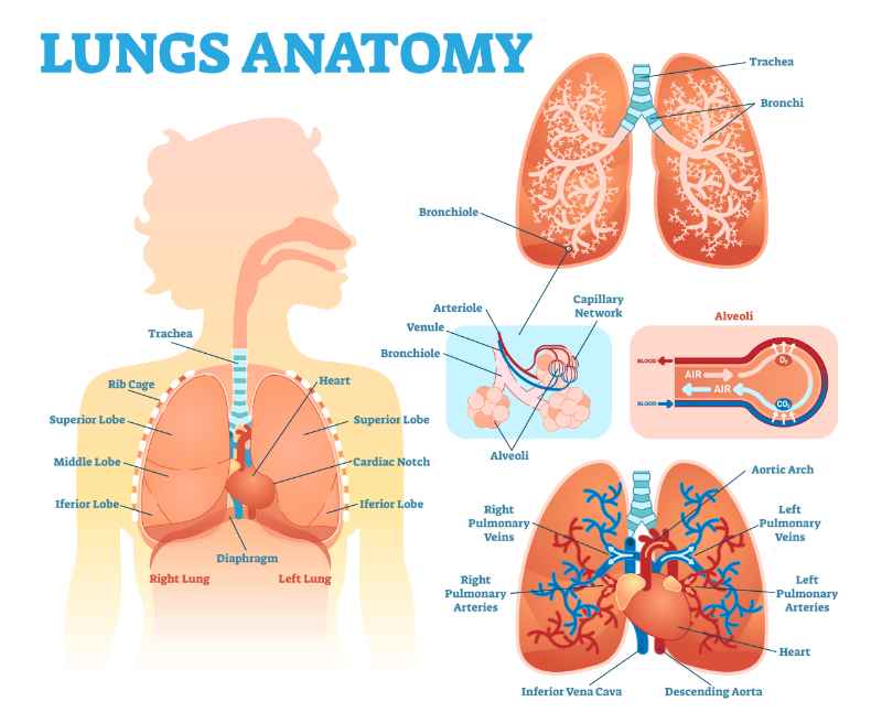
The respiratory system is an amazing network of organs that allows us to breathe; it includes the nose, mouth, throat, trachea, and lungs. When we inhale, air enters through the nose or mouth and travels down the throat and into the trachea. The trachea branches into two tubes called the bronchi, which lead to the lungs. Inside the lungs, the bronchi divide into smaller tubes called bronchioles, which eventually end in tiny air sacs called alveoli. It is in the alveoli where the exchange of oxygen and carbon dioxide takes place. Oxygen enters the bloodstream while carbon dioxide is expelled when we exhale; this continuous process keeps us alive and ensures our body gets oxygen (Patwa & Shah, 2015).

When a person has a pneumothorax, it means that there is air in their plural space or between the lung and the chest wall; this disrupts the normal functioning of the respiratory system. The presence of air in the pleural space can cause the lung to collapse partially or completely; this is considered a pneumothorax and can result in symptoms such as chest pain, shortness of breath, and a rapid heart rate (McKnight & Burns, 2023).

graphic showing anatomy of the lungs

Anatomy of the Lungs

Identify the Different Types of Pneumothoraces

There are a few different types of pneumothorax, each with its own underlying causes. Here are the four main types (Zarogoulidis et al., 2014):

  1. Spontaneous pneumothorax: This occurs without any apparent cause and is often seen in young, tall, and thin individuals. It can happen when a small air sac in the lung, called a bleb, ruptures, causing air to leak into the pleural space.
  2. Traumatic pneumothorax: This type of pneumothorax is caused by a chest injury, such as a rib fracture or a penetrating wound. The injury allows air to enter the pleural space.
  3. Tension pneumothorax: This is a serious and potentially life-threatening type of pneumothorax. It occurs when air enters the pleural space and cannot escape. The trapped air puts pressure on the lining, causing it to collapse and compress other structures in the chest, like the heart and major blood vessels.
  4. Iatrogenic pneumothorax: This type of pneumothorax is caused by medical procedures, such as lung biopsies, chest tube insertion, or mechanical ventilation. It can happen when the lung is accidentally punctured during these procedures.

Signs and Symptoms of Pneumothoraces

As healthcare professionals, it is important to be aware of the signs and symptoms of pneumothorax. Some common indicators include (McKnight & Burns, 2023):

  1. Sudden, sharp chest pain: Patients may describe a sharp or stabbing pain in the chest, often on one side; the pain may worsen with deep breathing or coughing.
  2. Shortness of breath: Patients may experience a sudden onset of difficulty breathing or a feeling of breathlessness. This can be due to the collapse of the lung and the resulting decrease in lung function.
  3. Tachypnea: Patients may exhibit tachypnea, which is an increased respiratory rate. This is the body's attempt to compensate for their reduced lung capacity.
  4. Cyanosis: In severe cases, patients may develop a bluish tinge to their skin, lips, or nails due to inadequate oxygenation.
  5. Decreased breath sounds: When auscultating the patient's chest, you may notice decreased or absent breath sounds on the affected side.
  6. Tachycardia: Patients may have an increased heart rate as the body tries to compensate for the decreased oxygen supply.

Other signs and symptoms that may be seen in pneumothorax include asymmetrical lung expansion and a hyperresonant percussion note.

In a tension pneumothorax, more serious symptoms may be seen and include jugular venous distension, low blood pressure, respiratory failure, and eventual cardiac arrest (McKnight & Burns, 2023).

It is important to note that the severity of symptoms can vary depending on the type and extent of the pneumothorax. Prompt recognition and appropriate management are crucial in ensuring the best outcomes for these patients.

Diagnostic Tools and Techniques

Healthcare professionals may utilize various diagnostic tools and techniques to confirm a suspected pneumothorax. The first is physical assessment. Thorough physical assessment, including auscultation of all lung fields, can provide valuable information. Auscultation of breath sounds can help identify a decrease in or absence of breath sounds on the affected side, which may indicate pneumothorax.

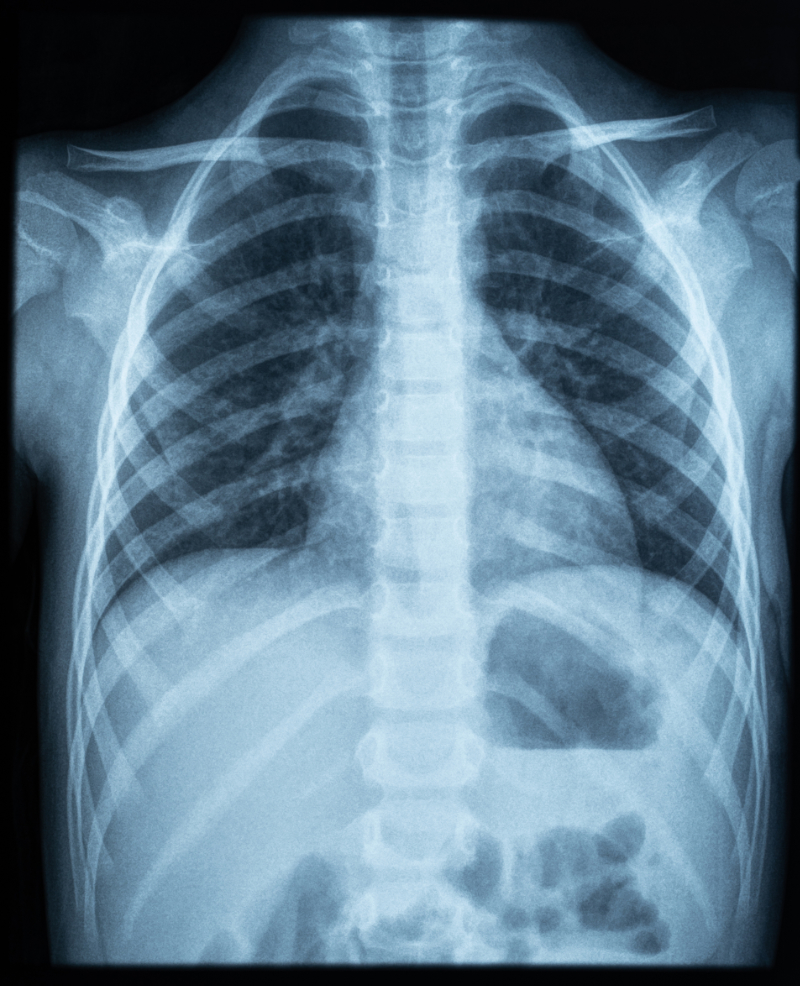
The first radiologic tool is a chest X-ray. A standard chest X-ray is often the initial diagnostic tool used to assess for a pneumothorax (Bhoil et al., 2021). It can help visualize the presence of air in the pleural space and determine the extent of lung collapse.

photo of chest x-ray

Chest X-ray

Another radiologic tool is a computerized tomography (CT) scan. CT scans may be ordered to obtain a more detailed image of the chest. CT scans can provide a clearer visualization of the pneumothorax and help identify any underlying disease and abnormality. The study by Burger et al. (2023) shows the importance of getting advanced imaging; this study presented a 13-month-old patient with what seemed to be a spontaneous pneumothorax, but after CT revealed a multilocular cystic lesion in the left hemithorax region.

The next radiologic tool is ultrasound. Ultrasound imaging can be used to detect the presence of pneumothorax at the bedside; it involves using sound waves to create real-time images of the chest and can be particularly useful in emergencies (Husain et al., 2012). The last diagnostic tool is arterial blood gases (ABG). ABGs may be performed to assess the patient's respiratory status and to determine the severity of the pneumothorax. It helps evaluate oxygen and carbon dioxide levels in the blood (Daley, 2024).

Potential Complications and Risks

Understanding the potential complications and risks associated with pneumothorax is essential. Most pneumothoraces can be managed effectively, but there are potential complications to be aware of. One such complication is tension pneumothorax, where air continuously enters the pleural space and cannot escape, causing increased pressure on the affected lung and potentially leading to cardiovascular compromise. Other risks include infection, bleeding, the development of adhesions, or scarring in the pleural space. Additionally, individuals with underlying lung conditions, such as COPD, are at a higher risk of recurrent pneumothoraces (McKnight & Burns, 2023). Recognizing these complications and promptly addressing them is crucial in providing appropriate care.

Treatment Options

Let's talk about the treatment options for pneumothorax. The approach to treating pneumothorax depends on the severity and underlying cause. Here are some common treatment options in descending order, with the conservative treatment plan listed first:

  • Observation: Healthcare professionals may choose a conservative approach and monitor the patient closely for small, uncomplicated pneumothoraces. The patient should not have any prior lung disorders, and breathlessness should not be present. Observation involves regular assessments, pulse oximetry, and serial ABGs to ensure the pneumothorax does not worsen (Li et al., 2014; Zarogoulidis et al., 2014).
  • Needle decompression: In some cases, needle decompression may need to be performed to remove the excess air from the pleural space. This procedure involves inserting an angiocath (size depends on the facility) into the chest to release the trapped air and relieve pressure on the lungs (Wernick et al., 2015).
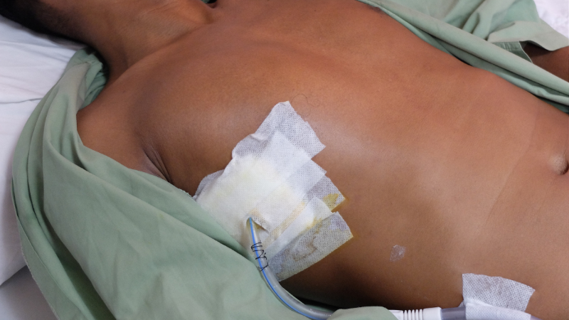
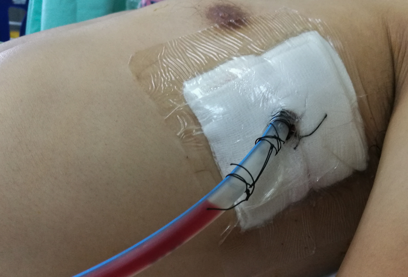
  • Chest tube (also called intercostal drain) insertion: This is a more definitive approach often used when a patient does not respond to decompression or aspiration. A chest tube may be inserted for larger or more severe cases. This procedure involves placing a flexible tube through the chest wall to drain the air and re-expand the lung. The test tube is connected to a drainage system for continuous air removal (Ravi & McKnight, 2022). Similar to a chest tube is a much smaller device known as a pigtail catheter. These devices are appropriate for smaller pneumothoraces and where hemothorax is not considered. This procedure is considered much less traumatic and more appropriate for children and older adults. A quantitative study by Panza et al. (2020) assures readers that "Pigtail catheters are a safe and effective alternative to traditional chest drains for infants with pneumothorax."

photo of patient with chest tube

Chest Tube

photo of patient with chest tube

Chest Tube from a Different Angle

  • Pleurodesis: In recurrent or persistent pneumothoraces, pleurodesis may be considered. This procedure involves creating adhesions between the lung and the chest wall to prevent the recurrence of pneumothorax. It can be done surgically or through chemical agents (Ali & Surani, 2023).
  • Surgery: In certain cases, surgical intervention may be necessary. Thoracotomy or video-assisted thoracoscopic surgery (VATS) may be performed to repair the lung and prevent future pneumothoraces. Many different types of thoracotomies can be performed, depending on the location and the technique (Lazopoulos et al., 2015). VATS is indicated for pneumothoraces when there is a persistent air leak, it is a recurrence, and if there is a suspicion of malignancy (Paliouras et al., 2015). However, there are risks and complications associated with surgeries that have to be watched closely.

The choice of treatment depends on factors such as the size of the pneumothorax, the patient's symptoms, and underlying health conditions. It is important to assess each case individually and determine the most appropriate treatment approach.

 Pigtail DrainageChest Tube Drainage
IndicationsNon-coagulable and non-viscid drainageThick, viscid, coagulable drainage
Size12-14 French (Fr)28-40 Fr
AdvantagesSimple, quick, small, less painfulLess kinking and clogging, less obstruction, acceptable for post-traumatic drainage, the ability for increased flow
DisadvantagesMore kinking, obstructions, and clogging, more thrombogenic, less flow because it's smaller in size, requires an ultrasoundPainful insertion with increased tissue disruption and larger insertion hole, more bleeding with possible injury to adjacent tissue, displacement/dislodgment, local infection
(Elsayed et al., 2018)

Interventions and Care Strategies

Healthcare providers have a critical role for patients with pneumothorax. There are several key interventions to consider, and here are a few of them, starting with monitoring respiratory status. Regularly assess the patient's respiratory rate, oxygenation saturation, and breath sounds while observing for signs of respiratory distress such as increased work of breathing or decreased oxygen levels. Another key intervention is positioning. Assist the patient in finding a comfortable position that promotes optimal lung expansion; this may include elevating the head of the bed or supporting the affected side to facilitate lung re-expansion. Oxygen therapy is another intervention. Administer supplemental oxygen as prescribed to maintain adequate oxygenation, monitor oxygen saturation levels, and adjust the oxygen flow as needed (McKnight & Burns, 2023). Pain management is also an essential intervention. Assess and manage the patient's pain related to pneumothorax, including administering analgesics as prescribed and using non-pharmacological pain relief strategies, such as positioning and relaxation techniques. Caring for the chest tube should also be performed. If a chest tube is inserted, ensure proper placement, monitor and document drainage, and assess for complications or signs of infection. Follow sterile technique during dressing changes and maintain a closed drainage system (Merkle & Cindass, 2023). Also, know how to troubleshoot the drainage system and what emergency supplies may be needed at the bedside (such as hemostat clamps or Vaseline dressings). Another key role in the care strategy is education and support. Provide patient and family education about pneumothorax, its management, signs of complications, and prevention information. Offer emotional support and address any concerns or questions they may have. Last is the collaboration with the healthcare team. Communicate and collaborate with other healthcare professionals involved in the patient's care, such as respiratory therapists, physicians, and surgeons to ensure comprehensive and coordinated care.

Individualized care is crucial, so tailor your interventions based on patient-specific needs or conditions. Regular assessments, effective communication, and patient education play a vital role in promoting positive outcomes.

Interprofessional Collaboration

Interprofessional collaboration is important in managing pneumothoraces. It involves healthcare professionals from different disciplines working together to provide comprehensive care. Here's why it matters:

Early detection and diagnosis- collaboration between healthcare providers such as nurses, respiratory therapists, and physicians helps recognize the signs and symptoms of pneumothorax, leading to prompt diagnosis and timely interventions.

Treatment planning- different professionals bring their unique expertise to the table. For example, nurses assist with patient assessment and monitoring, respiratory therapists provide respiratory support and manage oxygen therapy, physical therapists ensure early mobilization, and physicians make treatment decisions and perform procedures like chest tube insertion if needed. Collaboration ensures a well-rounded treatment plan.

Continuity of care- collaboration ensures continuity of care as patients move through different stages of treatment and recovery. Healthcare professionals work together to provide consistent and coordinated care, preventing gaps in care and promoting positive patient outcomes.

Patient-centered care- interprofessional collaboration places the patient at the center of the care. By working together, healthcare professionals can address the patient's individual needs, preferences, and goals, providing holistic care that considers all aspects of their well-being.

Latest Research and Advancements

Pneumothorax management has seen some exciting advancements in recent years. Here are the top five notable research findings and their advancements.

  1. Less invasive procedures: Minimally invasive techniques like VATS and thoracic ultrasound-guided chest tube insertion have gained popularity. Compared to traditional open surgeries, these procedures offer smaller incisions, reduced pain, faster recovery, and shorter hospital stays (Cleveland Clinic, 2022).
  2. Digital chest drainage systems: Traditional chest drainage systems are being replaced by digital systems that monitor air leaks, drainage volume, and patient progress. These systems allow for more accurate and timely adjustments and treatment plans (Wang et al., 2019).
  3. Pleurodesis techniques: Pleurodesis is the procedure that promotes adhesion between the lung and chest wall to prevent recurrent pneumothorax. Recent studies have explored novel techniques like autologous blood patch pleurodesis and chemical pleurodesis using agents like talc slurry or doxycycline. These techniques have shown promising results in reducing the risk of recurrence (Ali & Surani, 2023).
  4. High-flow nasal cannula (HFNC) therapy: HFNC therapy is a non-invasive method of delivering high-flow oxygen to patients. Recent research suggests that HFNC therapy may be a viable alternative to traditional oxygen therapy and pneumothorax management, as it can improve oxygenation and reduce the need for invasive intervention (Currie et al., 2007).
  5. Enhanced imaging techniques: Advanced imaging modalities like CT and ultrasound have improved the accuracy of diagnosing pneumothoraces. These techniques allow for better visualization of lung parenchyma and air leaks, aiding in prompt and accurate diagnosis.

Remember, these advancements are constantly evolving and ongoing. Research continues to shape the field of pneumothorax management. It's always exciting to see how medical science is progressing to improve patient outcomes.

Conclusion

A pneumothorax is a serious and sometimes life-threatening condition that healthcare providers should be aware of and understand how to effectively manage to provide the patient with the best optimal outcomes. The respiratory system is an important body system; when it is affected, other systems, such as the cardiac system, can suffer. Symptoms of pneumothorax are usually apparent; however, diagnostics should be run to ensure there are no other issues, especially if the patient has a pneumothorax due to trauma. X-rays, CT scans, and ultrasounds are all viable options. Depending on the severity of the pneumothorax, there are various treatment options. It is important for healthcare professionals to determine the best possible treatment options for their patients, from observation and oxygenation to invasive surgical procedures. Through the patient's journey with this condition, it is pertinent that healthcare providers collaborate with each other, such as respiratory therapists, to ensure the patient receives optimal care.

Select one of the following methods to complete this course.

Take TestPass an exam testing your knowledge of the course material.
OR
No TestDescribe how this course will impact your practice.

Implicit Bias Statement

CEUFast, Inc. is committed to furthering diversity, equity, and inclusion (DEI). While reflecting on this course content, CEUFast, Inc. would like you to consider your individual perspective and question your own biases. Remember, implicit bias is a form of bias that impacts our practice as healthcare professionals. Implicit bias occurs when we have automatic prejudices, judgments, and/or a general attitude towards a person or a group of people based on associated stereotypes we have formed over time. These automatic thoughts occur without our conscious knowledge and without our intentional desire to discriminate. The concern with implicit bias is that this can impact our actions and decisions with our workplace leadership, colleagues, and even our patients. While it is our universal goal to treat everyone equally, our implicit biases can influence our interactions, assessments, communication, prioritization, and decision-making concerning patients, which can ultimately adversely impact health outcomes. It is important to keep this in mind in order to intentionally work to self-identify our own risk areas where our implicit biases might influence our behaviors. Together, we can cease perpetuating stereotypes and remind each other to remain mindful to help avoid reacting according to biases that are contrary to our conscious beliefs and values.

References

  • Ali, M., & Surani, S. (2023). Pleurodesis. In StatPearls. StatPearls Publishing. Visit Source.
  • Bhoil, R., Kumar, R., Kaur, J., Attri, P. K., & Thakur, R. (2021). Diagnosis of Traumatic Pneumothorax: A Comparison between Lung Ultrasound and Supine Chest Radiographs. Indian journal of critical care medicine : peer-reviewed, official publication of Indian Society of Critical Care Medicine, 25(2), 176–180. Visit Source.
  • Burger, B. J., Pertzborn, M., Bielamowicz, K., & Ghazala, Z. (2023). Spontaneous pneumothorax or no pneumothorax at all?. Breathe (Sheffield, England), 19(3), 230045. Visit Source.
  • Cleveland Clinic. (2022). Video-Assisted Thoracic Surgery (VATS). Cleveland Clinic. Visit Source.
  • Currie, G. P., Alluri, R., Christie, G. L., & Legge, J. S. (2007). Pneumothorax: an update. Postgraduate medical journal, 83(981), 461–465. Visit Source.
  • Daley, B. (2024). Pneumothorax Workup. Medscape. Visit Source.
  • Elsayed, A., Alkhalifa, R.O., Alodayni, M.A., Alanazi, R.S., Alkhelaiwy, L.M., Zalah, M.A., Alnaeli, G., Alorabi, T.K., Al-Qarni, M., & Al-Otaibi, S. (2018). Implication of pigtail catheter vs chest tube drainage. International Journal Of Community Medicine And Public Health, 5(9), 3686-3890. Visit Source.
  • Husain, L. F., Hagopian, L., Wayman, D., Baker, W. E., & Carmody, K. A. (2012). Sonographic diagnosis of pneumothorax. Journal of emergencies, trauma, and shock, 5(1), 76–81. Visit Source.
  • Lazopoulos, A., Barbetakis, N., Lazaridis, G., Baka, S., Mpoukovinas, I., Karavasilis, V., Kioumis, I., Pitsiou, G., Papaiwannou, A., Katsikogiannis, N., Mpakas, A., Tsakiridis, K., Lampaki, S., Karavergou, A., Kipourou, M., Lada, M., Zarogoulidis, K., & Zarogoulidis, P. (2015). Open thoracotomy for pneumothorax. Journal of Thoracic Disease, 7(Suppl 1), S50–S55. Visit Source.
  • Li, Z., Huang, H., Li, Q., Zarogoulidis, K., Kougioumtzi, I., Dryllis, G., Kioumis, I., Pitsiou, G., Machairiotis, N., Katsikogiannis, N., Papaiwannou, A., Madesis, A., Diplaris, K., Karaiskos, T., Zaric, B., Branislav, P., & Zarogoulidis, P. (2014). Pneumothorax: observation. Journal of thoracic disease, 6(Suppl 4), S421–S426. Visit Source.
  • McKnight, C. L., & Burns, B. (2023). Pneumothorax. In: StatPearls [Internet]. Treasure Island (FL): StatPearls Publishing. Visit Source.
  • Merkle, A., & Cindass, R. (2023). Care of a Chest Tube. In: StatPearls [Internet]. Treasure Island (FL): StatPearls Publishing. Visit Source.
  • Paliouras, D., Barbetakis, N., Lazaridis, G., Baka, S., Mpoukovinas, I., Karavasilis, V., Kioumis, I., Pitsiou, G., Papaiwannou, A., Karavergou, A., Lampaki, S., Katsikogiannis, N., Mpakas, A., Tsakiridis, K., Korantzis, I., Fassiadis, N., Zarogoulidis, K., & Zarogoulidis, P. (2015). Video-assisted thoracic surgery and pneumothorax. Journal of Thoracic Disease, 7(Suppl 1), S56–S61. Visit Source.
  • Panza, R., Prontera, G., Ives, K. N., Zivanovic, S., Roehr, C. C., Quercia, M., Schettini, F., Bianchi, F. P., Tafuri, S., Di Mauro, A., & Laforgia, N. (2020). Pigtail catheters versus traditional chest drains for pneumothorax treatment in two NICUs. European Journal of Pediatrics, 179(1), 73–79. Visit Source.
  • Patwa, A., & Shah, A. (2015). Anatomy and physiology of respiratory system relevant to anaesthesia. Indian journal of anaesthesia, 59(9), 533–541. Visit Source.
  • Ravi, C., & McKnight, C. L. (2022). Chest Tube. In: StatPearls [Internet]. Treasure Island (FL): StatPearls Publishing. Visit Source.
  • Wang, H., Hu, W., Ma, L., & Zhang, Y. (2019). Digital chest drainage system versus traditional chest drainage system after pulmonary resection: a systematic review and meta-analysis. Journal of cardiothoracic surgery, 14(1), 13. Visit Source.
  • Wernick, B., Hon, H. H., Mubang, R. N., Cipriano, A., Hughes, R., Rankin, D. D., Evans, D. C., Burfeind, W. R., Jr, Hoey, B. A., Cipolla, J., Galwankar, S. C., Papadimos, T. J., Stawicki, S. P., & Firstenberg, M. S. (2015). Complications of needle thoracostomy: A comprehensive clinical review. International journal of critical illness and injury science, 5(3), 160–169.
  • Zarogoulidis, P., Kioumis, I., Pitsiou, G., Porpodis, K., Lampaki, S., Papaiwannou, A., Katsikogiannis, N., Zaric, B., Branislav, P., Secen, N., Dryllis, G., Machairiotis, N., Rapti, A., & Zarogoulidis, K. (2014). Pneumothorax: from definition to diagnosis and treatment. Journal of thoracic disease, 6(Suppl 4), S372–S376. Visit Source.