Sudden cardiac arrest (SCA) and sudden cardiac death (SCD) refer to the sudden cessation of cardiac activity with hemodynamic collapse, typically due to sustained ventricular tachycardia (VT)/ventricular fibrillation (VF). These events mostly occur in patients with structural heart disease (that may not have been previously diagnosed), particularly coronary heart disease (CHD).
The event is referred to as SCA (or aborted SCD) if an intervention (e.g., defibrillation) or spontaneous reversion restores circulation, and the event is called SCD if the patient dies. However, the use of SCD to describe both fatal and nonfatal cardiac arrest persists by convention. The specific causes of SCA vary with the population studied and patient age. SCA most commonly results from hemodynamic collapse due to VF in the setting of structural heart disease.
The outcome following SCA depends upon numerous factors, including the underlying cause and the rapidity of resuscitation.
Most individuals suffering from SCA become unconscious within seconds to minutes due to insufficient cerebral blood flow. There are usually no premonitory symptoms. If symptoms are present, they are nonspecific and include chest discomfort, palpitations, shortness of breath, and weakness.
SCA = Sudden cardiac arrest
SCD = Sudden cardiac death
Abnormal Cardiac Rhythms | |||
Acronym | Meaning | Acronym | Meaning |
AF | Atrial fibrillation | PVC | Premature ventricular contraction |
AV | Atrioventricular block | SQTS | Short QT syndrome |
CPVT | Catechaminergic polymorphic ventricular tachycardia | SVT | Supraventricular tachycardia |
LQTS | Long QT syndrome | VF | Ventricular fibrillation |
NSVT | Nonsustained ventricular tachycardia | VT | Ventricular tachycardia |
PEA | Pulseless electrical activity | WPW | Wolff-Parkinson-White |
Cardiac/Noncardiac Diagnoses | |||
ACS | Acute coronary syndrome | MI | Myocardial infartion |
ARVC | Arrhythmogenic right ventricular | STEMI | ST elevation MI |
CHD | Coronary heart disease | NSTEMI | Non-ST elevation MI |
CHF | Congestive hear failure | PTSD | Posttraumatic stress disorder |
CVD | Cardiovascular disease | SIDS | Sudden infant death syndrome |
HCM | Hypertrophic cardiomyopathy | SUDEP | Sudden unexplained death in epilepsy |
HF | Heart failure | SUNDS | Sudden unexplained nocturnal death syndrome |
Cardiac Interventions/Management | |||
ACE | Angiotensin-converting enzyme | EMI | Electromagnetic interference |
AED | Automated external difibrillator | EP | Electrophysiologic |
CABG | Coronary artery bypass graft | ESU | Electrosurgery unit |
CCB | Calcium channel blocker | ETT | Exercise tolerance testing |
CMR | Cardiac magnetic resonance imaging | ICD | Implantable cardioverter-defibrillator |
CRP | C-reactive protein | LVEF | Left ventricular carioverter-defibrillator |
CRT | Cardiac resynchronization therapy | MRI | Magnetic resonance imaging |
CRT-D | Cardiac resynchronization therapy defibrillator | PCI | Percutaneous coronary intervention |
DFT | Defibrillation threshold testing | S-ICD | Subcutaneous coronary intervention |
ECT | Electrocardiogram | WCD | Wearable cardioverter-defibrillator |
Others | |||
EMS | Emergency medical services | AHA | American Heart Association |
BLS | Basic life support | HRS | Heart Rhythm Society |
ALS | Advanced life support | LV | Left ventricular |
CARES | Cardiac Arrest Registry to Enhance Survival | PA | Posteroanterior |
ACC | American College of Cardiology | IV | Intravenous |
Various criteria have been used to define SCA and SCD in the medical literature. Difficulties in deriving a specific definition include the following:
For these reasons, operational criteria for SCA and SCD have been proposed that do not rely upon the cardiac rhythm at the event. These operational criteria focus on the out-of-hospital occurrence of a presumed sudden pulseless condition and the absence of evidence of a noncardiac condition (e.g., central airway obstruction, intracranial hemorrhage, pulmonary embolism, etc.) as the cause of cardiac arrest.
SCA is the sudden cessation of cardiac activity so that the victim becomes unresponsive, with no normal breathing and no signs of circulation. If corrective measures are not taken rapidly, this condition progresses to sudden death. Cardiac arrest should signify a reversed event, usually by CPR, defibrillation, cardioversion, or cardiac pacing. Sudden cardiac death should not describe events that are not fatal.
The following observations illustrate SCA:
SCA usually occurs in individuals with some form of underlying structural heart disease, most notably CHD.
Although not specifically mentioned in most of these studies, heart failure (HF) is a relatively common cause of SCD. Although the risk of arrhythmic and non-arrhythmic death can be reduced with appropriate chronic HF therapy, the SCD risk remains elevated. Thus, virtually all SCD survivors with HF receive an implantable cardioverter-defibrillator (ICD).
Examples of CHD (ischemic heart disease) include the following:
Other forms of structural heart disease, both acquired and hereditary:
Several major diseases must be considered as possible causes of SCD in patients without evidence of structural heart disease. Many of these disorders are familial and therefore are associated with an increased risk of SCD in first-degree relatives.
Fifteen to 25% of cardiac arrests are noncardiac in origin.
Noncardiac disease triggers for SCA/SCD include:
"Warning" symptoms may precede the SCA event in many individuals. Still, symptoms may be unrecognized or minimized by them, thus limiting the discovery of the symptoms, especially in those who do not survive the event. In addition, individuals who have SCA and are often resuscitated have retrograde amnesia and thus do not remember events or symptoms that may have been present.
As symptoms are nonspecific and may reflect benign conditions, and as these symptoms may not necessarily occur before all episodes of SCA (insensitive), their presence may not be of value in helping offset or prevent episodes. A causal or temporal relationship between symptoms and SCD has not been established.
Several clinical characteristics and other factors are associated with an increased risk of SCA among individuals without prior clinically recognized heart disease. Most risk factors for CHD are also risk factors for SCA. These include:
Current cigarette smoking and the number of cigarettes smoked per day among current smokers are strongly related to the risk of SCA in patients with CHD.
Based upon the observations that the risk of SCA is particularly high among current smokers and declines rapidly after stopping smoking, smoking cessation should be viewed as a critical component of efforts to reduce the risk of SCA and a multitude of other complications.
SCA risk is transiently increased during and up to 30 minutes after strenuous exercise compared to other times. However, the actual risk during one episode of vigorous exercise is very low (1 per 1.51 million episodes). Furthermore, the magnitude of the transient increase in risk during acute exercise is lower among men who are regular exercisers than men for whom exercise is unusual.
The small transient increase in risk during exercise is outweighed by a reduction in SCA risk at other times. Regular exercise is associated with a lower resting heart rate and increased heart rate variability, associated with a reduced risk of SCD.
One exception to the lower overall risk associated with intensive exercise occurs in patients with certain, often unrecognized underlying heart diseases. Examples include hypertrophic cardiomyopathy, anomalous coronary artery of wrong sinus origin, myocarditis, and ARVC.
A family history of SCA, either alone or with myocardial infarction, is associated with a 1.5 to 1.8-fold increased risk of SCA. The increase in risk is not explained by traditional risk factors that tend to aggregate in families, such as hypercholesterolemia, hypertension, diabetes mellitus, and obesity.
The magnitude of the increase in risk associated with the presence of family history is modest compared to the two- to five-fold increase in risk associated with other modifiable risk factors such as physical inactivity and current cigarette smoking. Few studies have examined potential gene-environment interactions related to the risk of SCD. Nevertheless, interactions of mutations or polymorphisms in specific genes and environmental factors likely influence this risk.
As manifested in part by higher serum concentrations of CRP, chronic inflammation has been implicated as a risk factor for various cardiovascular diseases (including ACS and stroke). Elevated serum CRP is also associated with an increased risk of SCA.
Moderate alcohol intake (e.g., one to two drinks per day and avoidance of binge drinking) may decrease the risk of SCD. In comparison, heavy alcohol consumption (six or more drinks per day) or binge drinking increases the risk for SCD.
Clinical observations have suggested a possible relationship between acutely stressful situations and SCA risk. Major disasters, such as earthquakes and war, resulting in a rapid transient increase in SCA rate in populations. The level of educational attainment and social support from others may alter the risk associated with stressful life events.
Excessive caffeine intake has been investigated as a potential risk factor for SCA. In the limited data available, no significant association between caffeine intake and SCA has been found.
After myocardial infarction, elevated plasma nonesterified fatty acid (free fatty acid) concentrations were associated with ventricular arrhythmias and SCD. However, nonesterified fatty acids were not associated with SCD in the Cardiovascular Health Study, a population-based cohort of older adults. In a population-based case-control study among individuals without prior clinically recognized heart disease, SCA cases had higher concentrations of trans isomers of linoleic acid in red blood cell membranes. In contrast, higher dietary intake and higher levels of long-chain n-3 polyunsaturated fatty acids (eicosapentaenoic acid and docosahexaenoic acid) in plasma and the red blood cell membrane are associated with a lower risk of SCD.
Management issues for survivors of SCA include the following:
The evaluation begins immediately after resuscitation. The highest priority is to exclude any obvious reversible factors that may have led to the event (Table 1).
Condition | Common Associated Clinical Setting |
Acidosis | Diabetes, diarrhea, drug overdose, renal dysfunction, sepsis, shock |
Anemia | Gastrointestinal bleeding, nutritional deficiencies, recent trauma |
Cardiac Tamponade | Post-cardiac surgery, malignancy, post-myocardial infarction, pericarditis, trauma |
Hyperkalemia | Drug overdose, renal dysfunction, hemolysis, excessive potassium intake, rhabdomyolysis, major soft tissue injury, tumor lysis syndrome |
Hypokalemia* | Alcohol abuse, diabetes mellitus, diuretics, drug overdose, profound gastrointestinal losses |
Hypothermia | Alcohol intoxication, significant burns, drowning, drug overdose, elderly patient, endocrine disease, environmental exposure, spinal cord disease, trauma |
Hypovolemia | Significant burns, diabetes, gastrointestinal losses, hemorrhage, malignancy, sepsis, trauma |
Hypoxia | Upper airway obstruction, hypoventilation (CNS** dysfunction, neuromuscular disease), pulmonary disease |
Myocardial Infarction | Cardiac arrest |
Poisoning | History of alcohol or drug abuse, altered mental status, classic toxidrome (e.g., sympathomimetic), occupational exposure, psychiatric disease |
Pulmonary Embolism | Immobilized patient, recent surgical procedure (e.g., orthopedic), peripartum, risk factors for thromboembolic disease, recent trauma, presentation consistent with acute pulmonary embolism |
Tension Pneumothorax | Central venous catheter, mechanical ventilation, pulmonary disease (e.g., asthma, chronic obstructive pulmonary disease), thoracentesis, thoracic trauma |
* Hypomagnesemia should be assumed in the setting of hypokalemia, and both should be treated. **CNS: central nervous system. |
Excluding patients with an obvious noncardiac etiology (e.g., trauma, hemorrhage, pulmonary embolus, etc.), structural heart disease is present in up to 90% of patients with SCD.
All survivors of SCD should undergo a complete cardiac examination to determine the nature and extent of underlying heart disease. The initial history, physical examination, and laboratory tests may provide evidence of one of these disorders, but further testing is usually necessary to confirm a diagnosis.
The standard evaluation typically includes:
Coronary angiography and echocardiography may be part of an urgent initial evaluation in the appropriate clinical setting.
In selected patients, cardiac magnetic resonance imaging (MRI) and, rarely, myocardial biopsy are performed.
SCD survivors who have been resuscitated should be given a complete neurologic examination to establish the nature and extent of impairment resulting from the arrest. The physical examination, rather than imaging studies or other testing, is the most useful way of elucidating the patient's degree of neurologic function, mental impairment, and determining prognosis.
The optimal approach to the primary prevention of SCA varies among the following categories:
There are two approaches to reduce the risk of SCA in the general population:
Patients who have had an MI are at an increased risk of SCA. However, this risk varies significantly among post-MI patients according to several factors.
The approach to the prevention of SCA in post-MI patients includes the following:
Heart Failure and Cardiomyopathy
Regardless of the etiology, patients with HF and LV systolic dysfunction are at an increased risk of SCA.
Given the mounting evidence related to the primary prevention of SCA, it is now clear that primary care providers can influence the occurrence of these events. There are clinical recommendations for those at risk of SCA that are likely to reduce risk.
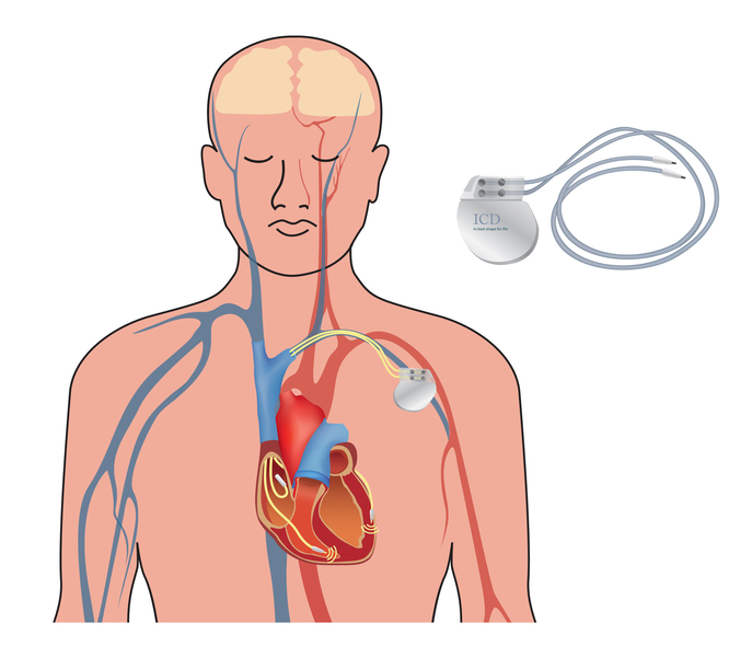
An ICD is the preferred therapeutic modality in most survivors of SCA. The ICD does not prevent the recurrence of malignant ventricular arrhythmias, but it effectively terminates these arrhythmias when they do recur.
ICD patients with frequent arrhythmia recurrences and device discharges may benefit from adjunctive therapies, such as antiarrhythmic drugs or catheter ablation.
The main indications for the use of an ICD can be divided into two groups (Russo et al., 2013):
Class | NYHA Functional Classification | Specific Activity Scale |
I | Patients with cardiac disease but without resulting limitations of physical activity. Ordinary physical activity does not cause undue fatigue, palpitation, dyspnea, or anginal pain. | Patients can perform to completion any activity requiring ≥7 metabolic equivalents, e.g., can carry 24 lb. up eight steps; do outdoor work (shovel snow, spade soil); do recreational activities (skiing, basketball, squash, handball, jog/walk 5 mph). |
II | Patients with cardiac disease resulting in slight limitation of physical activity. They are comfortable at rest. Ordinary physical activity results in fatigue, palpitation, dyspnea, or anginal pain. | Patients can perform to completion any activity requiring ≥5 metabolic equivalents, e.g., have sexual intercourse without stopping, garden, rake, weed, roller skate, dance fox trot, walk at 4 mph on level ground, but cannot and do not perform to completion activities requiring ≥7 metabolic equivalents. |
III | Patients with cardiac disease resulting in marked limitation of physical activity. They are comfortable at rest. Less than ordinary physical activity causes fatigue, palpitation, dyspnea, or anginal pain. | Patients can perform to completion any activity requiring ≥2 metabolic equivalents, e.g., shower without stopping, strip and make bed, clean windows, walk 2.5 mph, bowl, play golf, dress without stopping, but cannot and do not perform to completion any activities requiring >5 metabolic equivalents. |
IV | Patients with cardiac disease resulting in inability to carry on any physical activity without discomfort. Symptoms of cardiac insufficiency or of the anginal syndrome may be present even at rest. If any physical activity is undertaken, discomfort is increased. | Patients cannot or do not perform to completion activities requiring >2 metabolic equivalents. Cannot carry out activities listed above (specific activity scale III). |
*New York Heart Association
Implantable Cardioverter Defibrillator (ICD)
When the ICD senses an abnormal heart rhythm, it delivers an electrical shock to reset the heart to a normal rhythm.
Antiarrhythmic drugs are less effective than an ICD for secondary prevention of SCD. Consequently, their use in the setting of SCD is limited to an adjunctive role as described above, or in patients who do not want or are not candidates for an ICD (e.g., due to marked comorbidities or end-stage HF that make death likely).
Medications used to slow fast cardiac rhythms or stabilize irregular rhythms are known as antiarrhythmics. Antiarrhythmic medications are divided into classes by their primary mode of action.
Class I agents are subdivided into: Ia – Quinidine, Procainamide, Disopyramide Ib – Lidocaine, Tocainide, Mexiletine, Phenytoin Ic – Flecainide, Propafenone, Ecainide, Moricizine
Class II agents include: Metoprolol, Propranolol, Esmolol, Atenolol
Class III agents include: Amiodarone, Sotalol, Azimilide, Bretylium, Cloflium, Dofetilide, Tedisamil, Ibutilide, Sematilide
Class IV agents include: Verapamil, Diltiazem
Class V agents include: Magnesium, Digitalis (digoxin), Adenosine |
Despite advances in the treatment of heart disease, the outcome of patients experiencing SCA remains poor (Wong et al., 2014).
The reasons for the continued poor survival of patients with SCA are uncertain. Although some aspects of acute resuscitation have improved over time (increased bystander CPR and shortened time to defibrillation), these positive trends have been offset by adverse clinical features of patients presenting with SCA (such as increasing age and decreasing proportion presenting with VF). In addition, the response times of both BLS and ALS services have increased, possibly due to population growth and urbanization.
A pilot study comparing the feasibility of EMS transport to a regional cardiac arrest center (with increased transit time) versus transport to the closest hospital suggested no difference in 30-day mortality or major adverse cardiac events. These results should be considered hypothesis-generating for larger-scale studies (Patterson et al., 2017).
Survivors of SCA have variable susceptibility to hypoxic-ischemic brain injury, depending on the duration of circulatory arrest, the extent of resuscitation efforts, and underlying comorbidities.
There is an association between the mechanism of SCA and the outcome of initial resuscitation.
When the initially observed rhythm is asystole (even if preceded by VT or VF), the likelihood of successful resuscitation is low. Only 10% of patients with out-of-hospital arrests and initial asystole survive until hospital admission, and only 0 to 2% until hospital discharge. The poor outcome in patients with asystole or bradycardia due to a very slow idioventricular rhythm probably reflects the prolonged duration of the cardiac arrest (usually more than four minutes) and the presence of severe, irreversible myocardial damage.
Factors associated with successful resuscitation of patients presenting with asystole include:
Patients with SCA due to PEA (also called electrical-mechanical dissociation) have a poor outcome.
The outcome is much better when the initial rhythm is sustained ventricular tachyarrhythmia. Acute MI or myocardial ischemia is the underlying cause of VF for many of the patients who survive hospital discharge.
Survival is approximately 65% to 70% in patients with hemodynamically unstable VT. The prognosis may be better in patients found in monomorphic VT because of the potential for some systemic perfusion during this more organized arrhythmia. In addition, patients with VT tend to have a lower incidence of a previous infarction and a higher ejection fraction when compared with those with VF.
As many as one-third of cases of SCA are due to noncardiac causes. Trauma, nontraumatic bleeding, intoxication, near drowning, and pulmonary embolism are the most common noncardiac etiologies.
Despite the efforts of emergency personnel, resuscitation from out-of-hospital SCA is successful in only one-third of patients, and only about 10% of all patients are ultimately discharged from the hospital, many of whom are neurologically impaired (Chan et al., 2014).
The cause of death in-hospital is most often noncardiac, usually anoxic encephalopathy, or respiratory complications from long-term ventilator dependence.
In addition to later initiation of CPR and the presence of asystole or PEA (electromechanical dissociation), several other factors are associated with a decreased likelihood of survival with neurologic function intact following out-of-hospital SCA:
There are also several poor prognostic features in patients with SCA who survive until admission:
VF in the human heart rarely terminates spontaneously, and survival is therefore dependent upon the prompt delivery of effective CPR. Electrical defibrillation is the only way to reestablish organized electrical activity and myocardial contraction.
Increasing duration of VF has two major adverse effects:
It has been suggested that without CPR, survival from a cardiac arrest caused by VF declines by approximately 10% for each minute without defibrillation. After more than 12 minutes without CPR, the survival rate is only 2% to 5%.
Time to Resuscitation
These observations constitute the rationale for providing more rapid resuscitation in patients with out-of-hospital SCA. One approach is optimizing the EMS system within a community to reduce the response interval to less than eight minutes.
However, the response times of both BLS and ALS services have increased, possibly due to population growth and urbanization. Thus, bystander CPR and even defibrillation have been recommended and implemented in some settings. Such interventions permit more rapid responses than those provided by BLS or ALS personnel, with better survival as a result.
The administration of CPR by a layperson bystander (bystander CPR or bystander-initiated CPR) is important in determining patient outcome after out-of-hospital SCA. Survival after SCA is greater among those who have bystander CPR when compared with those who initially receive more delayed CPR from EMS personnel. In addition to improved survival, early restoration or improvement in circulation is associated with better neurologic function among survivors.
For adults with sudden out-of-hospital SCA, compression-only bystander CPR (without rescue breathing) appears to have equal or possibly greater efficacy compared with standard bystander CPR (compressions plus rescue breathing).
The importance of bystander CPR and support for compression-only bystander CPR comes from a combination of retrospective and prospective studies.
These observations were subsequently confirmed in larger studies (Nakahara et al., 2015).
Despite the benefits of bystander CPR, it is not always performed. Reasons for this include:
Interventions that appear to improve the rate of bystander CPR include verbal encouragement and instruction in CPR by EMS dispatchers and public campaigns to promote the delivery of bystander CPR.
Bystander CPR with only chest compressions improves survival to hospital discharge, compared with chest compressions with interruptions for rescue breathing, with an absolute improvement in mortality of 2.4% (Zhan et al., 2017).
Initial observational studies that evaluated the delivery of compression-only CPR versus standard CPR, including rescue breathing, found no significant differences in survival or long-term neurologic function between the two groups, suggesting that compression-only CPR could be safely delivered.
In a nationwide study of Japanese out-of-hospital cardiac arrest victims between 2005 and 2012, chest compression-only CPR improved the number of SCA victims receiving bystander CPR and the number of patients surviving with favorable neurological outcomes (Iwami et al., 2015). These findings hold promise for improving the delivery of bystander CPR. Further data are required to determine if bystander-delivered compression-only CPR (rather than standard CPR) will translate into better neurologic outcomes for patients with out-of-hospital cardiac arrest.
Several automated devices that deliver chest compressions have been developed in an attempt to improve upon chest compressions delivered by humans, as well as to allow rescuers to perform other interventions simultaneously.
VF's standard of care for resuscitation has been defibrillation as soon as possible. In the Seattle series of over 12,000 EMS-treated patients, 4,546 had witnessed VF. The defibrillation response interval was significantly correlated with survival to hospital discharge (odds ratio 0.88 for every one-minute increase in response time). Subsequent studies have shown similar benefits, with earlier defibrillation associated with improved survival (Nakahara et al., 2015).
Despite these findings, it has been suggested that outcomes may be improved by performing CPR before defibrillation, at least in patients in whom defibrillation is delayed for more than four to five minutes. An initial report from Seattle compared outcomes in two time periods. When an initial shock was given as soon as possible and subsequently when the initial shock was delayed until 90 seconds of CPR had been performed. Survival to hospital discharge was significantly increased with routine CPR before defibrillation, primarily in patients whose initial response interval was four minutes or longer.
However, in the largest study to date comparing shorter versus longer periods of initial CPR before defibrillation in 9,933 patients with SCA, patients were randomly assigned to receive 30 to 60 seconds versus 180 seconds of CPR before cardiac rhythm analysis and defibrillation (if indicated). There was no significant difference in the primary endpoint of survival to hospital discharge with satisfactory functional status.
Early defibrillation and CPR should be performed as recommended in the 2010 ACLS guidelines for SCA and ventricular tachyarrhythmia patients.
The use of AEDs by early responders is another approach to more rapid resuscitation. In most but not all studies, AEDs have improved survival after out-of-hospital cardiac arrest.
The OPALS study group has proposed two terminations of resuscitation rules for use by EMS personnel. The rule for BLS providers equipped with AEDs includes the following three criteria:
The ALS rule includes the BLS criteria, as well as two additional criteria219:
Validation of the predictive value of the BLS and ALS termination rules was performed with data from a retrospective cohort study that included 5,505 adults with out-of-hospital SCA.
However, the validity of these termination rules may be reduced with improvements in EMS and post-resuscitation care. One potential target for understanding and ameliorating current limitations to post-arrest care is the observed marked regional variation in prognosis following SCA.
The adequacy of CPR delivered to a victim of cardiac arrest and outcomes related to resuscitation efforts may depend on various factors (e.g., rate and depth of chest compressions, amount of time without performing chest compressions while performing other tasks such as defibrillation, etc.). The AHA 2010 Guidelines for Cardiopulmonary Resuscitation (CPR) and Emergency Cardiovascular Care emphasized early defibrillation (when available) and high-quality chest compressions (rate at least 100 per minute, depth of 2 inches or more) with minimal interruptions.
The effect of CPR quality has been evaluated in several studies (Brouwer et al., 2015):
End-tidal carbon dioxide levels have an excellent correlation with very low cardiac outputs when measured after at least 10 minutes of CPR. They may provide prognostic information, suggesting that the cardiac output maintained during CPR determines the outcome.
An increase in body temperature is associated with unfavorable functional neurologic recovery after successful CPR. The increase in temperature may be neurally-mediated and can exacerbate the degree of neural injury associated with brain ischemia. For the highest temperature within 48 hours, each degree Celsius higher than 37ºC increases an unfavorable neurologic recovery risk.
On the other hand, the induction of mild to moderate hypothermia (target temperature 32 to 34ºC for 24 hours) may benefit patients successfully resuscitated after a cardiac arrest, although studies have shown variable outcomes.
The incremental benefit of deploying EMS personnel trained in ACLS interventions (intubation, insertion of intravenous lines, and intravenous medication administration) on survival after cardiac arrest probably depends upon the quality of other prehospital services.
The risk of SCA increases with age, with older age being associated with poorer survival in some, but not all, studies of out-of-hospital cardiac arrests.
The incidence of SCA is greater in men than in women. The effect of gender on outcome has been examined in multiple cohorts, with the following findings:
The impact of preexisting chronic conditions on the outcome of out-of-hospital SCA was evaluated in a series of 1,043 SCA victims in King County, Washington, in the United States. There was a statistically significant reduction in the probability of survival to hospital discharge with increasing numbers of chronic conditions, such as:
The impact of comorbidities was more prominent with longer EMS response intervals.
The outcome of patients who experience SCA in the hospital is poor, with reported survival to hospital discharge rates of 6% to 15%. Several clinical factors have been identified that predict a greater likelihood of survival to hospital discharge:
Other factors have been identified that predict a lower likelihood of survival to hospital discharge:
Delays in providing initial defibrillation have been associated with worse outcomes. Delayed defibrillation (more than two minutes after SCA) occurred in 30% of patients and was associated with a significantly lower probability of hospital discharge survival.
Delayed defibrillation was more common with the black race, noncardiac admitting diagnosis, cardiac arrest at a hospital with fewer than 250 beds, an unmonitored hospital unit, and arrest during after-hours periods.
Multiple resuscitations involving CPR have also been associated with worse outcomes. Survival following in-hospital SCA treated with an AED has also been evaluated using data derived from the National Registry of Cardiopulmonary Resuscitation. Compared with usual resuscitative care, using an AED did not improve survival among patients with a shockable rhythm. It was associated with a lower survival to hospital discharge among patients with a non-shockable rhythm.
The AHA issued consensus recommendations regarding strategies for improving outcomes following in-hospital SCA. While the consensus recommendations focused on many of the same factors as out-of-hospital cardiac arrest (i.e., early identification of SCA, provision of high-quality CPR, early defibrillation when indicated), the authors commented on a lack of evidence specifically focused on in-hospital SCA, with many of the current guideline recommendations based on extrapolations of data from out-of-hospital SCA. Further data specifically focusing on in-hospital SCA are required before making additional recommendations.
Arterial hyperoxia early after SCA may have deleterious effects, perhaps due to oxidative injury. In a multivariable model, hyperoxia was an independent risk factor for death. Hypoxia was also an independent risk factor.
The reported long-term survival of resuscitated SCD survivors is variable and may depend upon multiple factors:
Mr. John Jomes is a 42-year-old married father of four, a stockbroker who was in a committee meeting at work at 0845 when he suddenly collapsed, falling forward and hitting his forehead on the table in front of him. EMS was quickly called while one of his work compatriots performed compression-only CPR. He regained consciousness, with some slight confusion noted.
EMS arrived 20 minutes later. EMS personnel related to ED personnel denied chest pain or SHOB. C/o slight neck pain with a frontal HA. BP remained 120-140/66-80, HR 35-66 sinus bradycardia to NSR with infrequent PVCs. Afebrile. Slight confusion persists. A soft neck brace remains on, with the patient remaining on a backboard.
In the emergency department, the patient denies any SHOB, CP. He continues to c/o slight posterior neck pain with a frontal HA. Past medical history negative for DM, HTN, or any cardiac/pulmonary issues. Negative past surgical history. The patient denies alcohol or drug usage or exposure to toxins. He takes Tylenol prn for aches and pains. States his mother died suddenly when she was 43 years of age and his uncle (his mother’s brother) in his 30s.
12-lead ECG shows sinus bradycardia to NSR with rare PVCs.
IV of 0.9 NS infusing at 100 ml/hr.
Continuous cardiac monitoring with pulse oximetry.
Following lab tests ordered and drawn: Comprehensive metabolic panel, CBC, Lipid profile, CK-MB, CK and MB, Troponins I and T, CRP, Coagulation studies, Toxicology screen.
Soft neck brace remains in place. Patient remains on a backboard.
PA and lateral chest x-ray ordered.
PA and lateral neck x-rays ordered.
12-lead ECG shows no evidence of cardiac ischemia, but the rhythm remains sinus bradycardia to NSR with rare unifocal PVCs.
All laboratory tests ordered are WNL or negative.
Chest and neck x-rays remain normal.
Cardiologist on-call to evaluate a patient in the emergency department.
Cardiologist to evaluate a patient for structural heart disease/arrhythmias/primary electrical diseases with appropriate diagnostic procedures ordered
Wife and other family members are not available at present to obtain further information on SCD in family history.
SCA and SCD refer to the sudden cessation of organized cardiac electrical activity with hemodynamic collapse.
SCD is the most common cause of cardiovascular death in the developed world.
The exact mechanism of collapse in an individual is often impossible to establish since, for the vast majority of patients who die suddenly, cardiac electrical activity is not being monitored at the time of their collapse. In studies, however, of patients with cardiac electrical activity monitored at the time of their event, VT or VF accounted for most episodes, with bradycardia or asystole accounting for nearly all of the remainder.
The risk factors for SCA are similar to those for CHD.
The approach to primary prevention of SCD varies according to a patient's clinical profile.
For the general population without known cardiac disease:
A heart-healthy lifestyle, including habitual physical activity, a heart-healthy diet, and abstinence or cessation of cigarette smoking, is recommended for the primary prevention of SCD.
Patients with known cardiac disease (e.g., prior MI, cardiomyopathy, or HF) are at an increased risk of SCA. The approach to the primary prevention of SCA in such patients includes the following:
The management of SCA includes acute treatment of the arrest, and for SCA survivors, a comprehensive evaluation and secondary prevention.
Secondary prevention of SCD, usually with an ICD, is appropriate for most SCA survivors.
Despite advances in the treatment of heart disease, the outcome of patients experiencing SCA remains poor. The reasons for the continued poor outcomes are likely multifactorial (e.g., delayed bystander CPR, delayed defibrillation, advanced age, decreased proportion presenting with VF.)
CEUFast, Inc. is committed to furthering diversity, equity, and inclusion (DEI). While reflecting on this course content, CEUFast, Inc. would like you to consider your individual perspective and question your own biases. Remember, implicit bias is a form of bias that impacts our practice as healthcare professionals. Implicit bias occurs when we have automatic prejudices, judgments, and/or a general attitude towards a person or a group of people based on associated stereotypes we have formed over time. These automatic thoughts occur without our conscious knowledge and without our intentional desire to discriminate. The concern with implicit bias is that this can impact our actions and decisions with our workplace leadership, colleagues, and even our patients. While it is our universal goal to treat everyone equally, our implicit biases can influence our interactions, assessments, communication, prioritization, and decision-making concerning patients, which can ultimately adversely impact health outcomes. It is important to keep this in mind in order to intentionally work to self-identify our own risk areas where our implicit biases might influence our behaviors. Together, we can cease perpetuating stereotypes and remind each other to remain mindful to help avoid reacting according to biases that are contrary to our conscious beliefs and values.