Sign Up
You are not currently logged in. Please log in to CEUfast to enable the course progress and auto resume features.

Course Library

Cerebrovascular Accident (CVA) (FL INITIAL Autonomous Practice -Differential Diagnosis)

1 Contact Hour including 1 Advanced Pharmacology Hour
Only FL ARNPs will receive credit for this course
Listen to Audio
CEUfast OwlGet one year unlimited nursing CEUs $39Sign up now
This course is only applicable for Florida nurse practitioners who need to meet the autonomous practice initial licensure requirement.
This peer reviewed course is applicable for the following professions:
Advanced Practice Registered Nurse (APRN)
This course will be updated or discontinued on or before Monday, September 22, 2025

Nationally Accredited

CEUFast, Inc. is accredited as a provider of nursing continuing professional development by the American Nurses Credentialing Center's Commission on Accreditation. ANCC Provider number #P0274.


Outcomes

≥ 92% of participants will know how to respond to a cerebrovascular accident.

Objectives

After completing this continuing education course, the participant will be able to meet the following objectives:

  1. Differentiate between a hemorrhagic and ischemic stroke.
  2. List the components in a proper stroke scale assessment.
  3. Delineate the importance of maintaining a stroke patients' blood pressure.
  4. Verbalize the long-term debility issues from having a stroke.
  5. List the criteria and contraindications with reperfusion therapy for the stroke patient.
  6. Recount the newest treatments available for the patient suffering from a stroke.
CEUFast Inc. and the course planners for this educational activity do not have any relevant financial relationship(s) to disclose with ineligible companies whose primary business is producing, marketing, selling, re-selling, or distributing healthcare products used by or on patients.

Last Updated:
  • $39 Unlimited Access for 1 Year
    (Includes all state required Nursing CEs)
  • No Tests Required
    (Accepted by most states & professions)
  • Instant Reporting to CE Broker
  • Instant Access to certificates of completion
Logo Audio
Now includes
Audio Courses!
Learn More
Restart
Restart
  • 0% complete
Hide Outline
Playback Speed

Narrator Preference

(Automatically scroll to related sections.)
Done
Cerebrovascular Accident (CVA) (FL INITIAL Autonomous Practice -Differential Diagnosis)
0:00
0:15
 
To earn a certificate of completion you have one of two options:
  1. Take test and pass with a score of at least 80%
  2. Attest that you have read and learned all the course materials.
    (NOTE: Some approval agencies and organizations require you to take a test and "No Test" is NOT an option.)
Author:    James Wittenauer (RN, MSN, MPA, RN-BC)

Case Study: Judy

Judy was a 76-year-old female who, with her husband, lives in a small city in a basement apartment within her daughter and son-in-law’s house. Judy has a history of diabetes and hypertension. While her daughter was away on a trip, Judy's son-in-law noticed Judy had a little bit of facial drooping and slurred speech. Upon checking her blood pressure, her son-in-law discovered Judy's blood pressure to be around 180/100 mm hg and urged Judy to go to the emergency room as he was concerned. She was having a stroke; she refused. Upon return from her trip, Judy's daughter took her to see her doctor, who then determined that Judy had an ischemic stroke.

After being diagnosed with an ischemic stroke, Judy was placed on Plavix. Her doctor also wanted her to go to rehab, which she refused. A few months after starting on Plavix, Judy fell. After the fall, she had an abrupt mental status change. She was brought to the local hospital, where she was diagnosed with a hemorrhagic stroke and was emergently transported to a higher level of care hospital where she had undergone emergency surgery.

After several months in rehabilitation, Judy came home. Her daughter and son-in-law took care of her for nine months until her death. She continued to have seizures and did not want any more life-prolonging treatments. Judy died in her home surrounded by her family.

The patient used in this case study, Judy, was not a made-up patient for this course. She was my mother-in-law.

Introduction to Cerebrovascular Accidents

A cerebrovascular accident (CVA), or stroke, is where the brain tissue is deprived of adequate perfusion, whether by hemorrhage or ischemia. Regardless of the cause, the state of the hypoperfusion results in brain damage to the patient. At times, the damage is minimal, with the patient having zero to a minimal residual effect. At other times, the damage is much more severe, resulting in expressive or interpretive aphasia, hemiparalysis, chronic pain syndrome, emotional debility, and co-morbidities.

The statistics in the United States surrounding CVAs are sobering. According to the most recent American Heart/Stroke Association Guidelines and information from the Centers for Disease Control (CDC), there were approximately 795,000 occurrences of strokes (Virani et al., 2020). Of that number, 610,000 were reported as new stroke cases, and 185,000 were reported as recurrent strokes (Virani et al., 2020). Of those 795,000 strokes, 87% were classified as ischemic strokes (Virani et al., 2020). The healthcare cost of strokes in 2014-2015 was $46 billion, also considered for direct healthcare costs, medications, and missed workdays (Virani et al., 2020).

Signs & Symptoms of Stroke

There are various signs and symptoms that the stroke patient will display. According to the American Stroke Association (2021), the signs and symptoms of a stroke can be broken down into various categories. The main signs and symptoms of a stroke are listed but not limited to Loss of balance, dizziness, vision changes including blurred vision, facial drooping, severe headache, weakness, numbness, aphasia, confusion, trouble with swallowing, memory changes, and involuntary eye movements. The ‘fastest’ way to tell if a person is experiencing a stroke is to utilize the FAST acronym (Amrican Stroke Association, 2021)

  • Facial Drooping: The patient will exhibit facial drooping of the eyes and/or lips.
  • Arm Weakness: Arm numbness and deficits in movement and motor function.
  • Speech Difficulties: Affected speech to include slurred speech and possibly aphasia.
  • Time: It is time for someone to call 911. When you notice these signs, it is time to call 911.

Diagnosis: Types of Strokes

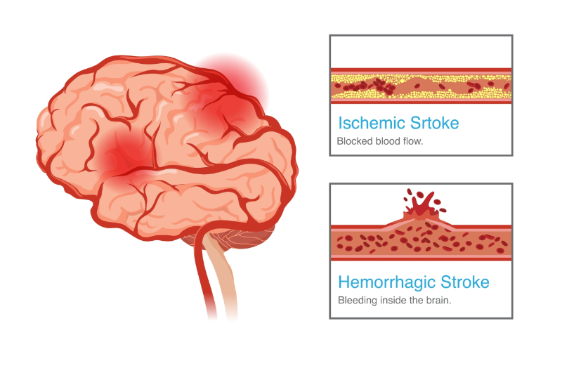
Although both kinds of strokes deprive the brain of perfusion, they are quite different and have their own treatment modalities (American Association of Neurological Surgeons [AANS], 2021).

Hemorrhagic Stroke

A hemorrhagic stroke is caused by an intracerebral or subdural bleeding episode within the brain (AANS, 2021). The resulting bleeding results in pressure on the neural tissue because there is only a limited space within the cranial cavity. The result is damage to the brain due to lack of perfusion to the tissue where there is the pressure exerted to it.

Ischemic Stroke

On the other hand, ischemic strokes, the most common type, happens in the same way that a myocardial infarction occurs in the heart. Due to a thrombotic event within the arterial vasculature which supplies the brain, the blockage robs that area of its’ needed oxygen and nutrients (AANS, 2021). As a result, brain damage ensues.

Prehospital Care

Prehospital care for a stroke can be just as vital as inpatient hospital care. According to the American Heart/Stroke Association Position Statement on the Nursing Care of the Stroke Patient (2021), it is up to the emergency medical services (EMS) personnel to not only properly assess and triage the stroke victim but it is also up to them to ascertain the onset of symptoms and to find out when the patient's “last known normal” (LKN) was (Ashcraft et al., 2021). This helps to identify which if any, reperfusion therapy the patient may benefit from. After performing the needed assessment, the EMS personnel will decide on preliminary treatment and which hospital to transport the patient to and initiate the transport. This decision is crucial as not every hospital is set up to deal with an acute stroke.

Once in the emergency department (ED), the ED staff will initiate the workup of the stroke patient to include an EKG, CT scan, the assessment for thrombolytic treatment, and a stroke assessment (Ashcraft et al., 2021). The non-contrast CT scan needs to be done before any other intervention (Ashcraft et al., 2021).

Stroke Scales

The stroke assessment is very important in assessing the severity of the stroke symptoms in the patient. Per the 2019 updated guidelines by the American Heart Association, there are many models of stroke scales to use, such as the prehospital scales (Powers et al., 2019):

  • The Cincinnati Prehospital Stroke Scale
  • The Los Angeles Pre-Hospital Stroke Scale
  • The Rapid Arterial Occlusion Scale

Other Scales that can be used are the acute assessment scales:

  • The Glasgow Coma Scale
  • The NIH Stroke Scale
  • The Modified NIH Stroke Scale
  • The Intracerebral Hemorrhage Scale

One of the most widely used stroke assessment scales is the National Institute of Health Stroke Scale or NIHSS for short. The scale is composed of eleven items. Each item is given a score of 0 to 3. The total score is used objectively by healthcare professionals to essentially quantify the impairment that results following a stroke (National Institutes of Health [NIH], 2003). The higher the score, the less impaired the patient is. The items include (NIH, 2003):

  1. Level of consciousness:
    • This area is broken up into three parts of responsiveness, the ability to answer orientation questions, and the ability to perform simple commands.
  2. Best gaze:
    • Best gaze assesses the patients' ability to keep their gaze on the provider.
  3. Visual:
    • This part assesses the presence or absence of the sense of sight.
  4. Facial Palsy:
    • Facial palsy looks for uneven facial expressions.
  5. Limb Motor-Arm:
    • This assesses the motor strength of the arms.
  6. Limb Motor-Leg:
    • This assesses the motor strength of the legs.
  7. Limb Ataxia:
    • This part assesses limb coordination.
  8. Sensory Perception:
    • This test assesses the ability of the patient to differentiate between the different sensations such as a pinprick or a light touch.
  9. Best Language:
    • This assessment is used to assess deficits in speech and for expressive aphasia.
  10. Dysarthria:
    • This assessment tests to see if the patient has slurred speech.
  11. Extinction and Inattention:
    • Commonly referred to as spatial neglect. This assessment looks at whether the patient can acknowledge or differentiate the space on the opposite side of the brain affected by the stroke.

The NIHSS should only be performed by someone certified in its use and should be done rather quickly without any coaching of the patient (NIH, 2003).

Treatment

Once the patient has been admitted to the hospital, depending on the severity of the stroke, the initial treatment used for the stroke, and any co-morbidities that might be present, different treatment modalities can be used.

As stated earlier, the first thing that needs to be done within the first few minutes is a non-contrast CT of the head to determine if the stroke is ischemic or hemorrhagic. After the CT has been interpreted, the decision to use thrombolytics can be made.

The use of thrombolytics is not without its risks. There are certain indications and contraindications to its use. According to Hughes et al. (2021), Alteplase is an indicated thrombolytic product. It is also typically used for clearing IV lines within three hours of symptom onset for myocardial infarction (MI) or between 3-4.5 hours of onset for deep vein thrombosis and ischemic stroke. However, the contraindications for the medication are far more numerous than the indications for administering it. There are both relative contraindications, which would make this treatment possibly inadvisable due to risk, and absolute contraindications, which make the treatment completely inadvisable due to risk.

Absolute contraindications include but are not excluded to (Hughes et al., 2021):

  • Significant trauma to the head within the past three months
  • Stroke within the past three months
  • Subarachnoid hemorrhage symptoms
  • An arterial puncture within the past seven days that cannot be compressed
  • History of:
    • Intracranial surgery
    • Hemorrhage
    • Arteriovenous malformation (AVM)
    • Aneurysm
    • Interspinal surgery
  • Active internal bleeding
  • Acute bleeding issues:
    • Platelet counts that are less than 100,000/mm3
    • An elevated partial thromboplastin time (PTT) at the normal upper limits from receiving heparin within 48 hours
    • Currently using an anticoagulant medication that results in an international normalized ratio (INR) of greater than 1.7 or a prothrombin (PT) of greater than 15
  • A blood glucose concentration of less than 50mg/dl
  • A multilobe infarction as displayed on a CT scan where the affected area is greater than one-third of a cerebral hemisphere

Although the maximum time limit for using Alteplace is 4.5 hours, newer findings suggest that the time limit may be increased. According to Oliveira-Filho and Samuels (2021), meta-analysis results from the EPITHET, EXTEND, and ECASS4 study trials suggest that patients could be treated between 4.5 to 9 hours after onset. Although these findings are preliminary, with additional future studies, this is promising information for treating ischemic stroke.

The Stroke Patient: Blood Pressure

In caring for the stroke patient, their blood pressure must be maintained and not permitted to drop too low as this may cause hypoperfusion of the brain tissue. According to Wajngarten and Silva (2019), there are specific criteria for maintaining a stroke patient's blood pressure dependent on the type of stroke and the impending treatment for that patient.

The blood pressure for an ischemic stroke patient needs to be maintained for a systolic level of less than 185 and a diastolic level of less than 110 if they are planning to receive a fibrinolytic intervention or for an intra-arterial intervention (Wajngarten & Silva, 2019).

For a patient with a hemorrhagic stroke with a systolic range of 150-220, the blood pressure can be lowered to a systolic level of 140 and is considered safe. The systolic blood pressure must be at this level before intervention can be taken. The authors note that more aggressive measures may be needed for cases with a systolic level of greater than 220, such as a vasoactive IV drip and constant monitoring (Wajngarten & Silva, 2019).

Blood pressure maintenance for patients who have had a stroke cannot be overstated to help prevent a further event. According to the American Heart Association (AHA), the control of hypertension is one of the secondary preventative measures discussed in preventing a further stroke, along with the other amendable risk factors of diabetes and hyperlipidemia (Wajngarten & Silva, 2019). The maintenance of blood pressure should be done in conjunction with the healthcare team as, according to Kim et al. (2020), self-monitoring and self-management appear ineffective and not cost-efficient.

Although there is no standard goal for optimal blood pressure control per Zonneveld (2018), Wajngarten and Silva (2019) suggested a reasonable approach for post-stroke blood pressure levels at less than 140/90, except for a recent lacunar stroke where blood pressure would benefit at less than 130/90.

Nursing Care for the Stroke Patient

Nursing care for the stroke patient includes different phases and, in those phases, there are different priorities.

Acute Phase

The first phase of care is considered the initial or acute phase in the emergency department or intensive care. This type of care prioritizes stabilizing the patient, completing the necessary diagnostic testing, preparing for interventions, and providing handoff to the next phase of care. The nursing interventions done for this include such measures as (Wajngarten & Silva, 2019):

  • Measuring blood sugar and blood pressure
  • Obtaining information for medical history regarding the time of symptom onset
  • Completion of the NIHSS
  • Assisting with the patient for the CT scan
  • Obtain baseline labs
  • Initiating two intravenous (IV) lines

The patient will undergo treatment that may or may not include interventions. There is a chance that the patient may not be eligible for interventions, and if this is the case, the patient will be stabilized with conservative care. After conservative care has been initiated and the patient stabilized, the next phase of care will begin.

Rehabilitative Phase

After the acute phase of care, the next phase is usually the rehabilitative phase. This phase of care will be the most challenging for the patient and their loved ones. The rehabilitative phase is where the secondary interventions to help prevent stroke recurrence and rehabilitation will begin.

According to the American Stroke Association’s post-stroke patient outcomes policy statement (2019), the patient stands a better chance of a decreased mortality and better functional outcome if admitted or transferred to a stroke center rather than a regular rehabilitation center. The reason for this is that the stroke center utilizes an interprofessional approach in the rehabilitation work for the patient. In fact, according to a study by Turner et al. (2015), the use of a stroke center versus admission to a general nursing unit results in a decrease in mortality after one year and a greater likelihood of the patient being able to be discharged home.

Post-Stroke Complications

The long-term problems relating to stroke and the debility that comes after are many and need to be considered. According to Chohan et al. (2019), the list of post-stroke problems includes but are not limited to:

Seizures

Seizures occur in 5 to 9 % of stroke patients and are more likely with the hemorrhagic variety (Chohan et al., 2019).

Urinary and Bowel Incontinence

Urinary and bowel incontinence can result from a neurogenic bladder caused by the stroke. It occurs with 25% of stroke patients at discharge and 15% at the one-year mark post-stroke (Chohan et al., 2019). Urinary incontinence at one month carries four-fold mortality over continent stroke survivors (Chohan et al., 2019). Bowel incontinence is also an occurrence with a higher percentage at 56%, which diminishes to 11% after three months (Chohan et al., 2019). Although manageable with a bowel regimen, bowel incontinence is associated with increased depression, poor body image, increased stress on the caretaker, and decreased participation in rehabilitation activities.

Cognitive Impairment

Cognitive impairment occurs in 10% of stroke patients initially and at a 30% rate at the end of the first year (Chohan et al., 2019). The risk of developing dementia post-stroke is increased by advanced age, diabetes, left hemispheric lacunar stroke, or previous stroke (Chohan et al., 2019).

Musculoskeletal Effects

There are various musculoskeletal problems that the stroke patient encounters such as increased spasticity, hemiplegic shoulder pain, wrist, and hand flexion (Chohan et al., 2019). All of these issues only add to the patients' discomfort, debility, and recovery. The shoulder pain, which is present in 9-40% of hemiplegic stroke victims, occurs 2-3 months after onset and results from four possible causes, including misaligned joint, spastic muscles, altered sensations from the stroke itself, or reflex sympathetic dystrophy (Chohan et al., 2019).

Psychosocial Effects

The psychosocial issues include depression, emotional lability, and mood changes (Chohan et al., 2019). Depression is very common but often missed. Some decreased mood is present after a stroke at an incidence of 70%, while 25-30% show extreme depression, which is especially more common in patients with an aphasic stroke (Chohan et al., 2019). Emotional lability or laughing or crying without any apparent reason is also referred to as a pseudobulbar effect caused by frontal cortical disruption (Chohan et al., 2019). The mood changes, often presenting as negative emotions such as apathy, anger, frustration, are due to both chemical changes in the brain because of the stroke coupled with the challenges the patient faces in living a post-stroke life (Chohan et al., 2019).

Newest Trends in Emergent Stroke Interventions

There have been newer developments in the intervention phase of the stroke patient. As mentioned by Rubin et al. (2014), a transformation occurred with the use of common contrast with CT angiography. This newer technique allows for the visualization of larger arteries' occlusion and the area of irreversible injury to be seen as well (Rubin et al., 2014).

The next improvement for treatment has been the increase in time allowance for the use of thrombolytics. Although mentioned earlier in the text, this is worth mentioning as different approaches are being considered for the use of thrombolytics. The first mention of a 4.5-9-hour window has been mentioned by Oliveiro-Filho and Samuels (2021). But according to Rehani et al. (2019), this window may be extended up to 24 hours. The increased time window depends on the amount of collateral circulation observed by advanced imaging studies. Although still in its early stages, the clinical trial shows promise in helping to prevent increased debility from a stroke.

The use of neuroprotectants is also being studied to help reduce the severity and improve survival. In a preliminary animal study by Zhang et al. (2021), when the neuroprotectant named NA-1 was delivered to mice by nanoparticle delivery, it reduced both the severity and mortality of a stroke. This was an improvement because when the substance was introduced without the use of a nanoparticle, the results were not as good (Zhang et al., 2021). It appears that this nanoparticle acts as an antioxidant, delivering the peptide exactly where it needs to go in acting to reduce the size of the stroke, reduce cerebral edema, and improve mortality (Zhang et al., 2021). Although in its infancy, this type of intervention could potentially make a huge difference in acute stroke intervention.

One of the biggest advancements being used currently is mechanical thrombectomy on patients with large vessel acute ischemic stroke, which according to Guo and Miao (2021) accounts for 20% of acute ischemic strokes. The procedure, usually done in an interventional lab, can be carried out with moderate sedation and can be performed up to 24 hours from the time of symptom onset (Guo & Miao, 2021). The problem with the procedure, aside from the risks and benefits inherent in any procedure, lies in the availability of providers to perform it, per Tawil and Muir (2017). But, as mentioned by Neale (2020), it is not only the right thing to do in ensuring that all prospective patients in the United States have access to the procedure, but it is also proving to be more cost-effective with better outcomes for the patient and overall reduced cost of care over the life of the patient.

Conclusion

Cerebrovascular accidents or strokes in the United States and across the world continue to be one of the biggest adversaries for both the patient, health care system, and for caregivers. However, it does not have to be. When the population works together to identify those experiencing a stroke, early intervention can begin sooner. When the health care system can get to the patient sooner, morbidity and mortality has the potential to decrease. Finally, when interventions can be made within a timely period, negative outcomes can be avoided, chances of long-term disability can be decreased, and overall patient outcomes post-stroke can be improved.

Select one of the following methods to complete this course.

Take TestPass an exam testing your knowledge of the course material.
OR
No TestAttest that you have read and learned all the course materials.

Implicit Bias Statement

CEUFast, Inc. is committed to furthering diversity, equity, and inclusion (DEI). While reflecting on this course content, CEUFast, Inc. would like you to consider your individual perspective and question your own biases. Remember, implicit bias is a form of bias that impacts our practice as healthcare professionals. Implicit bias occurs when we have automatic prejudices, judgments, and/or a general attitude towards a person or a group of people based on associated stereotypes we have formed over time. These automatic thoughts occur without our conscious knowledge and without our intentional desire to discriminate. The concern with implicit bias is that this can impact our actions and decisions with our workplace leadership, colleagues, and even our patients. While it is our universal goal to treat everyone equally, our implicit biases can influence our interactions, assessments, communication, prioritization, and decision-making concerning patients, which can ultimately adversely impact health outcomes. It is important to keep this in mind in order to intentionally work to self-identify our own risk areas where our implicit biases might influence our behaviors. Together, we can cease perpetuating stereotypes and remind each other to remain mindful to help avoid reacting according to biases that are contrary to our conscious beliefs and values.

References

  • American Association of Neurological Surgeons. (AANS). (2021). Cerebrovascular disease. Retrieved September 9th, 2021. Visit Source.
  • American Stroke Association. (2019). New recommendations for stroke systems of care to improve patient outcomes. American Heart Association (AHA). Retrieved September 15th, 2021. Visit Source.
  • American Stroke Association. (2021). Stroke symptoms. American Heart Association (AHA). Retrieved September 9th, 2021. Visit Source.
  • Ashcraft, S., Wilson, S., Nyström, K., Dusenbury, W., Wira, C., Burrus, T., & Nystr?m, K. (2021). Care of the patient with acute ischemic stroke (prehospital and acute phase of care): Update to the 2009 comprehensive nursing care scientific statement: A scientific statement from the American Heart Association. Stroke (00392499), 52(5), e164–e178. Visit Source.
  • Chohan, S., Venkatesh, P., & How How, C. (2019). Long-term complications of stroke and secondary prevention: An overview for primary care physicians. Singapore Medical Journal, 60(12), 616–620. Visit Source.
  • Guo, X., & Miao, Z. (2021). Advances in mechanical thrombectomy for acute ischaemic stroke from large vessel occlusions. Stroke and Vascular NeurologyVisit Source.
  • Hughes, R., Tadi, P., & Bollu, P. (2021). TPA therapy. StatPearls Publishing [Internet]. Visit Source.
  • Kim, L., Wilson, E., Davison, W., Clark, A., Myint, P., & Potter, J. (2020). Self-monitoring and management of blood pressure in patients with stroke or TIA: An economic evaluation of TEST-BP, A randomised controlled trial. PharmacoEconomics - Open, 4(3), 511–517. Visit Source.
  • National Institutes of Health. (NIH). (2003). NIH stroke scale [PDF]. Visit Source.
  • Neale, T. (2021). Mechanical stroke thrombectomy cost-effective in late time windows. TCTMD.com. Visit Source.
  • Oliveira-Filho, J., & Samuels, O. (2021). Approach to reperfusion therapy for acute ischemic stroke. UpToDate. Retrieved June 10th, 2021. Visit Source.
  • Powers, W., Rabinstein, A., Ackerson, T., Adeoye, O., Bambakidis, N., Becker, K., Biller, J., Brown, M., Demaerschalk, B., Hoh, B., Jauch, E., Kidwell, C., Leslie-Mazwi, T., Ovbiagele, B., Scott, P., Sheth, K., Southerland, A., Summers, D., & Tirschwell, D. (2019). Guidelines for the early management of patients with acute ischemic stroke: 2019 update to the 2018 guidelines for the early management of acute ischemic stroke: A guideline for healthcare professionals from the American Heart Association/American Stroke Association. Stroke (00392499), 50(12), e344–e418. Visit Source.
  • Rehani, B., Ammanuel, S., Zang, Y., Smith, W., Cook, D., Hetts, S., Josephson, S., Kim, A., Hemphill, J., & Dillon, W. (2019). A new era of extended time WINDOW acute stroke interventions guided by imaging. SAGE Journals. Visit Source.
  • Rubin, G., Leipsic, J., Joseph Schoepf, U., Fleischmann, D., & Napel, S. (2014). CT angiography after 20 years: A transformation in cardiovascular disease characterization continues to advance. Radiology, 271(3), 633–652. Visit Source.
  • Tawil, S., & Muir, K. (2017). Thrombolysis and thrombectomy for acute ischaemic stroke. Clinical Medicine Journal. 17(2), 161–165. Visit Source.
  • Turner, M., Barber, M., Dodds, H., Dennis, M., Langhorne, P., & Macleod, M. J. (2015). The impact of stroke unit care on outcome in a Scottish stroke population, taking into account case mix and selection bias. Journal of Neurology, Neurosurgery, and Psychiatry. 86(3), 314–318. Visit Source.
  • Virani, S., Alonso, A., Benjamin, E., Bittencourt, M., Callaway, C., Carson, A., Chamberlain, A., Chang, A., Cheng, S., Delling, F., Djousse, L., Elkind, M., Ferguson, J., Fornage, M., Khan, S., Kissela, B., Knutson, K., Kwan, T., Lackland, D., & Lewis, T. (2020). Heart disease and stroke statistics-2020 update: A report from the American Heart Association. Circulation, 141(9), e139–e151. Visit Source.
  • Wajngarten, M., & Silva, G. S. (2019). Hypertension and stroke: Update on treatment. European Cardiology, 14(2), 111–115. Visit Source.
  • Zhang, S., Chen, Z., Deng, G., Kimberly, W., & Simard, J. (2021). Neuroprotectant delivered to the brain in nanoparticles may improve stroke treatment, outcomes. EurekAlert!Visit Source.
  • Zonneveld, T., Richard, E., Vergouwen, M., Nederkoorn, P., de Haan, R., Roos, Y., & Kruyt, N. (2018). Blood pressure-lowering treatment for preventing recurrent stroke, major vascular events, and dementia in patients with a history of stroke or transient ischaemic attack. The Cochrane database of systematic reviews, 7(7), CD007858. Visit Source.