Sign Up
You are not currently logged in. Please log in to CEUfast to enable the course progress and auto resume features.

Course Library

Heart Failure Series: Part 3- Acute Heart Failure (FL INITIAL Autonomous Practice- Differential Diagnosis)

1.5 Contact Hours
Only FL APRNs will receive credit for this course
Listen to Audio
CEUfast OwlGet one year unlimited nursing CEUs $39Sign up now
This course is only applicable for Florida nurse practitioners who need to meet the autonomous practice initial licensure requirement.
This peer reviewed course is applicable for the following professions:
Advanced Practice Registered Nurse (APRN)
This course will be updated or discontinued on or before Tuesday, June 16, 2026

Nationally Accredited

CEUFast, Inc. is accredited as a provider of nursing continuing professional development by the American Nurses Credentialing Center's Commission on Accreditation. ANCC Provider number #P0274.


Outcomes

≥ 92% of participants will know the different presentations of acute Heart Failure.

Objectives

After completing this continuing education course, the participant will be able to:

  1. List the tools and tests used for the diagnosis of Heart Failure.
  2. Describe the Framingham Heart Failure Diagnostic Criteria.
  3. State the associated symptoms of acute Heart Failure.
  4. Explain what happens to the heart anatomically in cardiac remodeling.
  5. Differentiate Sudden Cardiac Death from Progressive Cardiac Pump Failure.
  6. Describe the acute dysrhythmias that are associated with Heart Failure.
CEUFast Inc. and the course planners for this educational activity do not have any relevant financial relationship(s) to disclose with ineligible companies whose primary business is producing, marketing, selling, re-selling, or distributing healthcare products used by or on patients.

Last Updated:
  • $39 Unlimited Access for 1 Year
    (Includes all state required Nursing CEs)
  • No Tests Required
    (Accepted by most states & professions)
  • Instant Reporting to CE Broker
  • Instant Access to certificates of completion
Logo Audio
Now includes
Audio Courses!
Learn More
Restart
Restart
  • 0% complete
Hide Outline
Playback Speed

Narrator Preference

(Automatically scroll to related sections.)
Done
Heart Failure Series: Part 3- Acute Heart Failure (FL INITIAL Autonomous Practice- Differential Diagnosis)
0:00
0:15
 
To earn a certificate of completion you have one of two options:
  1. Take test and pass with a score of at least 80%
  2. Attest that you have read and learned all the course materials.
    (NOTE: Some approval agencies and organizations require you to take a test and "No Test" is NOT an option.)
Authors:    Alyssa King (DNP, APRN, CPNP-PC, PMHNP-BC, CLC, CNE) , David Tilton (RN, BSN)

Introduction

The previous course, Heart Failure Series: Part 2- Chronic Heart Failure, covered a review of the circulation of blood through the heart, an in-depth description of left-side Heart Failure versus right-sided Heart Failure, as well as an examination of Biventricular Heart Failure. This course, the third course in this series, will cover an in-depth explanation of the more acute forms of Heart Failure as well as the diagnostic criteria of Heart Failure.

Development of Heart Failure

As mentioned and emphasized in the former course in this series, Heart Failure is a chronic process.

Lifestyle choices and the effects of other predisposing disease processes set up conditions that gradually increase cardiac damage with intervals of acute crisis. By the time a person is diagnosed with Heart Failure, using the traditional definition, the chances are good that the heart has been losing pumping capacity little by little for quite some time.

The heart is not in this process alone. The rest of the body also tries to compensate for the loss of cardiac output (Judeo Christian, n.d.):

  • Vasoconstriction in the systemic blood vessels occurs to elevate blood pressure, thus trying to structurally adjust and make up for the heart’s centralized loss of power.
  • The body diverts blood away from less acutely critical tissues and organs to support flow to the most vital organs: the heart, lungs, and brain.

These compensations bring us back to ejection fractions (EF), as mentioned in the first course in the Heart Failure series. Ventricular stroke volumes (ejection fractions) are a key diagnostic tool in Heart Failure. By measuring the heart’s actual functional capability, exact information can be obtained for diagnosis and later comparison, which will allow us to see how much progress is being made by treatments being offered.

As a quick review from the first course in the Heart Failure series to assist in applying the following information regarding clinical signs and symptoms of acute Heart Failure, please review Table 1.

Table 1: Heart Failure & Ejection Fraction (Gibson et al., 2021; Bozkurt et al., 2021)
HF with reduced ejection fraction (HFrEF)Symptomatic HF with LVEF ≤ 40%
HF with mildly reduced ejection fraction (HFmrEF)Symptomatic HF with LVEF 41-49% (previously referred to as HF with mid-range ejection fraction)
HF with preserved ejection fraction (HFpEF)Symptomatic HF with LVEF ≥ 50% (formerly known as diastolic failure)
HF with improved ejection fraction (HFimpEF*)This is a new classification which is distinctly defined as symptomatic HF with a baseline LVEF ≤ 40%, a ≥10-point increase from baseline LVEF, and a subsequent second measurement of LVEF > 40%

As the heart’s left ventricle is the one cardiologist’s test first by preference, finding an LVEF of less than 40% is a strong sign of Heart Failure.

Be aware, however, that a strong LVEF, greater than 75%, is a tell-tale indicator of a different yet potentially devastating cardiac syndrome known as hypertrophic cardiomyopathy (Sharrack & et al., 2022).

Diagnosis of Heart Failure

No single test exists for Heart Failure diagnosis as it is not one disease. It is a clinical syndrome. Any process that causes functional or structural damage to the cardiac system can start the cardiovascular changes leading to Heart Failure. Diagnosis has historically been based on clinical presentation, client history, and the results of laboratory and imaging studies, including ejection fraction testing.

All of these modalities used as diagnostic tools are good; do not disregard them. It is important to emphasize, especially in those suspected to be in acute Heart Failure, rapidly obtaining a serum natriuretic peptide level (B-type natriuretic peptide [BNP] or N-terminal pro-B-type natriuretic peptide [NT-proBNP]). These indicators of myocardial damage may not be enough to diagnose Heart Failure on their own. Yet, they do allow a quick rule-out so that another disease process can be diagnosed.

BNP is a hormone secreted from cardiac ventricular myocytes in response to myocardial stretch and stress (Novack & Zevitz, 2021). Its release activates a cascade of pathways, resulting in an overall protective effect on the myocardium (Novack & Zevitz, 2021). A higher BNP level corresponds with greater myocardial tissue damage, allowing an objective measurement that gives a clinical picture of myocardial function when combined with EF.

Due to other conditions, BNP and the other natriuretic peptides used for measurement and tracking damage may be elevated. Therefore, it is important to use ejection fractions as well to ensure it is Heart Failure that you, as a health professional, are tracking (Novack & Zevitz, 2021).

A diagnosis of Heart Failure can be ruled out with (National Institute for Health and Care Excellence [NICE], 2021):

  • BNP less than 100 ng/L
  • NT-proBNP less than 300 ng/L

Should peptide level results be higher than this, it is important to obtain an ejection fraction measurement as quickly as possible for this patient. Remember the new Universal Definition: a high BNP and an EF of less than 40% are diagnostic for Heart Failure.

The Framingham Criteria

There is nothing wrong with doing things old school. It is even better when both new diagnostics and definitions can incorporate the tried and true. One of the best diagnostic criteria for Heart Failure from past years is the Framingham Criteria. This diagnostic criterion consists of a list of symptoms and procedures where scoring positive for either two major criteria or one major with two minor criteria is diagnostic for Heart Failure (Domitru, 2022).

Table 2: Framingham Heart Failure Diagnostic Criteria
(Family Practice Notebook [FPN], 2022; Domitru, 2022)
Major Criteria include the following:
  • Cardiomegaly
  • Paroxysmal Nocturnal Dyspnea (PND)
  • Weight loss of 4.5 kg in 5 days in response to treatment
  • Jugular Vein Distention (JVD)
  • Rales
  • Acute Pulmonary Edema (PE)
  • Hepatojugular reflux
  • S3 gallop – Third heart sound gallop rhythm
  • Central Venous Pressure greater than 16 cm of water
  • Circulation time of 25 seconds or longer
  • Radiographic cardiomegaly
  • Autopsy proof of Pulmonary Edema, visceral congestion, or cardiomegaly
Minor Criteria (accepted only if they cannot be attributed to another medical condition) are as follows:
  • Nocturnal cough
  • Dyspnea on ordinary exertion
  • Pleural effusion
  • Tachycardia (rate of 120 bpm)
  • Hepatomegaly
  • Bilateral ankle edema
****Positive Heart Failure diagnosis requires two major criteria or one major with two minor criteria.

Diagnostics

The following tests have been found helpful in diagnosing Heart Failure.

The current gold standard testing is (Domitru, 2022):

  • Ejection Fraction testing by Echocardiogram (or another EF test)
  • B-type natriuretic peptide (BNP) levels
  • N-terminal pro-B-type natriuretic peptide (NT-proBNP) levels

Procedures that are often diagnostic include (Domitru, 2022; Dallas, 2021):

  • EKG
  • Chest X-Ray
  • Stress Test
  • Holter Monitor
  • Cardiac Catheterization with Angiogram
  • Cardiac MRI

Laboratory studies that can also assist in diagnosis (Domitru, 2022):

  • Thyroid Function Series (not just a TSH)
  • Complete blood cell (CBC) count
  • Iron studies
  • Urinalysis
  • Electrolyte levels
  • Renal and liver function studies
  • Fasting blood glucose levels
  • Lipid profile

Acute Heart Failure

Though less common than a chronic, insidious presentation, Heart Failure can strike rapidly and unexpectedly. Events like a heart attack, myocarditis, cardiac inflammation from rapid arrhythmias, and other acute events that tax and weaken the heart muscle start rapid structural changes.

Be aware that events may also occur in clients with chronic stable Heart Failure, causing a rapid worsening of their condition. This, too, is considered acute Heart Failure.

The symptoms of acute Heart Failure include (Assid, 2021):

  • Fatigue
  • Rapid weight gain
  • Shortness of breath
  • Persistent coughing or wheezing
  • Edema
  • Lack of appetite or nausea

Diagnosis and treatment are similar in acute and chronic Heart Failure, only with more urgency and more aggressive care with that of acute conditions. Be aware that as early as possible, obtain Heart Failure biomarkers! You will need those for an accurate assessment of damage progression or remission. Biomarkers important to assess for Heart Failure include cardiac troponins and natriuretic peptides and emerging biomarkers including adiponectin, mi-RNA, sST2, Gal-3, MR-proADM, OPG, CT-proAVP and H-FABP (Pourafkari et al., 2019).

Cardiac Remodeling in Heart Failure

Remodeling, in general, is the process by which the body changes the shape or function of an organ or bodily structure. In Heart Failure, a structural change occurs in the shape of the left ventricle, originating from triggers that the cells receive from the neurohormones. These structural changes result in the ventricular chamber becoming rounder and larger in size. Remodeling directly affects the heart rhythm (electrical conduction) and pumping strength. It is often assumed that larger muscles make us stronger, much like a bodybuilder. However, this is not true for the heart muscle. In the enlarged heart, many changes occur, including changes in the chemicals produced by the body (Buddy Balifu, n.d.).

That brings us to the subject of neurohormones. These chemical messengers cause the release of even more messengers through gene expression. The most common expression is an atrial natriuretic peptide (ANP) (Buddy Balifu, n.d.). An elevated concentration of neurohormones such as ANP in the heart during Heart Failure worsens it. ANP’s main function is to cause the body to eliminate salt through the kidneys. It is one of the body’s natural diuretics. Under normal conditions, cells in the atrium of the heart make a trace amount of ANP. However, in Heart Failure, cells in the ventricles also begin to make ANP. This surplus of natural diuretic production is normal in unborn children but not adults (Buddy Balifu, n.d.).

Stimulated enlargement stretches the heart’s walls (remodeling) in Heart Failure. Long-term overstimulation of heart cells results in a cascade of effects that should only happen during the normal time when these cells are supposed to stretch, which is in the developing fetus. Their effects lead to larger heart cells that are also weaker. This hypertrophy is devastating in the long term. It results in heart cells that do not function properly, reduced ejection fraction (HFrEF), and eventually an increased rate of cardiac cell die-off.

Acute Sudden Death with Heart Failure

The two most common end-of-life processes in Heart Failure are Sudden Cardiac Death (SCD), often referred to as sudden death (SD), and Progressive Cardiac Pump Failure. The chance of SCD increases for individuals with both Heart Failure and a known concurrent cardiac dysrhythmia. The longer or more often a person is in the fluid overload condition that accompanies Heart Failure, the more likely they are to develop an unstable heart rhythm (Buddy Balifu, n.d.). As the struggling heart enlarges, it causes electrical pathways in the heart to be over-stretched, thus predisposing the client to cardiac dysrhythmias. The substances in the body that control electricity (electrolytes) also get out of balance with the fluid shifts and edema-driven tissue damage occurring. As a result, if Sudden Cardiac Death does not occur, Heart Failure will progressively worsen until the lungs fill with fluid, drowning the patient. Therefore, having a comprehensive assessment and diagnosis is important to slow or halt the progression of the patient’s condition.

In these acute cases, how is death due to Heart Failure defined? The following are the respective definitions (Yow et al., 2021):

  1. Sudden Cardiac Death (SCD) – Death within one hour of onset of cardiovascular collapse in a previously stable person.
  2. Progressive Cardiac Pump Failure – Cardiac death following symptomatic or hemodynamic deterioration.

Acute Dysrhythmias

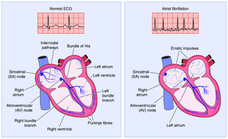
The disorganization of heart electrical activity often found with Heart Failure results in nonproductive or erratic heart muscle contractions, mentioned earlier and known as dysrhythmias. Of these, atrial fibrillation is the most common (Image 1). The atria’s uncoordinated activity prevents the atrial volume from fully contributing to ventricular end-diastolic volume, in turn decreasing stroke volume and cardiac output. The effect of atrial fibrillation on the ventricles includes the loss of synchrony of the atria and the ventricles contracting when they should, a rapid heart rate, and an irregular rhythm of ventricular contraction. Because the diastolic filling time constantly changes, the stroke volume can vary widely. The atria’s uncoordinated activity puts an unbearable burden on the failing heart.

Image 1: Atrial Fibrillation

image of atrial fibrillation graphic

Unable to cope with chaotic pressure changes, the lungs are prone to fill with fluid. Angina or chest pain from the oxygen deficit in the heart muscle itself may occur. Acutely life-threatening dysrhythmias such as ventricular tachycardia or ventricular fibrillation can result, rendering the heart completely unproductive.

Ventricular tachycardia or ventricular fibrillation are the preludes to Sudden Cardiac Death (Image 2). The individual experiencing either one may never feel it coming. A dysrhythmia, sometimes described as a primary dysrhythmia, deprives the heart, mind, and body of needed oxygenated blood. Without external intervention, such as cardiopulmonary resuscitation (CPR), the failing heart inevitably dies.

Image 2: Ventricular Tachycardia vs. Ventricular Fibrillation

ventricular tachycardia v.s. ventricular fibrillation image

Mitigating Chronic and Acute Heart Failure

Despite the progressive nature of Heart Failure, those who have it can lead happy, productive lives. Symptomatic control with medication, lifestyle changes, and even surgical treatments can all help to slow, or even for a time, halt the progression. That makes Sudden Cardiac Death more irritating when it takes away the opportunity for quality of life. Heart Failure patients do not have to wait passively to see if sudden death will strike. There are ways to minimize the occurrence of sudden death. It all starts with a good universal definition so that diagnostic work can recognize Heart Failure early, followed by early individualized treatment.

Controlling electrolyte shifts is a major factor in dysrhythmia control in Heart Failure patients. Basic lab work will be required on a routine basis, and a thorough cardiac workup is also an early must. Of particular interest are the results of the 12-lead EKG, both during stress and non-stress, as this will show the heart’s electrical activity. We will discuss further mitigation strategies as well as both non-pharmacological and pharmacological treatment for Heart Failure in the final course in this series.

Conclusion

This course covered an in-depth explanation of the more acute forms of Heart Failure as well as the diagnostic criteria of Heart Failure and Sudden Cardiac Death. For more information regarding the treatment of Heart Failure, please continue forth within the Heart Failure series to Heart Failure Series: Part 4- Treatment.

Select one of the following methods to complete this course.

Take TestPass an exam testing your knowledge of the course material.
OR
No TestAttest that you have read and learned all the course materials.

Implicit Bias Statement

CEUFast, Inc. is committed to furthering diversity, equity, and inclusion (DEI). While reflecting on this course content, CEUFast, Inc. would like you to consider your individual perspective and question your own biases. Remember, implicit bias is a form of bias that impacts our practice as healthcare professionals. Implicit bias occurs when we have automatic prejudices, judgments, and/or a general attitude towards a person or a group of people based on associated stereotypes we have formed over time. These automatic thoughts occur without our conscious knowledge and without our intentional desire to discriminate. The concern with implicit bias is that this can impact our actions and decisions with our workplace leadership, colleagues, and even our patients. While it is our universal goal to treat everyone equally, our implicit biases can influence our interactions, assessments, communication, prioritization, and decision-making concerning patients, which can ultimately adversely impact health outcomes. It is important to keep this in mind in order to intentionally work to self-identify our own risk areas where our implicit biases might influence our behaviors. Together, we can cease perpetuating stereotypes and remind each other to remain mindful to help avoid reacting according to biases that are contrary to our conscious beliefs and values.

References

  • Assid, P. (2021). What is acute heart failure? Retrieved February 22, 2022. Visit Source.
  • Buddy Balifu. (n.d.). Vardenafil (generic Levitra). Retrieved February 22, 2022. Visit Source.
  • Domitru, I. (2022). Heart failure. Medscape. Retrieved February 22, 2022. Visit Source.
  • Family Practice Notebook. (2022). Framingham heart failure diagnostic criteria. Retrieved February 22, 2022. Visit Source.
  • Gibson, G., Blumer, V., Mentz, J., & Lala, A. (2021). Universal definition and classification of Heart Failure: A step in the right direction from failure to function. American College of Cardiology. Retrieved February 20, 2022. Visit Source.
  • Judeo Christian. (n.d). Congestive Heart Failure overview. Visit Source.
  • National Institute for Health and Care Excellence. (NICE). (2021). Acute heart failure: Diagnosis and management. National Institute of Health National Library of Medicine. Retrieved February 22, 2022. Visit Source.
  • Novack, M., & Zevitz, M. (2021). Natriuretic peptide b type test. National Institute of Health. StatPearls. Retrieved February 28, 2022. Visit Source.
  • Pourafkari, L., Tajilil, A., & Nader, N. (2019). Biomarkers in diagnosing and treatment of acute heart failure. Biomarkers in Medicine. 13 (14). Visit Source.
  • Sharrack, N., Poenar, A., Simms, A., Greenwood, J., & Plein, S. (2022). Acute myocarditis mimicking hypertrophic cardiomyopathy in Marfan Syndrome and morphologically abnormal mitral valve. Journal of the American College of Cardiology: Case Reports. 4 (2). 105-110. Visit Source.
  • Yow, A., Rajasurya, V., & Sharma, S. (2021). Sudden cardiac death (SCD). StatPearls Publishing. Visit Source.