≥ 92% of participants will know how to implement treatment protocols for individuals experiencing a cerebrovascular accident (CVA), thereby minimizing brain damage and improving neurological outcomes.
CEUFast, Inc. is accredited as a provider of nursing continuing professional development by the American Nurses Credentialing Center's Commission on Accreditation. ANCC Provider number #P0274.
≥ 92% of participants will know how to implement treatment protocols for individuals experiencing a cerebrovascular accident (CVA), thereby minimizing brain damage and improving neurological outcomes.
Upon completion of this course, the participant will be able to:
A cerebrovascular accident (CVA), known as a stroke, is a medical emergency that occurs when the blood supply to part of the brain is interrupted or reduced, causing cell death within minutes. The resulting neurological damage can lead to long-term disability or death.
Stroke is the leading cause of death and the third leading cause of long-term disability worldwide (Centers for Disease Control and Prevention [CDC], 2024b). According to the CDC, every 40 seconds, someone in the United States has a stroke, and every 3 minutes and 14 seconds, someone dies from one (CDC, 2024b).
Understanding the anatomy and physiology of cerebral circulation, the distinction between a CVA and a transient ischemic attack (TIA), and the public health impact of stroke is foundational to providing effective care.
Anatomy and Physiology of Cerebral Circulation:
The brain has a high metabolic demand, requiring oxygen and glucose from the bloodstream to function properly. The primary vessels feeding the brain include the internal carotid arteries and the vertebral arteries. The internal carotid arteries provide blood to the anterior and middle parts of the cerebrum. The vertebral arteries join to form the basilar artery, which supplies the posterior part of the cerebrum, the brainstem, and the cerebellum.
Transient Ischemic Attack:
A TIA is a temporary ischemic event that resolves completely. Up to 15% of people who have a stroke had a TIA a few days or weeks beforehand (American Stroke Association, n.d.-b). Therefore, a TIA should never be ignored.
CVA vs. TIA:
Approximately one-third of people who experience a TIA will have a stroke within one year if they do not receive treatment. Therefore, a TIA should always be treated as a medical emergency (National Institute of Neurological Disorders and Stroke [NINDS], 2025).
A significant portion of strokes is preventable. By understanding and managing key risk factors, individuals can substantially reduce their chances of experiencing a CVA. These factors are categorized as either modifiable or non-modifiable risks.
Modifiable Risk Factors:
These are factors that an individual can actively control or treat through lifestyle changes, medical management, or a combination of both (Panuganti et al., 2023).
Non-Modifiable Risk Factors:
These are factors that cannot be changed but can alert individuals to a higher underlying risk, prompting more aggressive management of modifiable factors.
Recognizing the signs and symptoms of a stroke quickly is the first and most critical step in the chain of survival. The sooner a stroke is identified and treatment is initiated, the greater the chance of a good recovery.
Common Stroke Symptoms (American Stroke Association, n.d.-b):
The FAST acronym is a tool to recognize the most common signs of a stroke quickly.
Atypical Stroke Presentations and Their Significance (American Stroke Association, n.d.-b):
While the FAST acronym is an excellent screening tool, not all strokes present with these classic symptoms.
Atypical presentations are more common in women and older adults. Atypical symptoms can include:
These symptoms can be subtle and may not fit the traditional FAST model.
Once a stroke is suspected, a rapid diagnosis is essential for developing an effective treatment plan. Diagnostic imaging is used to differentiate between an ischemic stroke and a hemorrhagic stroke. An ischemic stroke requires blood-thinning agents to dissolve the clot, while a hemorrhagic stroke requires measures to stop the bleeding.
The first-line imaging for a suspected stroke is a non-contrast computed tomography (CT) scan of the brain. This scan is fast and effective at detecting bleeding. If there is no visible bleeding, an ischemic stroke is the presumptive diagnosis.
A CT scan may not show signs of an ischemic stroke for the first few hours. It is performed to rule out a hemorrhage and allow for the administration of thrombolytic therapy.
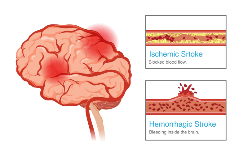
Strokes are broadly classified into two main categories based on their pathophysiology: hemorrhagic and ischemic. Since the two types of strokes require two different treatments, it is critical to differentiate between the two early (Feigin et al., 2025).
Types of Stroke
Hemorrhagic strokes account for approximately 13% of all strokes but are responsible for a disproportionately higher number of stroke-related deaths (CDC, 2024b).
There are two major types of hemorrhagic stroke: Intracerebral hemorrhage (ICH) and subarachnoid hemorrhage (SAH).
ICH: This is the most common type of hemorrhagic stroke. It occurs when a blood vessel within the brain tissue ruptures and spills blood into the brain itself. The accumulating blood forms a hematoma, which increases pressure inside the skull (intracranial pressure or ICP) and compresses brain tissue, leading to cell death. The most common cause is chronic, uncontrolled hypertension, which weakens the small arteries deep within the brain.
SAH: This type of stroke occurs when a blood vessel ruptures in the subarachnoid space, the area between the brain and the tissues that cover it. The space is normally filled with cerebrospinal fluid (CSF). When blood enters this space, it irritates the meninges (the layers of tissue surrounding the brain) and causes a rapid increase in ICP. SAH is often caused by the rupture of a cerebral aneurysm or an arteriovenous malformation (AVM).
The primary causes of hemorrhagic stroke are:
The clinical presentation of a hemorrhagic stroke often differs from that of an ischemic stroke and is typically more dramatic and severe. Common symptoms include:
The management of a hemorrhagic stroke is focused on stopping the bleeding, reducing ICP, and providing supportive care.
Aggressive and rapid control of blood pressure is critical to prevent further bleeding and expansion of the hematoma. Medications are used to lower blood pressure to a target range that minimizes the risk of bleeding while maintaining adequate blood flow to the brain.
The presence of blood in the brain can irritate brain tissue and increase the risk of seizures. Patients are often placed on seizure precautions, and anti-seizure medications may be administered.
An ischemic stroke occurs when a blood vessel supplying the brain becomes blocked, preventing blood flow. This is the most common type of stroke, accounting for approximately 87% of all cases (CDC, 2024a).
Thrombotic Stroke:
This type of stroke occurs when a blood clot (thrombus) forms within a blood vessel in the brain, blocking blood flow (Feigin et al., 2025).
Embolic Stroke:
This occurs when a blood clot or other debris forms elsewhere in the body, most often in the heart, and travels through the bloodstream to the brain. The clot (embolus) then becomes lodged in a smaller cerebral artery, blocking blood flow.
Lacunar Stroke:
This is a small ischemic stroke that occurs when a tiny, deep artery in the brain becomes blocked. These strokes often affect the internal capsule, thalamus, or basal ganglia. They are typically caused by long-standing, poorly controlled hypertension and diabetes, which damage the small penetrating arteries. While they may have a better prognosis, they can still cause significant neurological deficits.
The primary causes of ischemic stroke are closely linked to the underlying pathophysiology.
The acute management of an ischemic stroke is focused on restoring blood flow to the affected area of the brain as quickly as possible (Mac Grory et al., 2021).
The primary acute intervention for an ischemic stroke is thrombolytic therapy, most commonly with alteplase (tissue plasminogen activator [tPA]) (Mac Grory et al., 2021). This medication dissolves the blood clot, restoring blood flow. This therapy is highly time-sensitive and must be administered within a specific time window from the patient's "last known well" time.
Frequent and accurate neurological assessments are the cornerstone of nursing care in the acute phase. The nurse must monitor for signs of neurological deterioration, such as changes in level of consciousness, pupil response, and worsening motor or sensory deficits.
Long-term management of ischemic stroke focuses on preventing a recurrence. This includes aggressive management of hypertension, diabetes, hyperlipidemia, and AFib. Patients are often prescribed antiplatelet agents or anticoagulant medications to prevent future clot formation.
Mr. Robert H., a 67-year-old male with a history of hypertension, hyperlipidemia, and controlled type 2 diabetes mellitus, was attending an outpatient physical therapy session for chronic low back pain. During a set of seated TheraBand rows, he suddenly paused, dropped the band, and stated, "Something feels weird." The physical therapist immediately noticed right-sided facial droop, slurred speech, and weakness in the patient's right arm, along with confusion about the exercise instructions.
Recognizing the sudden onset of these symptoms as possible signs of a CVA, the physical therapist quickly applied the FAST assessment: facial asymmetry, arm weakness, and slurred speech were all present, with the onset occurring just moments earlier. The physical therapist directed a colleague to call 911 and report a suspected acute stroke while remaining with the patient to monitor breathing, consciousness, and safety. The patient was kept seated to prevent a fall. Emergency medical services (EMS) arrived within minutes, and the physical therapist provided the exact time of symptom onset, observed deficits, past medical history, and recent vital signs (blood pressure 188/96 millimeters of mercury [mmHg], heart rate 94, O₂ saturation 97%). Mr. H. was transported to the nearest stroke-ready hospital, where imaging confirmed an ischemic stroke in the left middle cerebral artery territory. Thanks to the rapid recognition and activation of EMS, he received tPA within the critical treatment window. Three months later, Mr. H. returned to physical therapy for post-stroke rehabilitation.
The acute stroke response is a highly coordinated effort that begins in the pre-hospital setting and continues through the emergency department and inpatient care. The goal is to rapidly assess, diagnose, and treat the patient to minimize brain damage.
The chain of survival begins with the public's recognition of stroke symptoms and the immediate activation of EMS.
EMS personnel are often the first healthcare professionals to see a stroke patient. Their role is to:
The "last known well" time is the time the patient was last seen at their normal baseline without any neurological symptoms. This is the starting point for the treatment clock.
Most acute stroke therapies have a strict time window from symptom onset.
EMS pre-notification is a crucial step that helps bypass delays in the emergency department. By providing the hospital with information about the patient's symptoms, "last known well" time, and estimated time of arrival, the hospital can activate the stroke team (neurologist, radiologist, nurses, CT technologists) before the patient even arrives. This reduces the time from arrival to CT scan and, ultimately, to treatment.
Standardized stroke scales and assessment tools are used to evaluate the severity of a stroke and guide treatment decisions (Mac Grory et al., 2021).
Hospitals can achieve certification as a Primary Stroke Center or a Comprehensive Stroke Center by meeting standards set by organizations like The Joint Commission. Ambulances and other emergency services take patients who are suspected of having a stroke to a center with one of these certifications. These certifications confirm that the hospital has the necessary resources, protocols, and expertise to provide timely and effective stroke care.
Some of these protocols include:
Door-to-CT:
Door-to-tPA:
National Institutes of Health Stroke Scale (NIHSS): A Detailed Review of the Components and Scoring:
NIHSS is a standardized, 15-item neurological examination used to quantify the severity of a stroke. The scale evaluates various neurological functions, and the scores for each item are summed to provide a total score, ranging from 0 to 42. A higher score indicates a more severe stroke. Some of these categories are split up differently, but the context is the same.
Glasgow Coma Scale:
The Glasgow Coma Scale (GCS) is a more general tool used to assess a patient's level of consciousness. It is helpful in patients with a hemorrhagic stroke or a stroke that has significantly affected their mental status. The GCS evaluates three main areas: eye opening, verbal response, and motor response. The scores are summed, with a maximum of 15 (fully awake and alert) and a minimum of 3 (unresponsive).
Cincinnati Prehospital Stroke Scale:
The Cincinnati Prehospital Stroke Scale is a rapid, three-item assessment tool used by EMS to screen for a possible stroke. It is essentially an abbreviated version of the NIHSS. The three components are:
If any one of these signs is present, the patient has a high probability of having a stroke and should be transported to a stroke center.
Once a stroke is confirmed as ischemic and the patient is a candidate for acute therapy, the goal is to restore blood flow to the brain as quickly as possible.
There are strict contraindications for tPA, primarily related to the risk of bleeding. These include a history of recent surgery or head trauma, a history of intracranial hemorrhage, and high blood pressure that cannot be controlled.
The nurse's role is critical in the administration of tPA. This includes frequent neurological assessments, close monitoring for signs of bleeding, and strict blood pressure management. The nurse must also be prepared to manage any adverse reactions, such as an anaphylactic response.
Mechanical thrombectomy is a surgical procedure to physically remove a large blood clot from a major cerebral artery (Mac Grory et al., 2021). It involves a small incision in the groin, where a catheter is threaded through the blood vessels to the site of the clot in the brain. A specialized device is then used to grab and remove the clot.
This procedure is indicated for patients with a large vessel occlusion in the anterior circulation of the brain who can be treated within a time window of up to 24 hours from their "last known well" time.
After a thrombectomy, the patient is closely monitored in an intensive care or stroke unit. Nursing care focuses on frequent neurological assessments, managing blood pressure, and monitoring the site of the catheter insertion for bleeding.
After initial stroke therapies, medical management is necessary to prevent and manage post-stroke residual complications. This includes:
Blood pressure control: In the setting of an ischemic stroke, blood pressure is often elevated as a compensatory mechanism to maintain blood flow to the brain. Overly aggressive lowering of blood pressure can be harmful. The goal is to maintain a blood pressure within a specific target range to support cerebral perfusion without increasing the risk of hemorrhage.
Blood glucose management: Both very high and very low blood sugar levels can worsen neurological outcomes after a stroke. The goal is to maintain blood glucose levels within a normal range to prevent further brain cell damage.
Fever control: Fever is a common complication of stroke and can significantly worsen brain injury. The presence of a fever should be promptly identified and treated with antipyretic medications and cooling measures.
The first few days after a stroke are a critical period for patient stabilization and prevention of secondary complications. Interventions are focused on a variety of areas.
Performing frequent and accurate neurological assessments is the most important nursing intervention. The nurse should continue to monitor the patient's neurological status using tools like the NIHSS and GCS to identify any signs of deterioration, such as a new onset of weakness, speech difficulty, or a change in level of consciousness.
A stroke can leave a patient with profound physical and cognitive deficits, making them vulnerable to injury. Patients with unilateral weakness or balance issues are at a high risk for falls. Nursing interventions include using bed alarms, keeping the call bell within reach, and ensuring the patient's environment is free of hazards.
Maintaining proper hydration is important, but fluid overload can worsen cerebral edema. The nurse monitors the patient's intake and output and laboratory values to maintain a stable fluid balance.
Patients with motor deficits may be unable to reposition themselves, putting them at high risk for developing pressure injuries (bedsores). The nurse must implement a turning and repositioning schedule and use specialized mattresses to protect the patient's skin.
The damage caused by a stroke can lead to a variety of medical, neurological, and psychological complications. Early identification and management of these complications are essential for a good recovery.
Medical Complications:
Neurological Complications:
Cerebral edema can be a life-threatening complication, especially after a large ischemic stroke. The swelling increases ICP, compressing brain tissue and potentially causing herniation, which is a displacement of the brain tissue.
Brain tissue that has been damaged by a stroke can become irritable and prone to seizure activity. Anti-seizure medications may be prescribed if a seizure occurs.
Psychological Complications:
The physical, cognitive, and social changes that result from a stroke can be overwhelming, leading to a high incidence of depression and anxiety. These conditions can significantly hinder a patient's motivation for rehabilitation.
Mr. John Smith, a 68-year-old male, is found by his wife slumped on the living room couch. His wife observes that the right side of his face is drooping, and when he tries to speak, his words are slurred and difficult to understand. She also notices that he is unable to lift his right arm when asked. His wife states that Mr. Smith was "acting completely normal" approximately 30 minutes ago, before she went to check the mail. Recognizing these sudden changes, she immediately calls 911. Mr. Smith has a known medical history of uncontrolled hypertension and type 2 diabetes mellitus, for which he takes daily medications, though he sometimes forgets to take them regularly. He also has a history of AFib, which is managed with a blood thinner.
Assessment:
Upon arrival, EMS personnel conduct a rapid initial assessment.
EMS quickly applies the Cincinnati Prehospital Stroke Scale. They observe a clear facial droop on the right side, right arm drift when asked to raise both arms, and abnormal speech (dysarthria). Based on these findings, a stroke is highly suspected.
Mr. Smith's wife confirms that he was last seen without symptoms 30 minutes ago. This "last known well" time is immediately documented and communicated to the hospital.
His blood pressure is recorded as 190/110 mmHg, and his heart rate is irregularly irregular at 110 beats per minute.
Pre-Notification:
EMS immediately pre-notifies the nearest designated Comprehensive Stroke Center, providing a brief report on Mr. Smith's symptoms, "last known well" time, and estimated time of arrival.
Upon arrival at the emergency department, the stroke team is activated and ready.
Mr. Smith is immediately moved to a dedicated stroke bay.
A neurologist performs a comprehensive neurological examination, including the NIHSS. His initial NIHSS score is 14, indicating significant neurological deficits affecting motor function, speech, and facial symmetry.
Imaging:
A non-contrast CT scan of the brain is performed within ten minutes of arrival. The CT scan reveals no evidence of intracranial hemorrhage (bleeding). This finding confirms the likely diagnosis of an ischemic stroke. A CT angiography (CTA) is also performed to identify any large vessel occlusions that might be amenable to mechanical thrombectomy. The CTA confirms a large clot in the middle cerebral artery.
Blood tests are drawn, including blood glucose, coagulation studies, and a complete blood count. An electrocardiogram (ECG) is performed, confirming Afib.
Intervention:
Based on the rapid assessment and diagnostic findings, immediate interventions are initiated to restore blood flow to Mr. Smith's brain.
Given that Mr. Smith's "last known well" time is within the 4.5-hour window for thrombolytic therapy, and there is no evidence of hemorrhage on the CT scan, intravenous alteplase (tPA) is administered. The nursing staff closely monitors his neurological status, vital signs (especially blood pressure), and watches for any signs of bleeding.
Endovascular Intervention (Mechanical Thrombectomy): Because the CTA identified a large vessel occlusion and Mr. Smith meets the criteria, he is immediately prepared for a mechanical thrombectomy. This procedure is performed in the interventional radiology suite, where a catheter is guided to the clot in his middle cerebral artery, and the clot is successfully removed.
Medical Management:
His blood pressure is carefully managed with intravenous medications to keep it within a safe range (typically below 185/110 mmHg before tPA, and then lower after tPA) to prevent further brain injury or hemorrhagic transformation.
His blood glucose levels are monitored and managed to prevent hyperglycemia, which can worsen stroke outcomes. His temperature is monitored, and if a fever develops, it is promptly treated.
Throughout this acute phase, nursing staff perform frequent neurological assessments (every 15-30 minutes), ensure patient safety by implementing fall and aspiration precautions, and maintain strict fluid and electrolyte balance.
Discussion of Outcomes:
The rapid and coordinated acute stroke response significantly impacted Mr. Smith's outcome.
Within hours of the mechanical thrombectomy, Mr. Smith's neurological deficits began to improve. His facial droop became less pronounced, his speech became clearer, and he regained significant strength in his right arm and leg. His NIHSS score decreased to 4 within 24 hours, indicating a mild residual deficit.
Due to vigilant nursing care, Mr. Smith did not develop any pressure injuries. His blood pressure and blood glucose remained well-controlled, minimizing secondary brain injury. A bedside swallow screen was performed, and he was placed on aspiration precautions until a formal swallow evaluation by a speech-language pathologist confirmed he could safely swallow thin liquids and a regular diet.
After a few days in the acute care setting, Mr. Smith was medically stable enough to be discharged to an inpatient rehabilitation facility. The interdisciplinary team (including social work) collaborated with him and his family to ensure a smooth transition, setting realistic goals for his continued recovery. His family was educated on his medications and the importance of adherence.
Mr. Smith's case highlights how timely intervention, adherence to stroke protocols, and comprehensive interdisciplinary care can lead to significant recovery and improved long-term outcomes for stroke patients. His continued rehabilitation and diligent management of his risk factors will be key to preventing future strokes.
This course has provided a comprehensive overview of a CVA, from its fundamental definitions to the complex processes of acute care and long-term rehabilitation. We have learned to differentiate between ischemic and hemorrhagic strokes, recognize key signs and symptoms using tools like the FAST acronym and NIHSS, and understand the critical time-sensitive nature of stroke treatment.
The role of the healthcare professional is vital at every stage of the stroke response. From the rapid assessment to the vigilant monitoring by nurses and the coordinated efforts of the rehabilitation team, each action contributes to the patient's outcome.
CEUFast, Inc. is committed to furthering diversity, equity, and inclusion (DEI). While reflecting on this course content, CEUFast, Inc. would like you to consider your individual perspective and question your own biases. Remember, implicit bias is a form of bias that impacts our practice as healthcare professionals. Implicit bias occurs when we have automatic prejudices, judgments, and/or a general attitude towards a person or a group of people based on associated stereotypes we have formed over time. These automatic thoughts occur without our conscious knowledge and without our intentional desire to discriminate. The concern with implicit bias is that this can impact our actions and decisions with our workplace leadership, colleagues, and even our patients. While it is our universal goal to treat everyone equally, our implicit biases can influence our interactions, assessments, communication, prioritization, and decision-making concerning patients, which can ultimately adversely impact health outcomes. It is important to keep this in mind in order to intentionally work to self-identify our own risk areas where our implicit biases might influence our behaviors. Together, we can cease perpetuating stereotypes and remind each other to remain mindful to help avoid reacting according to biases that are contrary to our conscious beliefs and values.