≥ 92% of participants will know evidence-based wound management of the most common burn wounds.
CEUFast, Inc. is accredited as a provider of nursing continuing professional development by the American Nurses Credentialing Center's Commission on Accreditation. ANCC Provider number #P0274.
≥ 92% of participants will know evidence-based wound management of the most common burn wounds.
By the end of this educational program, the participant will be able to:
This course is a basic and general overview of wound healing and nursing management of the most common burn wounds, with a focus on management of minor and moderate (or first- and second-degree) thermal burns and outpatient burn wound management. While assessment and early treatment of all burns are stressed in this course, it will not cover specific medical or nursing care of major or severe (third-degree burns) in depth, including treating other serious related injuries such as inhalation burns, polytrauma burns, pediatric burn wounds, nor extensive acute care treatments in advanced burn centers.
What is a burn injury wound? A burn is an injury to the body caused by physical contact or extrinsic exposure to environmental materials such as solids, liquids, gases, electricity, radiation, or other means, which results in thermal, chemical, electrical, or radiation damage to cells. The skin is the body's largest organ, and burns are some of the most common preventable skin injuries (Haruta & Mandell, 2023).
The World Health Organization (WHO) estimated that 180,000 people die each year due to burns (WHO, 2023). The vast majority occur in low- and middle-income countries, and almost two-thirds occur in the African and South-East Asia Regions. In India alone, over 1 million people experience moderate or severe thermal burns annually. Nearly 173,000 Bangladeshi children are moderately or severely burned every year. In Bangladesh, Colombia, Egypt, and Pakistan, 17% of children with burns have a temporary disability, and 18% have a permanent disability. In many high-income countries, burn death rates have been decreasing, and the rate of child deaths from burns is currently over 7 times higher in low- and middle-income countries than in high-income countries (WHO, 2023).
Other WHO facts:
Skin and tissue injury, cell death, or charring is caused by external exposure to (by touching, inhaling, eating, or drinking) heat sources that raise the temperature of the skin and tissue higher than it can tolerate. Hot materials such as liquids and steam can cause scalding thermal burns (WHO, 2023).
Viscous liquids such as oil, which maintain skin contact longer, can cause burns faster because of the thermal conductivity of the oil versus water. Oil heats faster than water (Mier, 2022). Time of contact with the hot liquid, conductivity of the liquid, and temperature differences (including boiling points) all impact the potential scalding tissue damage that is likely with skin/tissue contact.
Hot solid or semisolid materials such as metals, plastics, glass, etc., and flames can also cause thermal burns when coming in contact with the skin. Hot air/smoke can cause thermal injury to the respiratory tract. The extent of the burn injury is largely dependent on the anatomical location of the burn, the size of the surface area, the depth of tissue exposed to the heat source, and the length of time of exposure before the skin and tissues were brought back to acceptable temperatures. The physical health and overall condition of the individual and body systems involved (e.g., respiratory involvement) will also impact the extent of the burn injury. Please note: Frostbite is a thermal skin/tissue injury due to exposure to extreme cold. See the Clinical Pearls box below for a brief basic overview of frostbite (Mayo Clinic Staff, n.d.).
Friction burns (e.g., rug burn, rub burn, road rash) are also a type of thermal burn. These burns are often under-recognized and occur when the skin of the body rubs or drags against another surface (e.g., asphalt, flooring, concrete, metal, etc.) hard or fast enough to generate heat. Friction forces sufficient enough to cause friction burn injury are often associated with other types of injury, such as damaging underlying structures beneath the friction burn, particularly orthopedic trauma (Tracy et al., 2023).
Approximately 3% of all burns are chemical burns. This type of burn causes skin and tissue injury due to exposure to (by touching, eating, drinking, inhaling, or injecting) caustic chemical materials such as solvents, detergents, acids, alkali, or vesicant (blistering agents like certain IV medications and chemotherapy) products coming into contact with the skin and/or eyes. Acids (sulphuric, hydrofluoric, hydrochloric, acetic, formic, nitric, phosphoric, phenolic, and chloroacetic acids) cause localized protein denaturation and necrosis to skin that comes in contact with them, and the acid burn injury is halted when the acid is rinsed off, removed, or neutralized.
This encompasses skin and tissue injury due to direct exposure to electrical current. This may include lightning (millions of volts), power lines, motor vehicle batteries, household (110 to 220 volts), and appliance electricity (either alternating current [AC] or direct current [DC]). Overall, AC is considered more dangerous than direct current DC. Examples of DC electricity are lightning and a car battery. Household current is an example of AC. Typically, the higher the voltage, the more potential damage. How the electrical current enters the body, what parts of the body the current passes through, the length of exposure to the current, and the voltages all impact what type of burns will result and the extent of body damage (Mier, 2022; Żwierełło et al., 2023).
This encompasses skin and tissue injury due to prolonged exposure to ionizing radiation, such as radioactive substances, X-rays, or radiation treatments. Radiation burns may be associated with medical treatments or therapies such as radiation therapy for breast cancer. Alpha and beta rays usually cannot penetrate deep into the skin. The severity of damage depends on the type of radiation, the surface area of the body exposed to the radiation, and the length of time of exposure. Brachytherapy involves implanting radioactive seeds (e.g., Iodine-125, Iodine-123, Au-198), which emit radiation over a period of time into the body near the cancer site (e.g., breast cancer, prostate cancer, thyroid cancer). Typically, if these seeds are purposefully left in place permanently, the radioactivity will have decayed completely (stops producing any radioactivity) within an expected amount of time, depending on the isotope (Mier, 2022; Żwierełło et al., 2023).
Frostbite results from exposure to extreme cold and freezing temperatures. It may occur in an instant if bare skin is exposed to extreme cold, such as freezing metal. Frostbite may occur even in skin covered with gloves or clothing. Most common body parts affected by frostbite: Fingers, toes, nose, ears, cheeks, chin, and penis. Signs and symptoms include:
Mild to moderate frostbite causes slight changes in skin color. The skin may begin to feel warm. This is a sign of serious skin involvement. If you treat frostbite by rewarming it at this stage, the surface of the skin may look patchy. The affected area may sting, burn, and swell. A fluid-filled blister may form within 12 to 36 hours after rewarming. This stage is also called superficial frostbite. Please note: Changes in skin color may be difficult to see in darker skin tones. Deep frostbite significantly impacts all layers of the affected skin and tissues below. The affected skin turns white or blue-gray. Large blood blisters may appear 24 to 48 hours after rewarming. The final degree of tissue damage may take weeks to manifest. Weeks after the injury, tissue may turn black and hard as it dies. Assessment and treatment: People with frostbite may also have hypothermia. Hypothermia is a serious condition when the body loses heat faster than it can produce or maintain it. Symptoms of hypothermia include shivering, slurred speech, and being sleepy or clumsy. Symptoms in infants may include cold skin, a change in skin color, and very low energy. Anyone with symptoms of hypothermia or frostbite should seek medical care as soon as possible. Prevention: Limit time outside in freezing temperatures, wear protective gear, do not drink alcohol if planning to be outdoors in freezing weather, stay hydrated, keep moving, and when traveling in cold weather, carry emergency supplies. Seek emergency care for (Mayo Clinic Staff, n.d.):
Treatment (Mayo Clinic, 2024):
|
According to the American Burn Association (ABA), there were 32,540 cases of inpatient hospitalizations at burn centers in 2023. 45% of these injuries were caused by a flash or flame. 66% of these were at a private residence, with the median age being 40 years old, and males comprising 66% of this sample (ABA, 2024).
Those at higher risk of burn injury include the following (Mier, 2022):
Thermal, chemical, electrical, and radiation burns (but not frostbite) are typically classified into three ‘degrees’ depending on the severity and tissue depth of the burn. These are:
First-degree burns/superficial partial-thickness:
Second-degree burns/superficial and deep partial-thickness:
Third-degree burns:
In some cases, burns are not classified by ‘degrees’ but rather described (like many other types of skin wounds) only as partial-thickness wounds (involving epidermal and partial dermal layers of the skin) or full-thickness wounds (involving epidermis and dermis and deeper structures such as subcutaneous tissue, muscle, tendon, and even bones). Additionally, several measures to quickly determine minor, moderate, and major burn injury have been suggested (Szymanski & Tannan, 2023):
Minor: These are first- or second-degree partial-thickness burns involving less than 10% TBSA in children and less than 15% TBSA in adults. Burns do not involve the face or perineum, there is no threat of functional or cosmetic loss, and they most likely can be managed in an outpatient setting. Superficial, minor, first-degree burns tend to heal spontaneously.
Moderate: Second-degree partial-thickness burns with minimal threat to face and/or perineum, with a low risk of cosmetic impairment and TBSA 10%-15% in children or 15%-20% in adults OR any full-thickness burns > 2% but less than 10% TBSA. These burns will require admission to a healthcare facility, but do not always require a referral to a burn center.
Major: These include all full-thickness burns involving > 10% TBSA OR any partial-thickness burns > 20% TBSA in children or > 25% TBSA in adults, OR are any burns to the face or perineum or extremities, OR there is a significant risk for cosmetic impairment. All persons with these burns are best managed in a burn center. Most deep or major burns will require surgical interventions, including skin grafting and rehabilitation. Therefore, determining the depth and severity of burns is important but may also be challenging (Jaspers et al., 2019).
ABA recommends the following “Burn Severity Determination” (ABA, 2022):
Superficial:
Superficial partial-thickness:
Deep partial-thickness:
Full-thickness:
Assessment of superficial or minor burn trauma may be initiated in a home setting, community setting, or healthcare setting. The depth of superficial thermal injuries, such as a sunburn, should still be considered.
Assessment and treatment of burns ideally begin at the scene of the injury as soon as possible (Brekke et al., 2023; Shingleton et al., 2024; Szymanski & Tannan, 2023). Most clinical assessment methods of burn depth are based on visual observations (tissue appearance, color, and signs such as blistering, edema, complaints of pain, etc.) as well as tactile inspection of the burn area and surrounding skin (such as assessing the victim’s sensation, blanching, capillary refill time, skin temperature, etc.) (Jaspers et al., 2019). Shingleton et al. (2024) state, “The initial assessment of any burn casualty should follow the systematic approach laid out in the Advanced Trauma Life Support course (ATLS).” ABA also stresses the importance of hemodynamic stabilization and appropriate fluid resuscitation as soon as possible for burns exceeding 20% TBSA. It is important to note that burn wounds continue to evolve over the first 72 hours, so reassessment and frequent follow-up are important after the initial burn wound injury (Mier, 2022). Other factors related to burn injuries (such as sun and heat exposure), even superficial second-degree burns affecting less than 15% of TBSA (such as sunburn with blistering), should be treated outpatient. The potential for dehydration and adequate rehydration steps should be considered (Mier, 2022).
The fact that burn injuries may be continuously subject to change makes it extremely difficult to assess the total extent of tissue damage through visual inspection alone. It suggests the validity of clinical evaluation is limited, but remains necessary (Jaspers et al., 2019; Promny et al., 2019). Other methods that may be used to assess the area of burn wound tissue destruction, the need for surgical intervention or surgery readiness, and potential for healing include measures of blood flow, temperature, and oxygen saturation using technologies such as laser Doppler imaging (LDI) (Jaspers et al., 2019). However, more research is needed on this topic.
Individuals with major or severe burns are most at risk. Similar to the ABA’s “Major” burn criteria, Schaefer & Nunez Lopez (2023) suggest burns can be considered “severe” if they involve any of the following (and referral to a burn center should be considered for all of these burn injuries):
Additionally, other factors which increase the burn victim’s morbidity and mortality and should be evaluated/considered for advanced burn care and emergency treatment include traumatic injury concurrent with burn injury (e.g., multiple fractures), heat/steam/smoke/chemical inhalation injuries, and any baseline medical conditions like significant heart disease or lung disease which would increase the burn victim’s medical risk (Schaefer & Nunez Lopez, 2023). It is important to note that severe burns cause a systemic inflammatory and vasoactive response throughout the body during the first 24 hours post-injury, resulting in large fluid shifts and intravascular fluid loss. These responses typically peak around 6-8 hours after the injury. These shifts and vascular responses can lead to what is termed “burn shock” over a period of several hours. This is why aggressive intravenous (IV) fluid resuscitation and close monitoring (with caution to avoid overload) are imperative.
As a general rule of thumb, both children and adults with burns greater than 20% TBSA should undergo fluid resuscitation. Estimates of body size and TBSA burned are needed to calculate appropriate fluid resuscitation needs. Common formulas used to initiate IV fluid resuscitation include the Parkland Formula, the modified Brooke Formula, the Rule of Tens Formula, and the Galveston Formula (Schaefer & Nunez Lopez, 2023). We will not discuss the formulas since this is a “basics” (basic wound care for burns) course, and the focus is not on advanced burn care interventions.
Generally speaking, initial burn injury assessments should take place as soon after the burned individual is removed from the source of the burn (the burn process stopped or neutralized), and assessment points include (these are not necessarily in specific order, as the nature and severity of the injury may require assessment of several systems concurrently):
Rule of Nines
Important notes include the following:
Another way to estimate the TBSA is called the “Palmer Method." This method involves considering the size of the burned individual’s entire palmar surface (palm of one hand) as 1% TBSA and estimating how many palms it would take to cover all the areas of the burn(s) (ABA, 2022).
Palmer Method
Example:
The patient presents with an area of blistering from a scald burn, which is about two palm-sized areas on each forearm (2% TBSA each arm for a total of 4% TBSA for both arms), and three palm-sized blistering scald areas on one thigh (3% TBSA). This would quickly help you estimate the TBSA of this second-degree burn to be 7% TBSA. Calculating an accurate TBSA is important for determining many aspects of the patient’s care.
Knowledge of burn care is an essential basic trauma nursing skill. While not every nurse or health care professional may have trauma expertise, rapid initial assessment, and basic care at the scene of the injury, rapid transport to advanced care settings, and initial emergency room care can significantly impact the clinical outcomes of the victim with major burns. Survival is a major goal of burn care, coupled with “addressing and maximizing quality of life” (Mier, 2022). Brekke et al. (2023) state, “The quality of burn care is highly dependent on the initial assessment and care.” In a systematic review of 28 studies (6,461 patients), the authors report concern over poor agreement in the assessment of the TBSA estimates and burn depth in patients between healthcare facilities (referring hospitals, emergency services, and burn centers). This often results in a very high proportion of patients referred to burn centers with an overestimated TBSA (increases risk of fluid overload and other complications), especially in less severe burn injuries, and sometimes (though less frequently) an underestimation of TBSA in more extensive injuries (Brekke et al., 2023).
Mr. Smith is a 49-year-old medium olive-skinned male who presents to the primary care clinic with complaints of a “bad sunburn” he received while fishing off the coast of Florida two days ago. He has no known allergies, has no significant medical history, and takes no medications. He reports he was out in a boat for eight hours. The temperature was in the 90s all day, and it was sunny to partly cloudy. He reports he wore a hat but did not wear a shirt most of the time. His skin over non-sunburned areas is very dry. He complains of a mild headache. His anterior upper chest from the umbilicus upward and posterior torso from the waist up are red, warm, and painful. Both anterior forearms are also red, but not as painful. The skin in all of these areas is easily blanchable.
Additionally, the posterior torso has about 12 clear fluid-filled superficial-looking blisters ranging from 1-3 cm in diameter. Most of these are intact, but 2-3 are oozing a very small amount of clear serous fluid. The blisters cover an area approximately 8 cm x 4 cm over each shoulder area. Mr. Smith says his wife applied some generic skin care lotion last night when the blisters started appearing. He complains that wearing a shirt is extremely uncomfortable. His Vital signs are: Blood pressure 100/60 millimeters of mercury (mmHg), pulse 94 beats per minute, respirations 24 per minute, and temperature is 98.8 degrees Fahrenheit, orally.
Important notes include the following:
Burns often accompany other injuries (musculoskeletal, cardiovascular, polytrauma, blast injuries, lacerations, neurological, respiratory, etc.). Please note: Wound morbidity and patient mortality are greatly influenced by the TBSA affected by the burn injury (Żwierełło et al., 2023), but concomitant injuries to other areas of the body also greatly impact patient outcomes. For full-thickness burn injuries and partial-thickness burns involving > 10% TBSA, (or children under 10 years old) rapid assessment, determination of TBSA of the burn and other systems likely involved (genitals, face, respiratory system, extremities, and potential for cardiac involvement related to electrical burns), hemodynamic assessment and quick transport to advanced medical/burn care are imperative for best clinical outcomes (Brekke et al., 2023; Mier, 2022; Żwierełło et al., 2023). Several life-threatening emergencies related to burns themselves, which healthcare providers should be aware of and alert for even before referral to burn centers, include compartment syndrome and burn shock.
This causes increased pressure, which causes ischemia as blood vessels are compressed by this edema (Radzikowska-Büchneret al., 2023). This is a medical emergency, and “early recognition of the need for escharotomy and other decompressive therapies is imperative in order to avoid irreversible tissue ischemia and necrosis” (Butts et al.; Radzikowska-Büchner et al., 2023). The WHO recommends escharotomy or decompression procedures to relieve/prevent compartment syndrome to be done in the first 48 hours; however, it is reported that most clinicians believe early surgical intervention within 6 hours has better outcomes in reducing the risk of serious septic complications (including amputation). To relieve the constriction caused by increasing edema, eschar, and/or fluid overload (abdominal compartment syndrome), escharotomy, as a surgical procedure, is needed to relieve the constriction and restore perfusion and function to the affected tissues and organs. It is reported that a single incision in most cases will not provide adequate pressure relief from the constricting eschar. Therefore, more commonly, incisions are made bilaterally on the trunk (in the case of abdominal compartment syndrome) or laterally and medially on each affected extremity (Radzikowska-Büchner et al., 2023).
Severe burns, especially burns on an adult covering more than 20% TBSA, are associated with burn shock. The mechanisms of injury during this emergency includes (but are not limited to) these events which may escalate rapidly: decreased plasma volume by extravasation, decreased cardiac output, decreased urine output, hypovolemic shock, hypermetabolic response, breakdown of lipids (hyperlipidemia), breakdown of glycogen (hyperglycemia), breakdown of proteins, liver disfunction, edema formation, insulin resistance, gastrointestinal system dysfunction, mucosal atrophy (decreased absorptive capacity), endocrine response (increased stress hormones), alterations in cardiovascular system, changes in electrolyte balance, immune and inflammatory dysregulation, circulatory and microcirculatory impairment (inadequate tissue perfusion, insufficient supply of oxygen and nutrients, build up of metabolic wastes), increased vulnerability to bacteria, bacterial multiplication, sepsis, multiple organ failure, and imminent death. Fluid administration via IV (termed fluid resuscitation) is recommended for adults with > 20% TBSA burns (or > 10% in children). However, it should be carried out with caution and close monitoring and discontinued in patients without signs of hypovolemia, as it may exacerbate the formation of edema or cause excessive fluid overload (resulting in pulmonary and/or cerebral edema, compartment syndrome in the extremities, and/or development of acute kidney injury [AKI]) and increase mortality (Radzikowska-Büchner et al., 2023). Hence, there is a critical need for referral to specialized advanced burn care centers.
The main criteria for transport to or referral to a specialized advanced burn center care facility (or consultation with a burn specialist) are detailed on the ABA website.
Types of Injuries | Consultation | |
---|---|---|
Pediatric Injuries |
| |
Thermal Burns |
|
|
Inhalation |
|
|
Chemical |
| |
Electrical |
|
|
(ABA, 2022) |
Mrs. Jones was brought in by emergency medical services (EMS) to the emergency room. She is a 65-year-old Caucasian woman who fell asleep with a heating pad wrapped around her right ankle and the lower 1/3rd of her leg. She complained that she had twisted her ankle when she misstepped while climbing her stairs yesterday, and thought heat might help. Other medical history includes type II diabetes, well-controlled by diet, and borderline hypertension, for which she takes no medication. Her vital signs are: blood pressure 165/96 mmHg, pulse 104 beats per minute, respirations are 22, and oral temperature is 98.7 degrees Fahrenheit. She complains of severe pain in the affected leg over the entire area where the heating pad had covered. The left leg and all other skin surfaces are unremarkable. The lower right leg, from the middle of the shin to just below the toes (all the way around the entire leg), is very dark red, shiny, and very firm, and blisters are already appearing. Additionally, some skin has started to slough off over the area just above the ankle. The affected area is also already exhibiting edema compared to the left foot, and pedal pulses are not palpable on the right but are +2 on the left.
This is at least a deep second-degree burn (it may evolve over the next 72 hours to reveal some deeper structure damage, in which case it would be considered third-degree). It is a burn around the entire circumference of the lower leg and the ankle, making it a strong probability of compartment syndrome.
The involvement of a major joint, potential compartment syndrome, need for surgical intervention, potential for loss of function, age of the patient, and diabetes history should lead clinicians to refer to a burn center. Also, these burns should be counted in the TBSA calculations.
It has been at least four hours since the injury; she has edema and no pulse in that foot, and decompression may not be able to wait five more hours without risk of permanent damage. The emergency room provider/clinicians should consider surgical decompression or escharotomy procedure before transporting her to a burn center five hours away.
Skin is the largest organ in the human body, and its primary function is protection (primarily from environmental assaults, including microbial, chemical, radiation, and trauma). The skin is also involved in other essential body functions such as fluid and electrolyte balance, thermoregulation (retaining or releasing heat), immune function, nutrition and metabolism, and communication (expressing emotions, sensory such as touch, pain, heat/cold, and even cell signaling) (Mier, 2022; Wysocki, 2023). The skin typically weighs up to 15% of an adult's body weight (Wysocki, 2023). In some cases, the skin can be used as a route for medication administration (medications that are absorbed topically). Therefore, moderate to severe burn injuries (thermal, electrical, chemical, or radiation) will likely significantly affect the entire body and essential body functions.
The skin has three main layers (from outer layers to innermost layers):
To review human skin layers and major components with essential cellular functions, please refer to the following abridged table (adapted from Wound Series Part 1: Assessing and Diagnosing Chronic Wounds of the Lower Extremity) (Cowan, 2024; Wysocki, 2023).
Layers | Make up & thickness | Considerations | |
Epidermis The average turnover time of this skin layer ranges from 26 to 42 days, with complete regeneration lasting between 45 and 75 days. | The epidermis has five layers:
| Keratinocytes (stratified squamous epithelial cells). Averages 0.075 millimeters (mm) to 0.15 mm thickness over most of the body, except on the soles and palms, where it is thicker (0.4 to 0.6 mm), and eyelids and joints of the hands, which are much thinner. The epidermis is avascular and makes new skin cells (which are constantly being renewed). It helps hydrate your body and makes melanin (provides skin color). | In superficial burn injuries involving only the epidermis (a partial-thickness burn), with the dermal appendages intact, the appearance is typically intact skin, possibly slight swelling, with painful erythema that decreases after 2-3 days The damaged epidermis may peel off after 5-10 days, revealing intact skin underneath (not the same as blisters, which are moist when unroofed). Recovery should be rapid (< 14 days), and the risk of scarring is low (Markiewicz-Gospodarek et al., 2022; Żwierełło et al., 2023). |
[Basement Membrane Zone (BMZ) or Dermal-epidermal junction]* | *Note this is not a skin ‘layer’ but the junction between the dermis and epidermis. The BMZ has three distinct zones:
| One main function of the BMZ is to anchor the epidermis to the dermis. Since there are no blood vessels in the epidermis, this BMZ junction also facilitates nutrient flow for metabolites and other molecules into the epidermis. | This junction is affected in second-degree (partial-thickness) burns. This junction is where most blisters are also formed and are associated with dermatologic diseases and mechanical trauma (such as skin tears). In burns where the BMZ is disrupted and there is blistering (blistering may take up to 72 hours to appear), it would be considered a second-degree burn because the dermis would be exposed or involved, and the BMZ must be reformed. This will take longer to regenerate than the epidermis alone, and nutrient flow to the epidermis would be impacted. These burns, where the epidermis, BMZ, and possibly a very superficial portion of the top layer of the dermis may be impacted, are considered superficial partial-thickness wounds and may heal in 14-21 days (Markiewicz-Gospodarek et al., 2022). |
Dermis (also known as the corium) | The dermis consists of 2 layers: The papillary dermis:
| The dermis is the thickest part of the skin: it averages 2 mm over most of the body but can be up to 4 mm thick in some areas (the back/posterior trunk). The dermis of the eyelid is the thinnest at only 0.6 mm thick. Fibroblasts are the main cells found in the dermis and vary in size and number. Fibroblasts synthesize and secrete glycoproteins found in the dermis. Unlike the epidermis, the dermis is innervated (has nerve structures) and is vascular (has blood vessels). Because the dermis is vascular, it serves the body by assisting with the inflammatory response, hemostasis, thermal regulation, immune support, nutritional support, and wound healing. Other cells found in the dermal layers help the skin's immune system function properly, including lymphocytes, mast cells, and macrophages. | The dermis helps support the epidermis, protects the body from harm, produces hair and sweat, and allows the body to feel different sensations (sensory nerves in this layer). In wound healing, angiogenesis (forming new blood vessels from preexisting vessels) occurs in the dermis. Angiogenesis is stimulated in this layer by the presence of vascular endothelial growth factor (VEGF), which is secreted by keratinocytes in response to hypoxia/skin wounding. Burn injuries involving the dermis disrupt the essential functions listed in this table and are the most painful due to the involvement of nerve cells in this layer. The appearance of these burns is usually red, moist, and very painful. Burns involving the epidermis and into all layers of the dermis (but not all the way through the dermis and into subcutaneous tissue) are still second-degree burns (considered deep partial-thickness) wounds. These deeper second-degree burns impact the papillary and reticular dermis, which will take longer to regenerate (21-35 days or more) and may be complicated by epidermal necrosis, which may require surgical intervention. These deep partial-thickness wounds have a moderate risk of hypertrophic scarring (Markiewicz-Gospodarek et al., 2022; Radzikowska-Büchner et al., 2023; Żwierełło et al., 2023). |
Hypodermis (also known as subcutis or subcutaneous tissue) | The subcutaneous layer below the dermis attaches the dermis to underlying structures. The hypodermis, or superficial fascia, forms a subcutaneous layer below the dermis. It is largely comprised of adipose (fat) tissue. | This subcutaneous tissue helps regulate temperature, provides protection (insulation for the body, additional cushioning), and stores reserves of energy (fat). Sweat glands (both apocrine and eccrine) and growing hair follicles can extend into the hypodermis. The hypodermis also contains a reservoir of adipose-derived stem cells that help modulate skin repair and (in concert with other cells) affect paracrine and endocrine systems. This reserve supply of stem cells decreases with aging, and their ability to respond in skin repair is reduced in older adults. The hypodermis (subcutaneous tissue) also assists with the mobility of the skin over underlying structures (e.g., bones and joints). | These full-thickness wounds typically heal by surgical intervention (surgical excision, transplantation, and/or skin grafting). These burn wounds may take more than nine weeks to close (or longer), depending on the size and location of the burn, any complications such as infection present, and the overall health of the individual (Markiewicz-Gospodarek et al., 2022; Radzikowska-Büchner et al., 2023). |
Fourth-Degree Burns: It is common for any burn injury that extends into underlying structures below the hypodermis (such as muscle, tendon, or bone) to be classified as fourth-degree burns (Markiewicz-Gospodarek et al., 2022). Some clinicians may continue to call these third-degree burns or full-thickness burns or deep third-degree burns or deep full-thickness burns, but as deeper structures are involved, the impact on the host and the potential for complications increase. | |||
Please note: Pediatric populations have some unique skin characteristics and challenges related to burn wounds, but pediatric burns are not specifically a focus of this adult course.
Older adults, however, have some key skin differences related to aging (aged 65 and older) (Wysocki, 2023) that should be considered when managing burns and wounds in the older adult. A list of just a few of these includes:
The depth of the skin injury in any wound can help classify wounds as either partial-thickness (depth of injury only involves the epidermis and sometimes the dermis, but not beyond the subcutaneous tissue) or full-thickness (depth of injury involves the epidermis, dermis, and extends down to at least some subcutaneous tissue). It may also involve muscle, tendon, ligament, or bone (Betz, 2022; Bohn & Bryant, 2023). The extent of damage to specific layers of the skin potentially impacts many of the normal skin functions and should be considered when assessing and managing wounds (Wysocki, 2023).
Most partial-thickness wounds heal without scar tissue formation because they heal by re-epithelialization (Betz, 2022; Bohn & Bryant, 2023; Orsted et al., 2018). A superficial epidermal tissue loss is experienced, and this epidermal skin is regenerated by cells readily available at the skin's surface or within hair follicles (mainly keratinocytes). These partial-thickness wounds heal faster than full-thickness wounds, which involve greater tissue loss and more extensive cellular damage (requiring more complex mechanisms of repair, with many more cell types and chemical messengers involved in the process to coordinate the healing efforts within the wound bed) (Betz, 2022; Bohn & Bryant, 2023; Orsted et al., 2018).
Full-thickness wound healing takes place by one of three main mechanisms (Betz, 2022; Bohn & Bryant, 2023):
Wounds healing by primary intention tend to heal faster than wounds left open to heal by secondary intention. They may heal as fast as partial-thickness wounds because the wound edges have been approximated, and the body does not have to build new tissue or produce as much new tissue or extracellular matrix (scaffolding) or granulation tissue necessary for scar tissue formation. Since the body is repairing a defect similar to sewing together a torn garment, it takes fewer resources than manufacturing a patch, filling in the hole, and holding it all together (Betz, 2022; Bohn & Bryant, 2023).
Full-thickness burn injury wounds usually require surgical intervention to ensure proper wound healing. However, if the burn wound is a very small third-degree burn (less than 2% TBSA) over a fleshy part of the body, and does not involve muscle, tendon, bone, or ligament (or other significant structure underneath the subcutaneous tissue), the healthcare provider may elect a non-surgical approach to wound healing. This type of wound would heal by secondary intention (scar tissue formation and maturation).
In an acute partial-thickness (second-degree) burn injury wound over the upper 1/4 of the back/shoulders (such as Mr. Smith in our case scenario #1 who developed superficial clear fluid-filled blisters only on upper posterior back/shoulders on day two after sunburn), the pathway to healing is expected to follow progressive phases of wound healing, typically resolving within only a few days to two weeks.
The following phases of wound healing are typical in full-thickness wounds and often occur as overlapping phases.
Hemostasis & Inflammatory Phase
Proliferative Phase
The next overlapping phase is the proliferative phase. During this phase, if the expected pathways to healing occur, cytokines (chemical messengers) activate fibroblasts. These fibroblasts are the main players of this phase. Fibroblasts are largely responsible for synthesizing new extracellular matrix (ECM) or granulation tissue, largely made of collagen, to fill in the open defect/wound of any full-thickness wound. Other cells are also activated during this phase to produce growth factors and build new capillaries (neoangiogenesis) (Betz, 2022; Bohn & Bryant, 2023). Enzymes, including more than 15 matrix metalloproteinases (MMPs), secreted by fibroblasts, epithelial cells, neutrophils, and macrophages, play a major role during the inflammatory and proliferative phases, breaking down damaged proteins (such as collagen) and debris. Epithelial cells (particularly keratinocytes) are very active during the proliferative phase, helping to make the wound smaller and smaller and bringing the wound edges closer ("contracting" the wound edges). The proliferative phase typically lasts a few weeks (Betz, 2022; Bohn & Bryant, 2023). Most partial-thickness wounds will heal by re-epithelialization with minimal scarring, as will many full-thickness wounds healing by primary intention (e.g., sutured incision), which will completely close by re-epithelialization.
Maturation or Remodeling Phase
The maturation or remodeling phase occurs in full-thickness wounds after the wound is filled in with new granulation tissue and closed (epithelialized) by wound contraction. This phase is called the maturation or remodeling phase because the protein-rich ECM or new granulation tissue "scaffolding" within the closed wound (scar tissue) is continuously being broken down and replaced with stronger scar tissue over 12-18 months post-wound closure. The human body produces many types of collagens. The scar tissue within a closed full-thickness wound healing by secondary intention is comprised of various types of collagens, elastin, laminin, etc. Even after the scar maturation phase has concluded, the maximum tensile strength of the scar tissue of full-thickness wounds will reach only a maximum of 70-80% of the tensile strength of the surrounding tissue (Betz, 2022; Bohn & Bryant, 2023).
Furthermore, scar tissue contains no melanocytes, so the scar will require sunblock to protect against the sun's rays on any exposed body part. Likewise, scars over bony prominences will require additional protection against pressure-related injury for the rest of the individual's life. Thus, once a full-thickness scar has formed, it may always be considered a 'weak link' or vulnerable area in the body, needing protection, particularly from the sun and pressure-related damage (Bohn & Bryant, 2023). It is important to note that several factors associated with aging may impact this scar maturation phase and the final tensile strength of any full-thickness wound healing by secondary intention. Specifically, these factors include compromised fibroblast function, decreased elastin, flattening of the epidermal/dermal junction, and decreased macrophages in the dermis (Baumann et al., 2021; Betz, 2022).
In the past, clinicians have attempted to assign a specific timeframe for classifying an acute wound versus a chronic wound. Many clinicians have mistakenly thought that any wound that heals within 4-6 weeks is acute, and any wound that takes more than 6 weeks to close is chronic. That classification of acute versus chronic wounds is no longer supported by the scientific literature (Betz, 2022; Bohn & Bryant, 2023). The more accurate classification of a chronic wound is any wound that does not follow the expected pathway to healing (Betz, 2022; Bhoyar et al., 2023).
Mr. Hernandez is a 54-year-old male with no known allergies or significant medical history. He is well-groomed, about average height, and weighs 190 pounds (lbs). He is accompanied by his wife. He reports cooking outdoors on his grill three days ago when the flames flared up and burned an area of his medial left forearm, over the fleshy part (about six inches above his wrist). His wife immediately put aloe vera gel on the burn wound and wrapped it with gauze. He states the burned area was roughly the size and shape of a large egg (oval-shaped, approx. 2” wide x 3” long). He said it was very painful and red, but no blister was seen immediately. He says it is still rather painful to touch, and he has taken ibuprofen 200 milligrams (mg) two times a day, which helps with pain. He has “tried not to get it wet.” He has been reluctant to remove the bandage and comes to you in the outpatient clinic for bandage removal and wound assessment. There is evidence of some dried brownish exudate visible on the outer gauze over the area of the burn. You moisten the gauze with saline and gently remove it. When you do this, you unroof a blister the size of the entire burn, which you measure as 5 centimeters (cm) x 8 cm (about the size of ½ of his palm). The burn area is red, moist, shiny, and appears very shallow (less than 1 mm deep) and clean. The periwound skin is clear, dry, and intact; no edema noted. Patient is afebrile.
This skin blistered, indicating the epidermis and at least part of the dermis are involved (partial-thickness injury). This is a superficial second-degree burn (it has been over 72 hours, so it is not expected to reveal an evolution of any deeper burn injury after this time).
Not running cool water over it first, and refraining from washing it or changing the dressing for three days was not necessary. Appropriate wound care could include: wash gently with soap and water daily (may shower), pat dry, apply aloe vera gel and non-adherent cover dressing daily, continue ibuprofen every 6-8 hours as needed, and return in one week for re-evaluation. Evidence supports the use of aloe vera gel; it is cost-effective and working well so far, so there is no need to opt for more complicated dressings (make sure its “use by” date is not expired).
His burn is less than 1% TBSA since it was only ½ the size of one of his palms.
For all burns, the goals of treatment include the following (Cook et al., 2022):
It is recommended that individuals with chemical burns and electrical burns seek medical treatment in a healthcare facility. Also, while the WHO recommends that most chemical burns be flushed with water, experts point out that there are chemicals that, if exposed to the skin, should NOT be irrigated immediately with water. These types of compounds are not typically household items but may be in a workplace or other location. These include: elemental metals (e.g., calcium oxide, magnesium, phosphorus, potassium, and sodium), and phenols (not to be confused with lime fruits). When exposed to water, certain reactive metal compounds/chemicals and elemental metals may release hazardous byproducts or combust (explode or cause a fire). Examples include: cesium, lithium, magnesium, phosphorus, potassium, sodium, and titanium tetrachloride.
In most first-degree burns (such as typical sunburn), individuals may not seek medical treatment but rather, may self-treat at home with over-the-counter remedies (if at all). Some practical tips should include staying hydrated and seeking medical care if fatigue, nausea, dizziness, or headache with sunburn are noted. The patient should not apply ice to the sunburn; rather, run cool or room temperature water over the burn to soothe the pain. Also, keep the skin clean using a mild soap and water, and moisturize with a gentle lotion or aloe vera gel (avoid products with alcohol, perfumes, or dyes). Consider taking an over-the-counter medication such as ibuprofen at the first sign of sunburn to reduce pain and inflammation (as long as you do not have health reasons to avoid these medications) (Ermer-Seltun & Rolstad, 2022; Sharma et al., 2022; Travis, 2021).
When burns evolve from first-degree (intact skin) to second-degree burns (blistering and involvement of the BMZ and/or dermis), which they may do within the first 72 hours, the resulting non-intact skin and the potential risk for infection are increased. As a reminder, any second-degree burn involving more than 10% TBSA in patients under 10 or over 50 years old, or more than 20% TBSA in any other age group, and any second-degree burn that involves the face, hands, feet, major joints, perineum, or genitalia should be referred to a burn center. Please see the ABA Guidelines for Burn Patient Referral.
The same principles for topical therapy of other partial-thickness and full-thickness wounds are true for partial-thickness and full-thickness second- and third-degree burns. These include (Ermer-Seltun & Rolstad, 2022):
|
There are so many different wound products and dressing types/options available in modern wound care. Wound care products and dressings for burn wounds will influence the healing process. Wound characteristics dictate the topical management needs of the wound (Ermer-Seltun & Rolstad, 2022). For example, if the wound is a full-thickness, highly exudating wound, it indicates that an absorptive dressing is needed; if the wound bed is friable (easily traumatized, bleeds easily), it indicates that it needs a non-traumatic dressing (at least in the wound bed). However, if the wound is showing signs of high bioburden (hypergranulating, friable wound bed, increased pain, thicker exudate with change in color, some periwound erythema, and/or the wound that was improving is now appearing stalled or worsened), an antimicrobial dressing or product may be needed. If the patient cannot change their own dressings but rather will come to the clinic twice a week, it may need to be a product that is appropriate to leave in the wound for several days (or if the patient has home care, a dressing that home health could manage could be considered).
`This may not be an all-inclusive list of products currently available, as these are just examples. They are also not listed in any order of preference or efficacy.
Product | Typical Burn or Wound Type | Pros | Cons |
---|---|---|---|
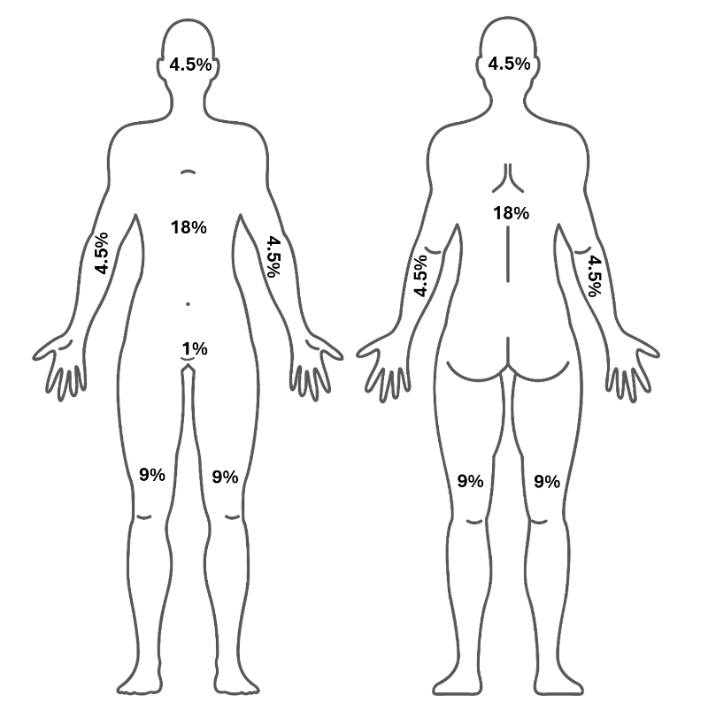
Petrolatum impregnated (e.g., Vaseline®) gauze covered with a second dry normal gauze layer secured by netting or tape. | Superficial partial-thickness burn wounds and deep dermal partial-thickness burns with scant to light exudate. |
|
|
Silicone-coated nylon dressings (with and without foam layer): soft silicone porous layer (Safetac®), which is transparent and flexible polyamide mesh (examples: Adaptic Touch® silicone dressing; Allevyn®; Biatain®); Equos 5-layer square with silicone adhesive (or Veratel® contact layer silicone wound dressing), Mepitel (fenestrated, thin, transparent porous and flexible polyamide mesh); Mepilex border wound dressing (polyurethane foam dressing with a Safetac® silicone contact layer and gently adherent border); Mepilex Ag (includes silver), etc. | Superficial partial-thickness burn wounds and deep dermal partial-thickness burns with light to moderate exudate (dependent on the ability of absorptive materials in specific silicone dressings). Some of these silicone sheets may also be used for status-post burns on healing skin to help prevent hypertrophic scarring (keloids). |
|
|
Polymeric hydrogel dressings: High water content (70-90%) gel dressing as a gel in a tube or as a flexible sheet, pad, or gel rope (e.g., Elastogel® sheets or pads, IntraSite®, Aqua clear®, Nu-gel®, Medagel®, etc.). May also be impregnated or in a combined product with silver (e.g., SilverSeal®, SilverSept®, etc.), or other antimicrobial. Hydrogels tend to be non-irritating and are metabolite-permeable. | Second-degree burns, some third-degree burns. Partial-thickness to full-thickness burns. Also, may be used on split-thickness skin donor sites. May also be used for status-post burns on healing skin to help prevent hypertrophic scarring (keloids). Suitable for all phases of wound healing and both acute and chronic wounds. |
|
|
Hydrofiber dressings: Typically made up primarily of sodium carboxymethyl cellulose. This fiber essentially turns into a gel when moistened or in contact with wound exudate. (Example: Aquacel®). May be impregnated with ionic silver as an antimicrobial dressing (Aquacel Ag® and Aquacel Ag® Burn Hydrofiber dressing). | Second-degree burns, some third-degree burns. Partial-thickness to full-thickness burns. Also may be used on split-thickness skin donor sites. Suitable for all phases of wound healing and both acute and chronic wounds. |
|
|
Hydrocolloid dressings (pads): Occlusive dressing that provides a moist wound environment. As the inner layer of the dressing comes into contact with exudate, a gel forms. | Superficial and deep dermal partial-thickness burns with scant to light exudate. |
|
|
Polyurethane film (polymer membranes) dressings: Typically, these transparent film dressings are coated with an adhesive on one side. These dressings are made in different thicknesses and are usually gas permeable (allows oxygen and moisture vapor transmission) but not permeable to liquid and environmental contaminants such as bacteria. | Suitable for second-degree burns, partial-thickness wounds with little or no exudate, and necrotic wounds (when autolytic debridement is desired). Also used to cover IV sites, donor sites, lacerations, and abrasions. |
|
|
Medicated wound dressings, gels, and ointments: These include various dressings, gels, and ointments that contain medications (some of which are specifically antimicrobial) to stimulate healing, assist with debridement, or help address wound healing impediments such as biofilm growth (e.g., surfactant wound care products). As far as the use of antimicrobials in burn wound care, it is important to note that a recent systematic review reports “it is necessary to emphasize that there is currently no ideal topical antimicrobial agent that can be recommended in all clinical scenarios” (Garcia Garcia et al., 2022). | |||
1% Silver sulphadiazine cream (SSD) (e.g. Silvadene®). This has been prescribed for more than 50 years, but there is some evidence to suggest some newer products may promote healing faster than SSD. Typically requires a secondary dressing. | Open superficial and deep dermal partial-thickness and some full-thickness wounds (second- and third-degree wounds). Often used to cover wounds. Please note: SDD is contraindicated in pregnant women close to term and in premature and newborn infants in the first two months of life. |
|
|
Collagenase ointment (enzymatic debriding agent) (Example: Santyl®). One of the only FDA-approved enzymatic debriding ointments (approved for adults). | Suitable for second-degree burns, partial- and full-thickness burns with necrotic tissue in wound bed. |
|
|
Nanocrystalline silver (NCS) in a flexible sheet or pad (such as Acticoat®, Acticoat Flex®, and Acticoat® surgical dressings). Antimicrobial (bactericidal) against over 150 organisms, including methicillin-resistant Staphylococcus aureus (MRSA), vancomycin-resistant Enterococcus (VRE), carbapenem-resistant Enterobacterales (CRE), gram-negative and gram-positive bacteria, yeasts, and fungi. | Second-degree burns, some third-degree burns (partial-thickness to full-thickness burns) with signs and symptoms of local wound infection and/or at high risk of wound infection. Also may be used on split-thickness skin donor sites. Suitable for all phases of wound healing and both acute and chronic wounds. Expect to see decreased signs and symptoms of infection within two weeks. |
|
|
Cadexomer Iodine, containing wound gels or pads (such as Iodosorb®), is a water-soluble modified starch polymer containing 0.9% iodine (available over-the-counter). | Second-degree burns, some third-degree burns (partial-thickness to full-thickness burns) with signs and symptoms of local wound infection and/or at high risk of wound infection. Also may be used on split-thickness skin donor sites. Suitable for all phases of wound healing and both acute and chronic wounds. Expect to see decreased signs and symptoms of infection within two weeks. |
|
|
3% Bismuth Tribromophenate and petrolatum impregnated gauze (e.g., Xeroform®), typically covered with a second dry normal gauze layer secured by netting or tape. | Superficial and deep dermal partial-thickness burns with scant to light exudate. |
|
|
Polyhexamethylene biguanide (PHMB) (typically 0.2% to 1%) impregnated gauze and wound dressing materials. Examples include AMD Gauze®, PuraPly®, and PuraPly AM® (cross-linked ECM in the form of a collagen sheet impregnated with 1% PHMB). | Second-degree burns, some third-degree burns (partial-thickness to full-thickness burns) with high bioburden signs and symptoms of local wound infection, and/or at high risk of infection. Also may be used on split-thickness skin donor sites. Suitable for all phases of wound healing and both acute and chronic wounds. Expect to see decreased signs and symptoms of infection within two weeks. |
|
|
(Bhoyar et al., 2023; Cook et al., 2022; Nedelec, et al., 2015; Radzikowska-Büchner et al., 2023) |
Many initial superficial burn wound therapies traditionally may consist of a non-adherent petrolatum impregnated (e.g., Vaseline®) gauze or a 3% Bismuth Tribromophenate in a petrolatum blend impregnated gauze (e.g., Xeroform®) or a palm tree oil-impregnated gauze covered with a second dry normal gauze layer secured by netting or tape. However, some evidence suggests petrolatum-based dressings may “hold in” the heat of burns. Therefore, other more efficacious therapies should be considered. Alternative, herbal, and integrative medicine applications are gaining use in clinical practice.
One such application is topical Aloe Vera Gel. This product is typically used on superficial epidermal and dermal partial-thickness (first-degree and second-degree) burn wounds. It is considered safe topically even for children and pregnant women (Catalano et al., 2024). There is ample evidence in meta-analyses and systematic reviews that consistently demonstrate superficial skin burns (first and second-degree) may heal 4-8 days faster when using Aloe Vera gel topically to treat the burns versus controls in clinical trials (Catalano et al., 2024; Maenthaisong et al., 2007; Sharma et al., 2022). Typical controls used as a comparison against Aloe Vera Gel in these studies included: saline gel, 10% povidone-iodine (Betadine®) and water, Calendula (plant often used in herbal topical therapies) ointment, petroleum jelly, 1% silver sulfadiazine cream (e.g., Silvadene®, Silverex®, Silverol®, Silveleb®, Silvazine®, BurnHeal®), dry gauze, etc. In several of these studies, participants reported less erythema and less pain in the topical Aloe Vera treatment arm. Aloe Vera is a cost-effective and minimal-risk topical wound treatment for first-degree and second-degree burns (Radzikowska-Büchner et al., 2023; Sharma et al., 2024).
A STSG is a surgical procedure to treat deep partial-thickness and full-thickness burns that are not likely to heal independently. They are also used for non-healing deep partial-thickness and full-thickness wounds resulting from trauma, surgical wounds, and other types of wounds, such as pressure ulcers. The desired benefits of STSGs are to re-epithelialize the wound quickly, protect underlying structures, reduce risk of infection, reduce scarring, improve fluid loss regulation, and improve aesthetics (appearance) and function (Braza et al., 2025).
In STSGs, a portion (typically an index-card-sized rectangle) of the person’s own skin (epidermis and part of the dermis) is ‘shaved’ off from a location on the burned individual’s body with healthy skin (typically using an instrument called a dermatome). This is done by a surgeon in a sterile environment after prepping the donor site and the burn wound receiving site. This thin layer of tissue (often only 0.015 to 0.018 inches thick) is usually meshed or fenestrated and then ‘grafted’ to the prepared site over the burn injury (note: the graft will fail if placed upside down). This STSG is sutured, stapled, or otherwise secured in place and covered with a special dressing to be left intact and undisturbed for 5-10 days (sometimes a negative pressure wound therapy device is used for this surgical dressing because the negative pressure being applied to the wound may encourage the STSG to re-epithelialize quicker/more completely. It is expected that STSGs will adhere to the grafted site within 5-7 days, so leaving it undisturbed during this time improves the chance of success. One drawback to the STSG procedure is that the donor site is now also a painful partial-thickness wound and must be cared for in addition to the STSG site. Appropriate dressing characteristics for STSG donor sites should include: a product/dressing that may be left in place for several days, encourages re-epithelialization, is soothing, non-adherent, promotes hemostasis, maintains a moist wound bed, but also manages exudates as needed, and is non-traumatic. The success rates of STSGs are reported to be 70-90% (Braza et al., 2025).
One of the main benefits of other skin grafts not taken from the patient’s healthy skin is avoiding making another wound in an already wounded patient. Biological, biosynthetic, or engineered products created from acellular components and other types of skin substitute applications are becoming more common and popular. However, the goals are the same – to close the wound, reduce scarring, fill in the defect, protect underlying structures, prevent infection, reduce fluid loss, and maintain function and appearance.
Currently, there are over 50 different wound care products that may be considered ‘skin substitutes’ (including biologic, synthetic, or biosynthetic), which may sometimes be used instead of autologous split thickness skin grafts (STSG). Examples of these skin substitutes include those listed in the table below. This may not be an all-inclusive list of products currently available, as these are just examples. They are also not listed in any order of preference or efficacy (Tavakoli & Klar, 2021).
Types and Examples | Types of wounds | Pros | Cons |
---|---|---|---|
Cryopreserved placental-membrane-based allograft (e.g., Grafix®). Grafix® typically comes in a cryogenic bag filled with a cryoprotectant solution. | Chronic wounds, deep partial-thickness to full-thickness burn wounds |
|
|
Cultured epithelial sheets or cultured epithelial autografts (CEA). Autologous CEA: Made from the patient’s own cultured keratinocytes (cells) from the epidermis. Allograft CEA: Made from donor cells from the skin of another unrelated human. | Suitable for partial- and full-thickness skin wounds that extend through epidermis and into the dermis or possibly part of hypodermis (subcutaneous tissue), but not for those which extend through the hypodermis and involve muscle, tendon, joint capsule, or bone. |
|
|
Epidermal templates (bioengineered epidermal tissue) Example: Apligraf® (“a bilayered bioengineered skin substitute [BBSS] composed of a bovine type I collagen lattice with a dermal layer of human fibroblasts and a layer formed by human keratinocytes, mimicking the normal structure of human skin”) (Tavakoli & Klar, 2021). | Suitable for partial- and full-thickness skin wounds that extend through epidermis and into the dermis or hypodermis (subcutaneous tissue), but not for those which extend through the hypodermis and involve muscle, tendon, joint capsule, or bone. |
|
|
Dermal templates (bioengineered dermal tissue) Example: Dermagraft® (a polyglactin scaffold containing cultured fibroblasts harvested from human neonatal foreskins). The bioabsorbable polyglactin scaffold degrades by the hydrolysis process in 20–30 days, but the fibroblasts by this time, have helped create a 3D dermal substitute. | Suitable for partial- and full-thickness skin wounds that extend through the epidermis and into the dermis or hypodermis (subcutaneous tissue), but not for those which extend through the hypodermis and involve muscle, tendon, joint capsule, or bone. |
|
|
Bioengineered composite allografts (dermo-epidermal skin equivalents) containing both major skin layers (epidermis and dermis). Typically, an allogenic dermis with autografted epidermal keratinocytes (examples: Alloskin®, from cadaver tissue). | Acute and chronic wounds – deep partial-thickness and full-thickness. |
|
|
Other: A 2-layer bioengineered ECM (example: OASIS® Wound Matrix - derived from a single layer of porcine small intestinal submucosa (SIS) (from pigs), which contains components similar to human dermis. It is normally incorporated and absorbed into the wound. It is minimally processed and sterilized. | Suitable for partial-thickness and full-thickness wounds, including second-degree burns and surgical wounds (donor sites/grafts, etc.). Apply after active bleeding, excessive exudate, acute swelling, and/or infection are controlled. |
|
|
There are over 3000 wound products used in current or modern wound care. Examples of other types of wound dressings include additional bioactive dressings, which contain materials such as collagen, elastin, hyaluronic acid, chitosan, alginate, etc. Biosynthetic skin substitutes and some bioactive dressings are used to reduce pain, accelerate wound healing, prevent, reduce, or minimize scarring by providing bioactive materials that attempt to mimic epidermal and dermal tissue and function. Ideally, when used mostly on superficial partial-thickness wounds (such as superficial second-degree burns), the expectation for some of these materials (particularly skin substitutes) is to heal the wound within 14 days. Using these materials on deep partial-thickness or full-thickness wounds may lead to higher infection rates.
In most wound care management circumstances, wet-to-dry dressings (gauze moistened with saline or other solution, placed in an open wound and removed when dry, thereby providing a mechanical debridement) are no longer considered evidence-based wound care. They are typically a very painful wound dressing when removed. Therefore, they are not usually appropriate for burn wound care.
In addition to surgical debridement, or as an alternative to surgical debridement, there is a growing body of evidence supporting the use of medical maggots with living larvae from Lucilia sericata (the green bottle fly) to debride necrotic and non-viable tissue in full-thickness and deep partial-thickness burn wounds. These larvae are grown under sterile conditions and closely controlled laboratory environments and shipped to clinicians when just a few days old, in quantities sufficient for the size of the wound. The larvae may be applied enclosed in sealed mesh bags or applied loosely in the wound and covered with saline, moistened gauze or other non-occlusive cover dressing. They are not appropriate for wounds that will be covered with occlusive dressings or on dependent surfaces where too much pressure will be applied (bottom of feet in persons walking or weight bearing over the wound, sitting surface areas when patients are likely to sit on them). They are typically left in the wound for up to three days, then removed (sealed in a plastic bag or doused first in alcohol and then disposed of in a biohazard bag along with other dressing materials).
When they first enter the wound, they are typically the size of a grain of rice, and after three days, they have typically grown to the size of a small jelly bean. The mechanism of action of these larvae is primarily through their larval secretions. These secretions are largely enzymatic, containing collagenase, proteases, and aminopeptidase (dissolves fibrin clots, breaks down type I and type III collagen, laminin, and fibronectin). The secretions also contain at least three amino acids, which have been demonstrated to promote the proliferation of human endothelial cells. The secretions also appear to promote granulation and neoangiogenesis (forming new blood vessels). The larval secretions are also bactericidal and bacteriostatic (can break down protective coatings that biofilm colonies produce and help to kill bacteria, fungi, and viruses that may inhabit those biofilm colonies). Reasons why this therapy may not be appropriate or should be discontinued include the patient's inability to tolerate it (complaints of pain) (Radzikowska-Büchner et al., 2023).
Additional burn treatments available in some hospitals and burn centers but not discussed in this course include hyperbaric oxygen therapy, negative pressure wound therapy, platelet-rich plasma, mesenchymal stem cell therapy, growth factor therapy, many nanomaterial dressings, and fish skin grafts (Radzikowska-Büchner et al., 2023).
In your clinical experience, you may come across severe burn wounds that were treated initially in a burn center or medical hospitalization but are now in need of chronic wound management. Some examples of these may be deep second-degree or any third-degree burn injury wounds, which were covered with skin grafts, but not all of the skin grafting was successful, and now there are one or more open, chronic wounds in need of topical therapy and long-term follow-up. The same principles of partial- and full-thickness chronic wound care apply in these cases. However, there may be additional considerations like the need for working with rehabilitation therapists/specialists and using compression garments (help reduce pain, itching, edema, etc.) and other wound applications (such as silicone sheeting) to reduce scarring or other devices to minimize contractures and provide additional protection to the affected areas of the body (Radzikowska-Büchner et al., 2023).
In summary, most superficial burn wounds may be treated in an outpatient setting unless they require emergency medical treatment or meet the criteria for referral to a burn center and/or surgical intervention. Most superficial burn wounds may be anticipated to heal following an expected pathway to closure and healing with appropriate wound care and dressings/treatments. Superficial partial-thickness burn wounds may heal with minimal scarring, while deeper partial-thickness burn wounds and full-thickness wounds, which are not surgically closed, are expected to heal by fibroblast proliferation, granulation tissue formation (to fill in the defect), and scar maturation. Appropriate monitoring of the wound healing process, exudate management, prevention of infection (or treatment of), promotion of adequate nutrition and hydration, and careful attention to aesthetics, minimizing scarring, and rehabilitation to maximize post-burn function are key factors in burn wound care.
CEUFast, Inc. is committed to furthering diversity, equity, and inclusion (DEI). While reflecting on this course content, CEUFast, Inc. would like you to consider your individual perspective and question your own biases. Remember, implicit bias is a form of bias that impacts our practice as healthcare professionals. Implicit bias occurs when we have automatic prejudices, judgments, and/or a general attitude towards a person or a group of people based on associated stereotypes we have formed over time. These automatic thoughts occur without our conscious knowledge and without our intentional desire to discriminate. The concern with implicit bias is that this can impact our actions and decisions with our workplace leadership, colleagues, and even our patients. While it is our universal goal to treat everyone equally, our implicit biases can influence our interactions, assessments, communication, prioritization, and decision-making concerning patients, which can ultimately adversely impact health outcomes. It is important to keep this in mind in order to intentionally work to self-identify our own risk areas where our implicit biases might influence our behaviors. Together, we can cease perpetuating stereotypes and remind each other to remain mindful to help avoid reacting according to biases that are contrary to our conscious beliefs and values.
There are several advanced healthcare certifications healthcare providers may consider if they anticipate having to provide advanced medical care for persons with severe burn injuries: