≥ 92% of participants will know how and why respiratory complications occur after a spinal cord injury and how these complications are mitigated.
CEUFast, Inc. is accredited as a provider of nursing continuing professional development by the American Nurses Credentialing Center's Commission on Accreditation. ANCC Provider number #P0274.
≥ 92% of participants will know how and why respiratory complications occur after a spinal cord injury and how these complications are mitigated.
After completing this continuing education course, the participant will be able to meet the following objectives:
Respiratory complications, acute and long-term, are the main cause of morbidity and mortality in patients who have had a spinal cord injury (SCI) (Bach et al., 2020a; Berlowitz et al., 2016; Garshick, 2021; Olivero et al., 2020; Reyes et al., 2020; Schilero et al., 2018a).
These complications include (but are not limited to) (Berlowitz et al. 2016; Garshick, 2021; Olivero et al., 2020; Reyes et al., 2020; Schilero et al., 2018a):
The degree of respiratory dysfunction is related to the extent and level of the neurological injury, and patients who have suffered a high cervical and thoracic injury are at the highest risk (Berlowitz et al., 2016; Garshick, 2020; Schilero et al., 2018a).
Respiratory dysfunction that leads to respiratory complications in SCI is complex and involves many factors; examples include (Garshick, 2021; Reyes et al., 2020):
Pulmonary complications are a common and well-known problem in SCI, but there is relatively little information about their management. Despite years of experience and research, there are still many unknowns in the treatment of SCI (Fehlings et al., 2017), and treatment recommendations are, at times, based on clinical experience and expert opinion. Management of care with appropriate interventions, prevention of pulmonary complications associated with SCI, and support for a return to the maximal level of functioning will be discussed.
The following muscles are those involved in the action of inspiration or inhalation of air (see Figure 1) (Randleman et al., 2021):
The following muscles are those involved in the action of expiration or exhalation of air (See Figure 1) (Randleman et al., 2021):
Figure 1
Muscles of Respiration
The process of inspiration involves the contraction of the diaphragm and the external intercostal muscles that allow the chest cavity to expand (Costanzo, 2018). At high levels of ventilatory activity, the accessory muscles are recruited to aid in this process (Costanzo, 2018). Expiration is a passive process, and normally it does not require the active use of the abdominal and the internal intercostal muscles. However, the abdominal and internal intercostal muscles are recruited when forced exhalation is needed, like during a cough or when exercising (Garshick, 2021). If a patient cannot generate a forced exhalation, i.e., a patient who has an SCI, an effective cough cannot be produced, and pulmonary secretions cannot be removed (Garshick, 2021). Also, the abdominal muscles and the internal intercostal muscles contribute to the normal functioning of the diaphragm, and when these muscles are not functioning, the efficiency of the diaphragm is negatively affected (Garshick, 2021).
Figure 2
Movement of the Rib Cage During Respiration
Movement of the Rib Cage During Inspiration (Figure 2):
Movement of the Rib Cage During Expiration (Figure 2):
You can use this mnemonic tool to help you remember what happens to the intercostal muscles during respiration:
The degree of respiratory failure associated with traumatic injuries to the spinal cord primarily depends on the level of the spinal lesion, and cervical and high-level thoracic injuries impair the function of the diaphragm, the intercostal muscles, the abdominal muscles, and the accessory muscles that are the primary movers of inspiration and expiration (Wang et al., 2020).
In addition, a complete SCI, defined as the absence of motor or sensory function below the injury (classified as American Spinal Injury Association (ASIA) score A), results in greater functional impairment than incomplete injuries (ASIA scores B–D). Other factors that are associated with pulmonary complications in SCI patients are age, preexisting medical illnesses, smoking, and associated major traumatic injuries (Garshick, 2021).
Spirometry (meaning the measuring of breath) is the most common of the pulmonary function tests (PFTs). Spirometry measures lung function, specifically the amount (volume) and/or speed (flow) of air that can be inhaled and exhaled. Spirometry generates charts that show the volume and flow of air with one inhalation and one exhalation.
There are four lung volumes and four lung capacities. A lung capacity consists of two or more lung volumes (Costanzo, 2018):
The four lung volumes are:
The four lung capacities are:
Forced expiratory volume in one second (FEV1): The volume of air exhaled at the end of the first second of forced expiration.
Individuals with SCI exhibit reduced lung volumes and flow rates because of respiratory muscle weakness. Changes in the spirometric measurements in SCI are dependent on injury level and posture (Schilero et al., 2018a).
The higher the level of injury, the greater the decrease in PFT parameters (Schilero et al., 2018). Patients who have an SCI are often in the supine/recumbent position, and FVC, FEV1, and VC are increased in this position (Schilero et al., 2018b). When a patient is erect, the abdominal contents fall forward, and the diaphragm flattens. This impairs the ability of the major muscle of inspiration in patients with quadriplegia/tetraplegia.
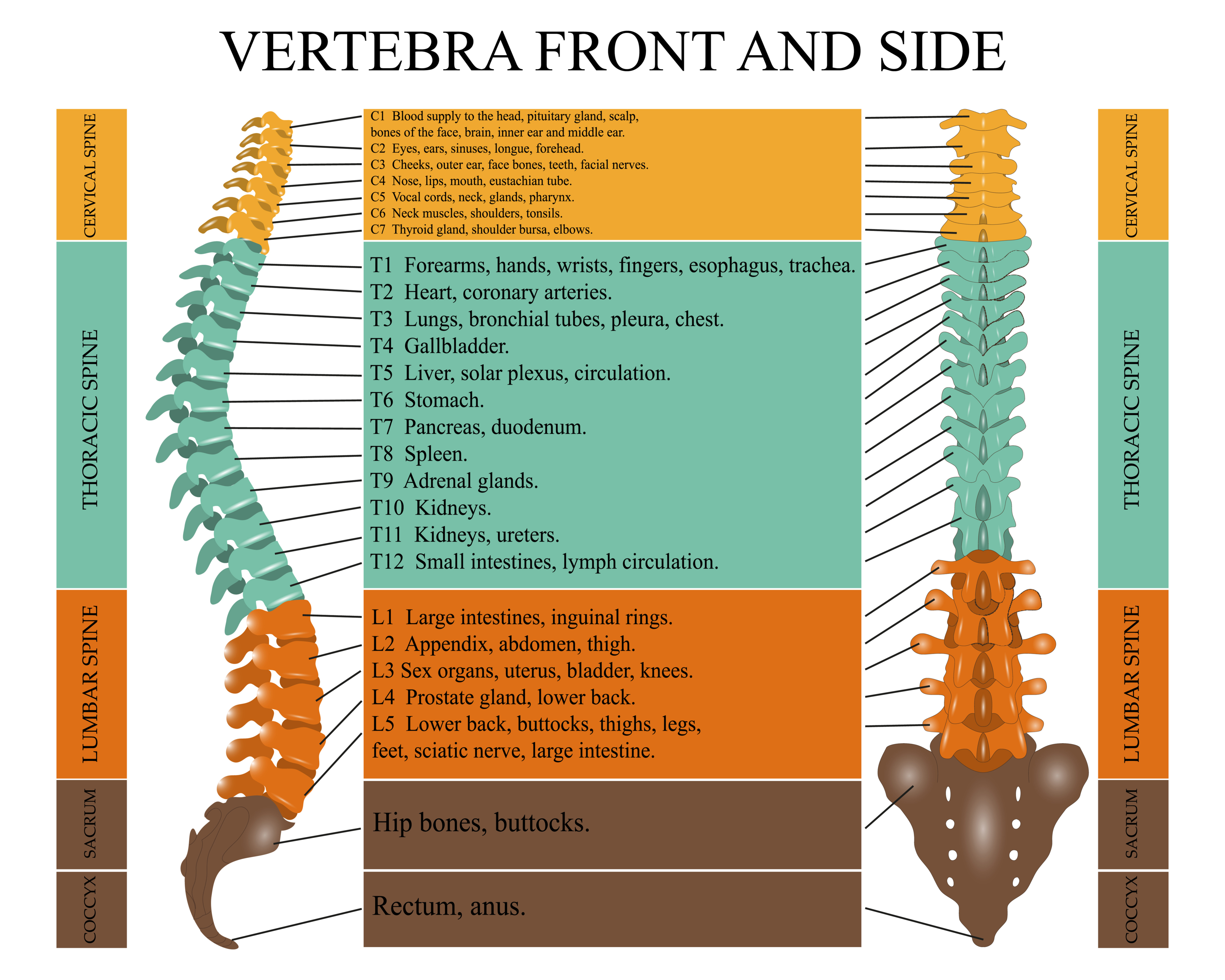
Injury to the cervical or thoracic spinal cord affects the spinal nerves that innervate respiratory muscles. The diaphragm, the major muscle of inspiration, receives its innervation from the third, fourth, and fifth cervical spinal segments (Figure 3).
Figure 3
Vertebra
The respiratory pattern of patients who have diaphragmatic paralysis is often a paradoxical movement of the abdomen: the abdominal wall retracts during inspiration and protrudes during the expiration phase (Vázquez et al., 2013; Hachmann et al., 2017):
After the initial stage of spinal shock has passed, patients with quadriplegia/tetraplegia may develop abnormal spinal reflexes and respiratory muscle spasticity (Garshick, 2021). This decreases the elasticity of the abdominal compartment (Vázquez et al., 2013). Spastic contractions of the abdominal muscles increase the load of the inspiratory muscles, and this increases the pressure that must be overcome for inspiration to be initiated (Vazquez et al., 2013).
Pulmonary physiologic changes due to SCI are related to the extent of neurological impairment. The American Spinal Injury Association Impairment Scale (AIS) is used to classify the degree of impairment that is based on strength in key muscles and a sensory examination (Table 1) (Hansebout & Kachur, 2018; Sánchez et al., 2020):
A |
|
B |
|
C |
|
D |
|
E |
|
*Muscle function is graded using the International Standards for Neurologic Classification of Spinal Cord Injury.
*For an individual to receive a grade of C or D (i.e., motor incomplete status), he/she must have either:
*Patients without an initial spinal cord injury do not receive an AIS grade.
The patient’s strength in key muscle groups is assessed and graded. A muscle grade of 5/5 is normal, and a grade of 3/5 means the muscle can be moved against gravity (Naqvi & Sherman, 2021):
Complete motor SCI includes: (See Table 1 above)
Incomplete motor SCI includes: (See Table 1 above)
With quadriplegia/tetraplegia, there is an injury to at least one of the eight cervical segments of the spinal cord. With paraplegia, there is an injury in the thoracic, lumbar, or sacral regions.
Respiratory dysfunction that causes pulmonary complications in SCI patients is complex and involves many factors, these include (Berlowitz et al, 2016; Garshick, 2021; Reyes et al., 2020):
Immediately after an SCI, flaccid paralysis can occur, affecting all the muscles that are caudal to the level of injury (Berlowitz et al., 2016; Hansebout & Kchur, 2018). This physiologic state is known as spinal shock or spinal paralysis (Berlowitz et al., 2016; Garshick, 2021; Hansebout & Kachur, 2018). Spinal shock may continue for days or many months, and during the recovery period, the patient’s pulmonary function is impaired, primarily due to impairment of the muscles of respiration (Berlowitz et al., 2016; Garshick, 2021; Hansebut & Kachur, 2018). Pulmonary function is also impaired because of a decrease in chest compliance, decreased airflow, hyper-responsiveness of the bronchial passages, and an abnormally blunted response to hypercapnia (Garshick, 2021).
After resolution of spinal paralysis, spastic paralysis occurs, muscle tone is increased, and pulmonary function should improve (Grashick, 2021; Vázquez et al., 2013). This improvement typically happens several days or several weeks after the injury (Garshick, 2021). Improvement in respiratory muscle performance after SCI largely occurs in the first year following injury.
The extent of ventilatory muscle impairment depends upon the degree and location of the injury, as well as the duration of time since the injury. The higher the level and more complete the injury, the more likely it is that there will be respiratory muscle dysfunction (Table 2).
Spinal Cord Level | Muscle Involvement | Effect on Respiration | Clinical Consequence |
C1-3 | Complete paralysis of all respiratory muscles | Vital capacity is only 5-10% of normal. Absent cough | Apnea and immediate death |
C3-6 | Varied Impairment of diaphragmatic contraction | Vital capacity 20% of normal. Weak and ineffective cough | Ventilation necessary in the acute stages. The majority will be weaned from mechanical ventilation |
C6-8 | Diaphragm and accessory cervical inspiratory muscles intact. Intercostals and abdominal muscles intact | Expiration is entirely passive. Secretion retention. No respiratory failure unless co-existing lung/chest injury/illness | |
T2-4 | Vital capacity 30-50% normal. Weak cough |
Ventilatory muscles innervated below the level of a complete SCI are completely nonfunctional (AIS A or B), while the degree of ventilatory muscle compromise is variable in patients with incomplete injuries (AIS C or D) (See Table 1 above):
Excess bronchial mucous production is caused by autonomic dysfunction that causes a predominantly parasympathetic stimulation of the lungs (Andrade et al, 2021; Bach et al, 2020a).
Lung and chest wall compliance is reduced in patients who have had an SCI (Garshick, 2021). This change is complicated and not completely understood, but it may be related to reduced lung volume and changes in surfactants (Garshick, 2021). Also, the joints, ligaments, and tendons of the rib cage will stiffen over time, limiting chest wall compliance (Bach et al, 2020b).
Patients who have a cervical or high thoracic spinal SCI and tetraplegia have increased airway resistance, expiratory airflow limitation, and bronchial hyperresponsiveness/bronchospasms (Andrade et al., 2021; Garschick, 2021; Hill et al., 2022).
The cause/causes of these abnormalities are likely due to (Andrade et al., 2021; Garshick, 2021; Hill et al., 2022):
The expiratory airflow limitation can be reversed with an inhaled anticholinergic, e.g., ipratropium, or an inhaled beta-2 agonist, e.g., albuterol (Garshick, 2021; Schilero et al., 2018a).
Individuals with quadriplegia/tetraplegia do not have a normal increase in the ventilatory drive when blood carbon dioxide levels are increased, and if supplementary oxygen is given to these patients, they can develop hypercapnia (Garshick, 2021; Bach et al., 2020a). The mechanisms for this are not completely understood, and this issue has not been extensively researched; it may be caused by a decrease in blood pressure when the patient is in a sitting position (Garshick, 2021).
Performing PFTs and calculations of the predicted PFT values need to be adjusted/modified when doing PFTs in patients who have had an SCI (Garshick, 2021).
Garshick (2021) notes: “Many SCI patients, particularly those with complete tetraplegia, cannot meet the acceptability and reproducibility standards for spirometry set by the American Thoracic Society (ATS).” (Note: Specific recommendations for measuring PFTs in SCI patients was not addressed in the most current (2019) ATS PFT measurement recommendations).
Approximately 90% of patients who have had a traumatic SCI will need intubation, and up to 40% of patients who had a complete cervical lesion will become ventilator-dependent (Schreiber et al., 2021).
The two most important markers that predict the need for intubation are the level of the injury and the ASIA classification (Yonemitsu et al., 2021; Hasenbout & Kachur, 2018; Bach, 2020a; Garshick, 2020):
The thickness and excursion of the diaphragm reflect the ability of this muscle to move and contract, and the diaphragm is one of the primary muscles of inspiration. Also, patients who have a high cervical or thoracic injury depend primarily on the diaphragm for ventilation (Vázquez et al., 2013).
Critically ill patients and patients who have had an SCI often develop diaphragmatic atrophy and limited diaphragmatic excursion (Guzel et al., 2022; Reardon et al., 2021; Soták et al., 2021; Zhu et al., 2021). Diaphragmatic dysfunction can negatively affect pulmonary function, and it is a major factor in the failure to wean patients from mechanical ventilation (Zhu et al., 2019; Soták et al., 2021; Qian et al., 2018).
Ventilator-induced diaphragmatic dysfunction (VIDD) is also associated with increased ICU stay, increased mortality, and prolonged mechanical ventilation (Reardon et al., 2021).
Ventilator-induced diaphragmatic dysfunction can occur very quickly after mechanical ventilation has begun. Levine et al. (2008) found that after 18-69 hours of mechanical ventilation, there was significant atrophy in the myofibers of the diaphragms of brain-dead patients.
Ventilator-induced diaphragmatic dysfunction (VIDD) is caused by mechanical ventilation (muscle inactivity because of mechanical ventilation) and factors like infections, malnutrition, sepsis, and the use of glucocorticoids and neuromuscular blockers (Reardon et al., 2021; Soták et al., 2021). Ultrasonography is the most effective technique for assessing the structure and function of the diaphragm in SCI patients and critically ill patients (Qian et al., 2018; Soták et al., 2021; Zambon et al., 2017; Zhu et al., 2021).
Currently, there are no well-proven and universally used methods for preventing VIDD. Phrenic nerve stimulation has been successful in animal experiments, and there is some promising research on its use in humans (Reardon et al., 2021; Sotak et al., 2021).
The start of weaning and the strategy to employ is determined by the following three factors:
There is relatively little research on weaning SCI patients from mechanical ventilation, and the studies on this topic that are available are often small, observational, and have study design problems (Garshick, 2020; Schreiber et al., 2021).
The criteria for suitability for weaning from Garshick (2020) and Bach (2020a), respectively, are outlined below in the bulleted lists. Regarding the criteria, Garshick (2020) wrote: “The accuracy of weaning predictors (e.g., rapid shallow breathing index) in predicting weaning success has not been specifically addressed in this patient population, and we typically follow standard clinical criteria for weaning . . .” Schreiber et al. (2021) wrote: “The probability of weaning success remains difficult to predict; no previous systematic review or meta-analysis has been conducted on the topic, and no societal guidelines or recommendations on weaning are available for this population.”
Weaning is done by using exercise-rest periods (Bach, 2020a; Füssenich et al., 2018; Garshick, 2020). The patient is disconnected from the ventilator, and continuous oxygen or compressed humidified air is delivered. He/she breathes spontaneously for a while, and then the patient is put back on full ventilatory support (Bach, 2020a). If the patient has a motor-complete cervical SCI, the patient should be weaned while recumbent; this will put the diaphragm higher and allow for a more effective contraction (Garshick, 2020).
The periods of spontaneous breathing are increased as tolerated, and the patient should be monitored for signs that spontaneous breathing is difficult, e.g., agitation, increased heart rate and/or respiratory rate, or a decrease in vital capacity (Garshick, 2020).
Manually assisted coughing, mechanical insufflation-exhalation, endotracheal suctioning, and other types of chest physiotherapy are important for preventing atelectasis during the weaning period (Garshick, 2020). Garshick (2020) recommends using chest physiotherapy techniques before each weaning period.
It is known that patients with cervical SCI have increased resistance in the airways due to a loss of sympathetic control.
The effectiveness of these medications as an adjunct to weaning has not yet been proven, and clinical experience is very limited.
Non-invasive ventilation (NIV) is defined as the delivery of positive pressure ventilation without the use of an invasive device (an endotracheal tube or a tracheostomy) and by way of a noninvasive device like a face mask, a nasal mask, or nasal prongs (Hyzy & McSparron, 2021b).
Non-invasive ventilation can be delivered by a standard or portable ventilator, and the interface device can be (Hyzy & McSparron, 2021b):
The two commonly used modes of delivery are bilevel NIV, commonly called bilevel positive airway pressure (BPAP – not BiPAP), and continuous positive airway pressure, CPCP (Hyzy & McSparron, 2021b).
Non-invasive ventilation is a well-established technique, and it can be used (Daoud et al., 2020; Hill & Kramer, 2022; Hyzy & McSparron, 2021a):
Non-invasive ventilation as a technique for weaning and (hopefully) decannulating SCI patients has many benefits. It can help avoid many of the complications of acute and long-term tracheostomy tube placement, and it has been used successfully (Guia et al., 2021; Ker et al., 2021; Toki et al., 2021). Most SCI patients who have ventilatory insufficiency should be considered for NIV weaning, and it is recommended to begin NIV weaning as soon as possible (Toki et al., 2021; Guia et al., 2021).
To be considered as a candidate for NIV weaning, patients should (Ker et al., 2021; Toki et al., 2021):
This weaning technique has been successfully used, but it has not been well studied. It is noted that there are no established guidelines/protocols for the indications for and timing of NIV weaning (Hill & Kramer, 2022; Guia et al., 2021; Ker et al., 2021; Toki et al., 2021).
Phrenic or diaphragmatic pacemakers are a form of respiratory support that may be used for the partial or complete withdrawal of mechanical ventilation in patients with SCI, thereby improving their quality of life (Wijkstra et al., 2022).
Electrostimulation of the phrenic nerve consists of triggering diaphragmatic contractions through direct electrical stimulation of the phrenic nerve in the neck and chest.
Candidates for electrostimulation include patients with (Marion, 2022; Wijkstra et al., 2022):
It is, therefore, necessary before implantation to perform phrenic nerve conduction studies (Vashisht & Chowdry, 2021). The pacing electrodes can be placed in the chest, the diaphragm, or the neck. After implantation, a period of diaphragmatic conditioning is needed to improve the muscle tone of the diaphragm, which may have atrophied due to lack of use (Marion, 2022).
Patients will often be on electrophrenic respiration for part of or the whole day, and then during the night, the patient will receive ventilator support (Vázquez et al., 2013).
Advantages of electrophrenic respiration include (DiMarco, 2013):
Regarding quadriplegic/tetraplegic patients who have an inadequate function of the phrenic nerves and in whom the implantation of phrenic/diaphragmatic pacemakers is not possible, there are experimental studies in animals that have suggested that high-frequency spinal cord stimulation (HF-SCS) may someday be an alternative approach for SCI patients (DiMarco & Kowalski, 2019).
Approximately 50% of tetraplegic patients who are ventilator-dependent will be able to achieve full-time ventilatory support with diaphragmatic pacing (DiMarco & Kowalski, 2019). It is important to note that diaphragmatic pacemakers only supplement the inspiratory function and do not replace the expiratory functions such as coughing or the removal of secretions. It is here where rehabilitation and external mechanical assistance have considerable importance.
Tracheostomy is a common procedure in patients with traumatic SCI, especially in cervical SCI or thoracic level with associated injuries.
Effective therapy in patients with cervical SCI consists of (Foran et al., 2022; Hansebout & Kuchar, 2018):
For patients with incomplete injuries, evidence of respiratory failure should prompt immediate airway intervention with endotracheal intubation. Patients who have been endotracheally intubated are at risk for respiratory and other complications, and in many cases, an elective tracheostomy is performed to prevent these issues when prolonged mechanical ventilation is likely (Foran et al., 2022; Mubashir et al., 2021; Handesbout & Kuchar, 2018) (Foran et al., 2022).
Tracheostomy can make weaning easier by decreasing airway resistance, it can make suctioning easier and more effective, it reduces the complications of long-term endotracheal intubation, and it is more comfortable than endotracheal intubation (Foran et al., 2022).
Risk factors that indicate the need for post-injury tracheostomy include (Hansebout & Kuchar, 2018; Long et al., 2022; Yu et al., 2022):
Specific to a cervical SCI cohort, the extent of the injury was the most important factor in determining the need for a tracheostomy (Branco et al., 2011).
In a retrospective study, patients with high-thoracic SCI have more respiratory complications (including the need for tracheostomy and intubation), even after adjustment for age, sex, and Glasgow Coma Scale (GCS), compared with patients with low-thoracic SCI or thoracolumbar fractures (Cotton et al., 2005). In this study, respiratory complications significantly increased the mortality risk in less severely injured patients.
Winslow et al. (2022) noted that respiratory complications are as important as the level of injury is contributing to the prediction of length of stay.
The literature has suggested that early tracheostomy (within the first 7 days) has many advantages, including (but not limited to) (Foran et al., 2022; Mubashir et al., 2021):
However, Mubashir et al. (2021) notes that evaluating the available evidence is difficult and “. . . it is challenging to make conclusive interpretations. Future prospective trials with a larger patient population are needed to fully assess short- and long-term outcomes of tracheostomy timing following acute SCI.”
Several recent systematic reviews also found that early tracheostomy does not reduce mortality and finds no effect on the duration of MV or intensive care stay in a general critical care population (Foran et al., 2022).
Both surgical and percutaneous tracheostomies (PTs) can be safely performed in the ICU (Kaczmarek et al., 2017). A PT is done by making a small incision and then a blunt dilation of the trachea to insert the tracheostomy tube.
Advantages of PT include (Kaczmarek et al., 2017; Ramakrishnan et al., 2019):
Patients who have had an SCI and will undergo spinal fusion surgery and a tracheostomy require special attention. The presence of a tracheostomy stoma could cause cross-contamination from the tracheostomy incision site to the spinal surgery incision site, and this has made the timing of the two procedures with each one an issue of concern (Galeiras et al., 2018). Performing the tracheostomy after the spinal surgery has typically been the approach, but the optimal time for separating the two procedures is not known. Although the research is limited, several studies found no infections or a very low rate of infection when a tracheostomy is done without waiting or with waiting several weeks after spinal surgery (Galeiras et al., 2018).
Tracheostomy reduces the work of breathing and reduces airway resistance, intrinsic positive end-expiratory pressure (PEEP), and peak inspiratory pressures (PIP) (Chen et al., 2019; Hyzy & McSparron, 2021a). Tracheostomy can also make ventilatory weaning easier (Hyzy & McSparron, 2021a).
Although pulmonary complications are a common and well-known problem in SCI, there is relatively little information about their management. Despite years of experience and research, there are still many unknowns in the treatment of SCI, and treatment recommendations are, at times, based on clinical experience and expert opinion (Fehlings et al., 2017).
Many patients who have an SCI cannot generate sufficient lower intrathoracic and expiratory pressure to produce an effective cough and clear secretions (Berlowitz et al., 2016; Nygren-Bonnier et al., 2018). This puts them at risk for atelectasis, impaired gas exchange, and infections (Nygren-Bonnier et al., 2018). One of the most important goals of treating respiratory dysfunction in SCI patients is managing secretions to prevent these complications (Reyes et al., 2020; Vázquez et al., 2013).
Many techniques can be used to manage and clear secretions, including (but not limited to (Bach et al., 2020; Blakeman et al., 2022; Luo, 2021; Spinou, 2020; Reyes et al., 2020; Vázquez et al., 2013):
Deciding what treatments should be used should be done on a case-by-case basis, and the need will be, obviously, greater for some patients. Bach et al. (2020) indicated that if an SCI patient does not have a tracheostomy and is medically stable, “. . . there is neither theoretic reason why, nor evidence that, routine chest oscillation, percussion, and vibration benefit them unless they have an acute pulmonary disease or are overwhelmed with peripheral secretions.”
Interventions for mobilizing secretions are essential for preventing mucus plugs, atelectasis, pneumonia, and respiratory failure and should be started early after the injury.
Postural drainage uses positioning to help move pulmonary secretions to the upper airway so that they can be removed by coughing or other methods (Luo, 2021; Vázquez et al., 2013).
The positioning of the patient with the affected lung area in the upper position allows for gravity to help in the drainage.
Different postural drainage positions can be used:
The position should be held for approximately 20 minutes, and postural drainage can be combined with percussion and vibration (Bach et al., 2020a). The goal is to drain the peripheral airway divisions (Bach et al., 2020a). To drain the six central airway divisions, an effective cough must be produced (Bach et al., 2020a).
Percussion and vibration consist of external manipulations of the chest to mobilize secretions.
The technique of pulmonary percussion is done by tapping on the chest with a cupped hand. A percussion vest and hand-held percussion devices can be used as well, and percussion can be combined with postural drainage (Reyes et al., 2020; Vázquez et al., 2013). Vibration is essentially the same technique, except that a vibrating force is applied. The 11th and 12th ribs, often called the floating ribs, should not be contacted during percussion and vibration (Vázquez et al., 2013).
Contraindications to percussion and vibration include (Vázquez et al, 2013):
An intact, normal cough is necessary to clear airway secretions. Patients who have had an SCI, for several reasons, often have a weak and ineffective cough, putting them at risk for pulmonary complications like atelectasis and pneumonia (Reyes et al., 2020; Vázquez et al., 2013). Assisted coughing techniques that target the inspiratory or expiratory phase to either assist a cough or stimulate a cough are commonly used as preventive therapy (Reyes et al., 2020; Spinou, 2020; Vázquez et al., 2013). Assisted coughing techniques that can be used are discussed below.
Assisted coughing techniques have been used for many years, and the research indicates that all the commonly used techniques can improve airway clearance, and adverse effects are uncommon (Morrow et al., 2021; Spinou, 2020). However, there is little evidence that can be used to compare techniques, and what evidence there is suggests that in terms of improving peak cough flow, the assisted cough techniques are similar (Morrow et al., 2021; Spinou, 2020).
This maneuver consists of abdominal thrusts directed posteriorly and towards the patient’s head (Bach, 2020). The patient inhales, coughs, and the abdominal thrust is done (Reyes et al., 2020). The effectiveness of manually assisted coughing can be improved with the prior administration of nebulized saline to thin the secretions. Contraindications to manually assisted coughing include (Spinou, 2020):
This procedure, also called Mechanical Insufflation-Exsufflation, is begun by applying positive pressure to the airway (insufflation) using a mechanical device and via a face mask, mouthpiece, tracheostomy to immediately afterward transform this positive pressure into negative pressure (exsufflation) (Bach, 2020; Reyes et al., 2020; Vázquez et al., 2013).
The lungs are fully inflated and then fully emptied within 4 to 6 seconds, and a sudden change of pressure occurs within 0.02. seconds is enough to effectively clear respiratory secretions (Bach, 2020).
There is some evidence that cough peak flow (the maximum amount of airflow generated during a cough) is improved when a manually assisted cough and mechanical insufflation-exsufflation are used together (Spinou, 2020). There is evidence that suggests that mechanical insufflation-exsufflation is better than manually assisted coughing for improving cough peak flow (Spinou, 2020). An anticholinergic or a beta2 agonist bronchodilator may increase the effectiveness of mechanical insufflation-exhalation if administered prior to simulated cough (Bach et al., 2020a).
Advantages in the use of an insufflation-exsufflation device during intensive and post-intensive care may be (Vázquez et al., 2013):
Contraindications to the use of insufflation-exsufflation devices include (Reyes et al., 2020; Vázquez et al., 2013):
Glossopharyngeal breathing has been used for decades as a way of improving cough effectiveness and pulmonary function (Nygren-Bonnier et al., 2018; Spinou, 2020; Reyes et al., 2020). The patient is instructed to do a total lung capacity maneuver and then to use his/her mouth and throat muscles to inhale as many gulps of air as possible, followed by a passive exhalation (Berlowitz et al., 2016; Nygren-Bonnier et al., 2018; Reyes et al., 2020).
Glossopharyngeal breathing can be difficult and uncomfortable to do, but it can improve peak cough flow, total lung capacity, and vital capacity (Nygren-Bonnier et al., 2018; Reyes et al., 2020). This technique can cause dizziness, syncope, and abnormal changes in blood pressure and heart rate (Nygren-Bonnier et al., 2018). Therefore, it is vital that patients must be instructed on how to do glossopharyngeal breathing correctly.
Airway suctioning is an important part of secretions management in patients who have an artificial airway (Blakeman et al., 2022). Blakeman et al. (2022) reviewed the available literature and made these recommendations for the practice of artificial airway suctioning:
Complications of suctioning include increased arterial pressure, increased heart rate, cardiac arrhythmias, and oxygen desaturation. Preoxygenation, sedation, and suctioning as needed may help avoid these complications (Blakeman et al., 2022).
Respiratory muscle training (RMT) has been successfully and safely used for patients who have had an SCI (Berlowitz & Tamplin, 2013; Boswell-Ruys et al., 2020; McDonald & Stiller, 2019; Reyes et al., 2020; Wang et al., 2021).
A wide variety of devices and techniques have been used for RMT, including (but not limited to) (Boswell-Ruys et al., 2020):
No evidence supports one type of RMT in favor of another. Although the current research indicates that RMT is a useful therapy for patients who have had an SCI, evidence of its support has come from small studies (Berlowitz & Tamplin, 2013; Boswell-Ruys et al., 2020; Bach et al., 2020a). RMT can also increase muscle strength. However, these gains can quickly be lost, so RMT needs to be ongoing, and the increase in muscle strength may not necessarily help prevent respiratory complications (Bach et al., 2020a).
Per EMS (1900): At approximately 18:00, 18-year-old Thomas Hutton dove face-first into the 3-foot end of a cement swimming pool after drinking beer all afternoon with his friends. He hit his forehead on the bottom of the pool. His friends pulled him out of the pool while calling 911. He has not regained consciousness since.
Upon arrival in the emergency department, EMS reports:
The patient is on a backboard with a semi-rigid cervical collar in place.
A 20-gauge peripheral IV was placed in the left antecubital with 1000 ml Normal Saline infusing at 100 ml/hr. Patient on 100% rebreather mask with oral airway inserted.
GCS continues at 3.
Chest x-rays show pulmonary infiltrates.
Cervical spine x-rays indicate an extension teardrop fracture at C2, with neck CT confirming the diagnosis.
Head CT shows a fracture of the frontal bone with a hematoma at the base of the occiput.
All laboratory tests WNL except:
Continuing GCS of 3 with imaging studies indicative of a severe head injury and an extension teardrop fracture at C2: A neurologist and neurosurgeon should be consulted.
Chest x-ray indicating pulmonary infiltrates and respiratory acidosis coupled with continuing hypertension, rapid, shallow respirations, and lower than expected SaO2 per oximetry: Emergent intubation indicated.
Pulmonary physiologic changes due to SCI are related to the extent of neurological impairment. The American Spinal Injury Association Impairment Scale (AIS) is used to classify the degree of impairment that is based on strength in key muscles and a sensory examination.
Immediately after SCI, flaccid paralysis affects all muscles caudal to the level of injury (i.e., spinal shock). Subsequent improvements in pulmonary function are due primarily to functional descent of the neurologic injury level as spinal cord inflammation resolves, enhanced recruitment of accessory ventilatory muscles, retraining of deconditioned muscles, and the evolution from flaccid to spastic paralysis.
The extent of ventilatory muscle impairment depends upon the degree and location of the injury, as well as the duration of time since the injury. The higher the level and more complete the injury, the more likely it is that there will be respiratory muscle dysfunction. Complete injury above C3 produces near-total ventilatory muscle paralysis because the phrenic nerve, which innervates the diaphragm, arises from the third to fifth cervical roots.
Immediately after an SCI, flaccid paralysis can occur, affecting all the muscles that are caudal to the level of injury. This physiologic state is known as spinal shock or spinal paralysis. This state may continue for days or months, and during this time, pulmonary function is impaired. This is primarily due to impairment of the muscles of respiration but also due to a decrease in chest compliance, decreased airflow, hyper-responsiveness of the bronchial passages, and an abnormally blunted response to hypercapnia.
After the resolution of spinal paralysis, spastic paralysis occurs. During spastic paralysis, muscle tone is increased, and pulmonary function should improve. This happens several days or several weeks after the injury. Improvement in respiratory muscle performance after SCI largely occurs in the first year following injury.
Assessment of pulmonary function in SCI patients is done with PFTs. Performing PFTs and calculations of the predicted PFT values need to be adjusted/modified when doing PFTs in patients who have had an SCI. Many SCI patients will have abnormal PFTs, but there is an improvement over the first few months-year after the injury.
After an SCI, the long-term course of pulmonary function can be a decline, stability, or improvement.
Approximately 90% of patients who have had a traumatic SCI will need intubation, and up to 40% of patients who had a complete cervical lesion will become ventilator-dependent. It is best to proceed with intubation under controlled circumstances rather than waiting until the condition becomes an emergency, as hypoxia in an SCI patient can adversely affect the patient’s neurological and many patients who have cervical spinal cord injury have a delayed onset of respiratory complications. Intubation is necessary for managing secretions, and SCI is often accompanied by many complications like trauma, coma, ARDS, and multi-organ system insufficiency. In these situations, non-invasive ventilation will not be effective. There are no specific criteria for ventilator settings that are specific to SCI patients.
If it is likely that the patient may be ventilator-dependent for a long time, a tracheostomy is recommended. Tracheostomy can make weaning easier by decreasing airway resistance, it can make suctioning easier and more effective, it reduces the complications of long-term endotracheal intubation, and it is more comfortable than endotracheal intubation. Tracheostomy can be done at the bedside (PT) or in the OR.
In regard to criteria for weaning eligibility, Garshick (2020) wrote: “The accuracy of weaning predictors (e.g., rapid shallow breathing index) in predicting weaning success has not been specifically addressed in this patient population, and we typically follow standard clinical criteria for weaning . . .” Schreiber et al. (2021) indicates that the probability of weaning success continues to be difficult to predict as there are no previous systematic review or meta-analysis has been conducted on the topic. Schreiber et al. (2021) also state that there are no societal guidelines or recommendations on weaning that are available and specific to this population of patients. Therefore, the typical weaning protocol is utilized. This protocol is alternating periods of exercise (the patient is disconnected from the ventilator) and rest (the patient is reconnected to the ventilator).
Conservative methods of preventing respiratory complications in SCI patients include:
Phrenic or diaphragmatic pacemakers are a form of respiratory support that may be used for the partial or complete withdrawal of mechanical ventilation in patients with SCI. Electrostimulation of the phrenic nerve consists of triggering diaphragmatic contractions through direct electrical stimulation of the phrenic nerve in the neck and chest.
Non-invasive ventilation (NIV) is defined as the delivery of positive pressure ventilation without the use of an invasive device and by way of a noninvasive device like a face mask, a nasal mask, or nasal prongs. Non-invasive ventilation can be delivered by a standard or portable ventilator. The two commonly used modes of delivery are bilevel NIV, commonly called bilevel positive airway pressure (BPAP – not BiPAP), and continuous positive airway pressure, CPCP. Non-invasive ventilation is a well-established technique that can be used to treat patients who are not intubated or do not have a tracheostomy. It can also be used for those who have acute or chronic respiratory dysfunction caused by a neuromuscular or chest wall disease, including SCI. In addition, it can also be used to wean SCI patients who are endotracheally intubated, those who have a tracheostomy and are ventilator-dependent, as well as for those requiring tracheal decannulation.
CEUFast, Inc. is committed to furthering diversity, equity, and inclusion (DEI). While reflecting on this course content, CEUFast, Inc. would like you to consider your individual perspective and question your own biases. Remember, implicit bias is a form of bias that impacts our practice as healthcare professionals. Implicit bias occurs when we have automatic prejudices, judgments, and/or a general attitude towards a person or a group of people based on associated stereotypes we have formed over time. These automatic thoughts occur without our conscious knowledge and without our intentional desire to discriminate. The concern with implicit bias is that this can impact our actions and decisions with our workplace leadership, colleagues, and even our patients. While it is our universal goal to treat everyone equally, our implicit biases can influence our interactions, assessments, communication, prioritization, and decision-making concerning patients, which can ultimately adversely impact health outcomes. It is important to keep this in mind in order to intentionally work to self-identify our own risk areas where our implicit biases might influence our behaviors. Together, we can cease perpetuating stereotypes and remind each other to remain mindful to help avoid reacting according to biases that are contrary to our conscious beliefs and values.