Sign Up
You are not currently logged in. Please log in to CEUfast to enable the course progress and auto resume features.

Course Library

Common Skin Conditions Across the Lifespan

1 Contact Hour including 1 Pharmacology Hour
Listen to Audio
CEUfast OwlGet one year unlimited nursing CEUs $39Sign up now
This peer reviewed course is applicable for the following professions:
Advanced Practice Registered Nurse (APRN), Athletic Trainer (AT/AL), Certified Nurse Midwife, Certified Nurse Practitioner, Certified Registered Nurse Anesthetist (CRNA), Certified Registered Nurse Practitioner, Clinical Nurse Specialist (CNS), Licensed Practical Nurse (LPN), Licensed Vocational Nurses (LVN), Midwife (MW), Nursing Student, Occupational Therapist (OT), Occupational Therapist Assistant (OTA), Physical Therapist (PT), Physical Therapist Assistant (PTA), Registered Nurse (RN), Registered Nurse Practitioner, Respiratory Therapist (RT)
This course will be updated or discontinued on or before Friday, February 18, 2028

Nationally Accredited

CEUFast, Inc. is accredited as a provider of nursing continuing professional development by the American Nurses Credentialing Center's Commission on Accreditation. ANCC Provider number #P0274.


Outcomes

≥ 92% of participants will know how to recognize and manage common skin conditions.

Objectives

After completing this continuing education course, the participant will be able to:

  1. Distinguish between skin conditions affecting different populations (pediatric and adult patients).
  2. Describe the clinical features of common dermatologic disorders.
  3. Summarize the diagnostic characteristics of common dermatologic disorders.
  4. Outline evidence-based treatment strategies for common skin conditions.
  5. Identify potential complications or comorbid conditions related to dermatologic disorders.
CEUFast Inc. and the course planning team for this educational activity do not have any relevant financial relationship(s) to disclose with ineligible companies whose primary business is producing, marketing, selling, re-selling, or distributing healthcare products used by or on patients.

Last Updated:
  • $39 Unlimited Access for 1 Year
    (Includes all state required Nursing CEs)
  • No Tests Required
    (Accepted by most states & professions)
  • Instant Reporting to CE Broker
  • Instant Access to certificates of completion
Logo Audio
Now includes
Audio Courses!
Learn More
Restart
Restart
  • 0% complete
Hide Outline
Playback Speed

Narrator Preference

(Automatically scroll to related sections.)
Done
Common Skin Conditions Across the Lifespan
0:00
0:15
 
To earn a certificate of completion you have one of two options:
  1. Take test and pass with a score of at least 80%
  2. Reflect on practice impact by completing a course evaluation.
    (NOTE: Some approval agencies and organizations require you to take a test and the course evaluation is NOT an option.)
Author:    Oniel D. Laucella (APRN, RN, AGPCNP-BC, APHN-BC, CWP, CPAHA)

Introduction to Skin Conditions

The skin is the largest organ of the human body and plays an essential role in protecting internal systems, regulating body temperature, and facilitating sensory input. Composed of the epidermis, dermis, and subcutaneous tissue, it serves as the first line of defense against environmental threats, including microbes, allergens, and ultraviolet radiation. Its function extends beyond physical protection to include immune surveillance, fluid retention, and thermoregulation (Gupta & Lipner, 2021).

Skin conditions are among the most frequent complaints in primary care and urgent care settings. Disorders such as acne, eczema, fungal infections, and bacterial skin infections affect individuals across all age groups. According to the American Academy of Dermatology (2025), approximately one-third of the U.S. population is affected by a skin disease at any given time. These conditions range in severity from minor irritations to complex, chronic illnesses that can significantly impair a patient's physical and emotional well-being.

Accurate diagnosis of skin disorders relies heavily on the clinician's ability to assess lesion morphology, distribution, and patient history. Common lesion types include macules, papules, vesicles, pustules, and plaques, each of which may suggest a different underlying pathology. Understanding the clinical presentation of these lesions allows nurse practitioners and other advanced practice providers to identify conditions early and implement appropriate interventions (Dinulos, 2020).

Prompt treatment is especially important in preventing complications. For example, diaper dermatitis in infants can lead to secondary fungal infections if not addressed quickly (Oakley, 2017). In adult patients, untreated bacterial infections such as those caused by methicillin-resistant Staphylococcus aureus can result in cellulitis or systemic infection, requiring hospitalization. Some skin findings, such as the malar rash seen in lupus or the velvety skin changes in acanthosis nigricans, may also serve as early indicators of systemic disease.

Healthcare providers play a vital role in identifying and managing dermatologic conditions in both pediatric and adult populations. A strong foundation in dermatology enables clinicians to reduce disease burden through early detection, patient education, and evidence-based treatment. Continuing education focused on skin conditions equips providers with the tools to deliver high-quality, informed care that improves outcomes and enhances patients' quality of life.

Assessment of Skin Lesions

Accurate assessment and classification of skin lesions are fundamental skills for healthcare providers. A comprehensive dermatologic assessment requires both visual and tactile evaluation, as well as an understanding of lesion morphology, distribution, and associated systemic signs. Identifying the type, configuration, and evolution of lesions allows clinicians to narrow the differential diagnosis and determine the most appropriate course of treatment (Dinulos, 2020).

Lesions are typically categorized as either primary or secondary. Primary lesions arise from previously normal skin (Shetty et al., 2022) and include macules, papules, nodules, plaques, vesicles, bullae, pustules, wheals, and cysts. Each type has distinct characteristics. For instance, a macule is a flat, non-palpable discoloration under 1 centimeter (cm) in diameter, whereas a papule is a small, raised lesion. Vesicles and bullae are fluid-filled, with vesicles being smaller than 1 cm and bullae exceeding that size. Pustules contain purulent fluid and are commonly associated with infections or inflammatory conditions like acne or impetigo.

Secondary lesions develop from the evolution of primary lesions or from external manipulation such as scratching or infection (Shetty et al., 2022). These include scales, crusts, erosions, ulcers, fissures, scars, excoriations, and lichenification. For example, lichenification is the thickening of the skin with exaggeration of normal skin markings, often seen in chronic eczema as a result of repetitive scratching. The identification of secondary changes can offer valuable insight into the chronicity and severity of a condition.

In addition to morphology, the distribution and pattern of lesions provide critical diagnostic clues. Lesions with an annular (ring-shaped) pattern may suggest tinea corporis or erythema multiforme, while linear lesions can result from contact dermatitis due to exposure to an irritant in a specific shape or path. A dermatomal distribution, typically unilateral and confined to a nerve root segment, is a hallmark of herpes zoster. Bilateral and symmetrical lesions are often seen in systemic conditions such as psoriasis or atopic dermatitis (Dinulos, 2020; Oakley, 2025).

A thorough skin assessment should also include a review of systemic symptoms such as fever, fatigue, or joint pain, which may suggest an underlying autoimmune or infectious etiology. Patient history, including recent exposures, medications, travel, and personal or family history of skin disorders, further supports accurate clinical decision-making. In cases of diagnostic uncertainty, advanced diagnostic tools such as dermoscopy, skin scrapings, or biopsy may be warranted.

Ultimately, structured skin assessments—conducted with attention to lesion type, distribution, evolution, and systemic context—enable providers to deliver timely and effective care. Early recognition and classification of skin lesions not only improves diagnostic accuracy but also facilitates targeted treatment and reduces unnecessary referrals or complications.

Common Pediatric and Adult Skin Conditions

Urticaria

Urticaria, or hives, is a common dermatologic condition affecting approximately 20% of individuals at some point in their lives (Powell et al., 2015). It manifests as transient, erythematous wheals that are intensely pruritic and often blanchable. Lesions typically resolve within 24 hours but may recur, especially in chronic urticaria, which persists for six weeks or longer. Triggers can include foods, medications (e.g., nonsteroidal anti-inflammatory drugs [NSAIDs], antibiotics), insect stings, infections, or autoimmune processes. Treatment involves identifying and eliminating the trigger, if known, and initiating pharmacological management. Second-generation H1 antihistamines (e.g., cetirizine, loratadine) are the first-line agents due to their reduced sedation profile (Schettini et al., 2023). For refractory cases, omalizumab or immunosuppressive agents such as cyclosporine may be used. Educating patients on trigger avoidance and stress reduction is essential for long-term control.

Diaper Dermatitis

Diaper dermatitis affects up to 35% of infants at some point, with Candida albicans overgrowth contributing to many persistent or treatment-resistant cases (Benitez Ojeda & Mendez, 2023). The condition presents as a well-demarcated, bright red rash often involving the skin folds, with characteristic satellite pustules and papules. It is frequently associated with prolonged exposure to moisture, stool, and urine, and can be exacerbated by recent antibiotic use, which disrupts normal flora. The cornerstone of management includes frequent diaper changes, air exposure, application of zinc oxide or petroleum-based barrier creams, and topical antifungals such as clotrimazole or nystatin. In more severe or recurrent cases, a short course of topical corticosteroids may be used under medical supervision. Prevention strategies, including the use of breathable diapers and avoiding over-washing with harsh soaps, help reduce recurrence (Benitez Ojeda & Mendez, 2023).

Acne Vulgaris

Acne vulgaris is the most common skin condition in adolescents, affecting nearly 85% of individuals aged 12 to 24 years (Zaenglein et al., 2016). It is a multifactorial disorder involving increased sebum production, follicular hyperkeratinization, Cutibacterium acnes colonization, and inflammation. Clinical manifestations include open and closed comedones, inflammatory papules, pustules, nodules, and, in severe cases, cysts and scarring. Lesions typically occur on the face, chest, and back. First-line treatment involves topical agents such as benzoyl peroxide, retinoids (e.g., adapalene), and antibiotics. Oral antibiotics (e.g., doxycycline) or hormonal therapy (e.g., oral contraceptives or spironolactone) may be indicated for moderate to severe cases. Isotretinoin is reserved for nodulocystic or treatment-resistant acne (Sutaria et al., 2023). Early and consistent management can significantly reduce physical and psychosocial impact.

Community-Acquired MRSA Skin Infection

Community-acquired methicillin-resistant Staphylococcus aureus (CA-MRSA) is a significant cause of skin and soft tissue infections, accounting for many infections seen in U.S. emergency departments (Loewen et al., 2017). It often presents as painful, red, swollen lesions resembling spider bites, abscesses, or cellulitis. Risk factors include crowding, poor hygiene, skin trauma, and participation in contact sports. Incision and drainage (I&D) remain the primary treatment for uncomplicated abscesses. Empiric antibiotics such as trimethoprim-sulfamethoxazole, doxycycline, or clindamycin are used in cases with systemic symptoms or high-risk features. Culture and sensitivity testing guide targeted therapy. Patient education on wound care, hygiene, and not sharing personal items is vital to prevent spread (Loewen et al., 2017).

Herpes Simplex Virus Type 1 (HSV-1)

HSV-1 is a prevalent infection, affecting many individuals (Johns Hopkins Medicine, n.d.). It typically causes orolabial herpes, presenting as grouped vesicles on an erythematous base around the lips or mouth, which later crust and resolve. Primary infection may be accompanied by fever, lymphadenopathy, and malaise. The virus becomes latent in the trigeminal ganglia and can reactivate due to stress, sun exposure, or immunosuppression. Antiviral therapy with acyclovir, valacyclovir, or famciclovir is effective in reducing the duration and severity of symptoms, especially when initiated during the prodromal phase (Ahmad et al., 2024). Chronic suppressive therapy may be considered in patients with frequent recurrences or in those who are immunocompromised.

Tinea Corporis (Ringworm)

Tinea corporis is a superficial dermatophyte infection of the skin, primarily caused by Trichophyton rubrum, and is especially common in children and athletes. Its prevalence varies by region but can affect up to 30% of the population in some tropical and subtropical areas, particularly where heat and humidity promote fungal growth (Yee et al., 2025). Tinea corporis presents as annular lesions with central clearing, scaly raised borders, and mild pruritus. Diagnosis is clinical but may be confirmed by KOH examination or fungal culture. Topical antifungal agents like terbinafine and clotrimazole are effective for localized infections, while systemic therapy (e.g., oral terbinafine) may be necessary for extensive or resistant cases. Patient education should emphasize hygiene, avoidance of shared personal items, and proper drying of skin after bathing.

Intertrigo

Intertrigo is a common inflammatory condition that affects skin folds, particularly in individuals with obesity, diabetes, or poor hygiene. It occurs when friction, heat, and moisture lead to skin breakdown, creating an ideal environment for secondary infection with Candida or bacteria. It presents as red, raw, and macerated plaques, often with itching or burning. Recent studies report that intertrigo affects approximately 16% to 38% of nursing home and long-term care residents, highlighting its high prevalence in vulnerable populations with limited mobility and comorbid conditions (Kottner et al., 2020). Management focuses on reducing moisture and friction using absorbent powders, barrier creams, and antimicrobial or antifungal agents. Maintaining a dry environment and treating coexisting infections are essential to prevent recurrence.

Scabies

Scabies, caused by the mite Sarcoptes scabiei, is a highly contagious parasitic infestation affecting people across all age groups and socioeconomic levels. Globally, scabies is estimated to affect more than 200 million people at any given time and over 400 million individuals annually, underscoring its significant public health burden (World Health Organization [WHO], n.d.). Symptoms include intense pruritus, especially at night, and characteristic burrows, typically found in web spaces, wrists, elbows, and genital areas. Diagnosis is clinical, though microscopic identification of mites or eggs may confirm the diagnosis. First-line treatment is permethrin 5% cream, applied overnight and repeated after one week. Oral ivermectin is used in resistant or crusted scabies. All close contacts should be treated simultaneously, and environmental cleaning is essential.

Balanitis

Balanitis is the inflammation of the glans penis and is most common among uncircumcised males. Current literature estimates that balanitis affects approximately 3% to 11% of men, with a notably higher prevalence in those who are uncircumcised due to the warm, moist conditions under the foreskin that promote microbial growth (Leber, 2025). Symptoms typically include erythema, edema, discharge, and irritation—often stemming from poor hygiene, exposure to irritants, or infections such as Candida albicans. Diagnosis is typically clinical. Treatment is tailored to the specific cause: antifungal creams for candidal balanitis, topical antibiotics for bacterial infections, and guidance on gentle hygiene, avoidance of irritants, and good glycemic control in diabetic patients. In refractory or recurrent cases, circumcision may be considered to prevent recurrence and improve genital hygiene (Leber, 2025).

Herpes Zoster (Shingles)

Herpes zoster is a reactivation of the latent varicella-zoster virus, typically affecting adults over age 50. The lifetime risk is approximately 30%, increasing with age (Centers for Disease Control and Prevention [CDC], n.d.). Shingles presents as a painful, unilateral, vesicular rash in a dermatomal distribution, often preceded by tingling or burning.Complications include postherpetic neuralgia, especially in older adults.Antiviral treatment (valacyclovir, acyclovir, or famciclovir) is most effective when started within 72 hours of symptom onset.The recombinant zoster vaccine (Shingrix) is highly effective in preventing both herpes zoster and postherpetic neuralgia and is recommended for adults aged 50 and older. Pain management and patient education on early recognition are key components of care (Patil et al., 2022).

Skin Conditions with Images, Descriptions, and Treatments

Urticaria

photo of urticaria skin condition

Image 1: Urticaria

Description of lesions: Multiple annular wheals ranging in size from 1 cm to 3 cm localized on the upper right back. The wheals are uniform, light pink in color, with a smooth surface and a blanched center surrounded by a red halo or flare (area of erythema). The wheals are superficial. The wheals are also raised and palpable, with some areas forming a coalescent arrangement. The lesions are asymmetrically distributed and accompanied by pruritus and edema.

Diagnosis: Urticaria (New) - ICD-10 L50.9

Treatment: Second-generation H1 antihistamine

Name: John Doe                    Date: January 15th, 2025

rx graphic


Loratadine 10 mg

Disp: #30 tablets

Sig. Take one tablet by mouth daily PRN for urticaria.

REFILLS: 0 (Zero)

Diaper Dermatitis

photo of diaper rash

Image 2: Diaper Rash

Description of lesions: Multiple irregular coalescing lesions localized at the upper pelvis/lower abdominal area. The primary lesions appear to be moist, erythematous plaques with maceration. The plaques are well-demarcated. The periphery of the lesions displays scaling and surrounding satellite papules and pustules with evidence of erosions dispersed bilaterally across the inner thighs. The area is noted to be tender and pruritic.

Diagnosis: Diaper Dermatitis (Candidiasis) (New) - ICD-10 L22

Treatment: Topical antifungal agent

Name: Jane Doe                    Date: February 15th, 2025

rx graphic


Nystatin (topical ointment) 100,000 units per gram

Disp: #1 tube of 15 grams

Sig. Gently massage ointment into the affected area every eight hours for seven days for diaper rash.

REFILLS: #1 (One)

Acne Vulgaris

photo of acne vulgaris skin condition

Image 3: Acne Vulgaris

Description of lesions: Multiple round erythematous papules and pustules present in a reticular arrangement on the nose, bilateral cheeks, and chin. The papules and pustules vary in size, measuring 2 millimeters (mm) to 5 mm in diameter. The papules present on the medial cheeks are coalescing, surrounded by more discrete, satellite erythematous papules with some erosion and excoriation. There are also scattered closed comedones with no visible keratin plugs. The lesions are accompanied by pain and tenderness.

Diagnosis: Acne Vulgaris (New) - ICD-10 L70.0 

Treatment: Oral tetracycline antibiotic and combination topical retinoid and antiseptic agent

Name: Jina Doe                    Date: March 11th, 2025

rx graphic


Doxycycline 100 mg capsules

Disp: #60

Sig. Take one capsule by mouth twice daily (every 12 hours) for three months for acne.

Refills: #3 (Three)


Adapalene/benzoyl peroxide topical gel 0.1%/2.5%

Disp: #1 bottle

Sig. Apply a thin layer to affected areas on face once daily for acne.

Refills: #3 (Three)

Community-Associated Methicillin-Resistant Staphylococcus Aureus

photo of mrsa

Image 4: CA-MRSA

Description of lesions: Deep, inflammatory, ruptured, erythematous nodule approximately 3 cm in diameter. The wound bed is dark red and moist. Wound edges are well-demarcated. Erythema and some desquamation were also noted in the peri-wound area.

Diagnosis: Community-Associated Methicillin-Resistant Staphylococcus Aureus (CA-MRSA) - (New) ICD-10 B95.62

Treatment: Tetracycline antibiotic

Name: Jackie Doe                    Date: April 12th, 2025

rx graphic


Minocycline 100 mg capsule

Disp: #28

Sig. Take one capsule by mouth every 12 hours for seven days for skin infection.

Refills: 0 (zero)

Herpes Simplex Virus (Type 1)

photo of hsv on face

Image 5: HSV Type 1

Description of lesions: One single 5 mm round-shaped, yellow and crusted pustule with a well-defined border in a circulate configuration localized to the left side.

Diagnosis: Herpes Simplex Virus (Type 1) (New) - ICD-10 B00.9

Treatment: Antiviral ointment

Name: Jasmina Doe                    Date: May 25th, 2025

rx graphic


Acyclovir ointment 5%

Disp: #1 tube of 15 g

Sig. Apply to the affected area every three hours, up to six times a day for seven days for outbreak.

REFILLS: #1 (One)

Tinea Corporis

photo of tinea corporis

Image 6: Tinea Corporis

Description of lesions: One single annular plaque, 2 cm in diameter and variegated in color (dark red to pink). Borders are well-defined and raised with crusting on the surface.

Diagnosis: Tinea Corporis (New) - ICD-10 B35.4

Treatment: Topical antifungal cream

Name: Justin Doe                    Date: June 5th, 2025

rx graphic


Clotrimazole 1% (topical cream)

Disp: #1 tube of 30g

Sig. Apply to the affected area twice daily for four weeks for skin infection.

REFILLS: #1 (One)

Erythema Intertrigo

photo of erythema intertrigo

Image 7: Erythema Intertrigo

Description of lesions: Red, glistening, coalesced plaques. The rash is localized along the intertriginous skin-fold area beneath the right breast.

Diagnosis: Erythema Intertrigo (New) - ICD-10 L30.4

Treatment: Topical antifungal agent

Name: Jiang Doe                    Date: July 17th, 2025

rx graphic


Clotrimazole cream 1%

Disp: #1 tube of 30 g

Sig. Apply to the affected area twice a day for two weeks for skin infection.

REFILLS: #1 (One)

Sarcoptes Scabiei (Scabies)

photo of sarcoptes scabiei scabies

Image 8: Sarcoptes (Scabiei (Scabies)

Description of lesions: Small erythematous crusted vesicles are seen with curved (S-shaped) burrowing (this is a characteristic location to find active scabies lesions). Secondary scaling and excoriation noted and caused by scratching. The scabies rash usually appears two to six weeks after exposure to parasitic infestation caused by Sarcoptes scabiei var. homitis. Patients with scabies complain of a rash with unremitting itching and an inability to stop scratching.

Diagnosis: Sarcoptes Scabiei (Scabies) (New) - ICD-10 B86

Treatment: Topical antiparasitic cream

Name: Jolio Doe                    Date: August 28th, 2025

rx graphic


Permethrin 5% topical cream

Disp: #1 tube

Sig. Apply cream to entire skin from chin down, including under fingernails and toenails. Leave on for eight hours. Repeat in 14 days if live mites are still present.

REFILLS: #1 (One)

Balanitis

graphic showing balanitis

Image 9: Balanitis

Description of lesions: Erythematous lesions on the glans and/or the foreskin of the penis. Swelling and irritation of the glans are present. Causes include skin disorders, infection, poor hygiene, uncontrolled diabetes, and harsh soaps. Symptoms include pain, redness, and a foul-smelling discharge from under the foreskin. Treatment depends on the cause, but may require antibiotics, steroid creams, or antifungal creams. The most common cause is Candida; pending swab, initial treatment will be an antifungal.

Diagnosis: Balanitis (New) - ICD-10 N48.1

Treatment: Topical antifungal cream

Name: Jamal Doe                    Date: September 7th, 2025

rx graphic


Miconazole nitrate 2% topical cream

Disp: #1 tube

Sig. Apply cream to affected area twice daily (every 12 hours) for three weeks.

REFILLS: #1 (One)

Herpes Zoster (Shingles)

photo of shingles

Image 10: Herpes Zoster (Shingles)

Description of lesions: Multiple 0.5 mm grouped fluid-filled dome-shaped vesicles and bullae 1 mm or greater in diameter on erythematous, edematous plaques. The fluid inside the vesicles and bullae may be clear or hemorrhagic in nature. There are areas noted where the vesicles and bullae have evolved into pustules with some erupting purulent material, which later go on to form crusted erosions. The lesions are found to be in a dermatomal pattern. New lesions continue to appear for up to one week and are preceded by pain, itching, or tingling in the area where they will develop. The lesions occur most commonly in older adults and immunocompromised people.

Diagnosis: Herpes Zoster (Shingles) (New) - ICD-10 B02.9

Treatment: Oral antiviral agent

Name: Juanita Doe                    Date: October 27th, 2025

rx graphic


Valacyclovir 1000 mg tablets

Disp: #21 tablets

Sig. Take one tablet by mouth every eight hours for seven days for shingles.

REFILLS: #1 (One)

Other Skin Conditions Across the Lifespan

Pediatric and Adolescent Conditions

Atopic Dermatitis (Eczema)

  • Chronic, relapsing inflammation common in infancy and childhood.
  • Strong genetic and allergic component.

Molluscum Contagiosum

  • Viral infection causing umbilicated papules.
  • Common in children and immunocompromised individuals.

Hand-Foot-and-Mouth Disease

  • Caused by Coxsackievirus; involves painful oral ulcers and vesicular rash on palms and soles.

Cradle Cap (Infantile Seborrheic Dermatitis)

  • Greasy, scaly patches on the infant's scalp; benign and self-limiting.

Impetigo

  • Highly contagious, often seen in children. Presents as honey-colored crusts around the nose and mouth.
  • Cause: Staphylococcus aureus or Streptococcus pyogenes.

Warts (Verruca Vulgaris)

  • Common in school-aged children due to frequent minor trauma and exposure.
  • Cause: Human papillomavirus (HPV).

Adult and General Population Conditions

Psoriasis (Plaque Psoriasis)

  • Autoimmune condition causing thick, scaly plaques on extensor surfaces.
  • Associated with arthritis in some adults (psoriatic arthritis).

Contact Dermatitis (Allergic/Irritant)

  • Acute or chronic reaction to allergens or irritants (e.g., nickel, latex, poison ivy).

Urticaria (Hives)

  • Transient wheals due to histamine release from allergens or unknown triggers.
  • It can be acute or chronic.

Seborrheic Dermatitis

  • Common on scalp, face, and trunk; "dandruff" variant in mild forms.

Vitiligo

  • Autoimmune depigmentation disorder that can affect individuals of all ages.

Lichen Planus

  • Pruritic, purple, polygonal papules—often associated with autoimmune disease.

Folliculitis

  • Inflammation of hair follicles, often caused by shaving, hot tubs, or tight clothing.
  • Cause: Staphylococcus aureus, Pseudomonas, or fungal organisms.

Cellulitis

  • A deeper bacterial skin infection more common in adults with skin trauma, edema, or comorbidities.
  • Cause: Streptococcus pyogenes, Staphylococcus aureus.

Older Adult & Geriatric Conditions

Actinic Keratosis

  • Premalignant rough, scaly lesions caused by sun damage.

Seborrheic Keratosis

  • Benign "stuck-on" lesions; common in older adults.

Basal Cell Carcinoma (BCC)

  • It is the most common skin cancer, especially on sun-exposed areas.

Squamous Cell Carcinoma (SCC)

  • The second most common skin cancer; may arise from actinic keratoses.

Xerosis (Age-Related Dry Skin)

  • Common in older adults due to decreased sebaceous activity.

Erythrasma

  • Often occurs in older or obese individuals with diabetes or limited mobility. Presents as reddish-brown patches in skin folds.
  • Cause: Corynebacterium minutissimum

Case Study: Pain That Comes in a Stripe

Patient Description

Mr. Joseph H., a 64-year-old retired postal worker, presented to New Dawn Primary Care Clinic for his scheduled appointment with Nurse Practitioner Amanda. He reports a 3-day history of localized neuropathic pain and sensory disturbances localized to the left mid-back. He described the discomfort as sharp, burning, and shock-like sensations, which he rated as 7 out of 10 in severity. The pain had been persistent, interfering with his sleep and daily routines. Within 24 hours of the initial symptoms, he developed clusters of painful, fluid-filled blisters in the same region. Mr. H. denied fever, headache, or systemic symptoms. He has a medical history of hypertension and type 2 diabetes mellitus, managed with lisinopril and metformin. Notably, he had never received the shingles (herpes zoster) vaccine, despite meeting CDC age-based eligibility recommendations. He expressed regret about this, stating he was unaware of the vaccine's availability and importance.

The patient lives alone in a private apartment and reports no recent illness, known exposures, or travel. He maintains regular follow-up with his primary care provider for chronic disease management, but reported missing his last wellness visit. His diabetes has been moderately controlled, with a recent HbA1c of 7.5%. He denied any known immunosuppressive conditions or medications. The nurse practitioner recognized that Mr. H.'s age, diabetic status, and lack of vaccination placed him at increased risk for reactivation of latent varicella-zoster virus (VZV), leading to shingles. The timing, pain characteristics, and sudden vesicular eruption raised a strong clinical suspicion for herpes zoster.

Clinical Presentation

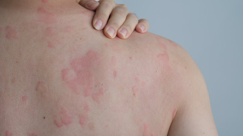
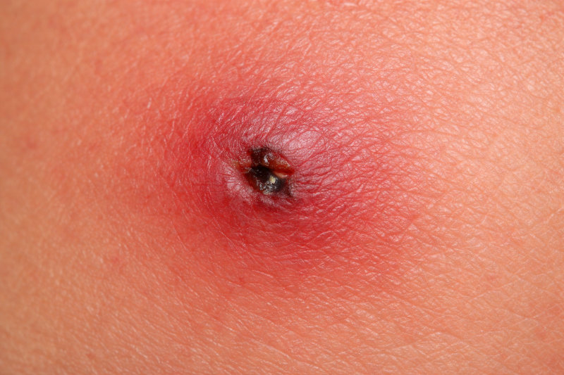
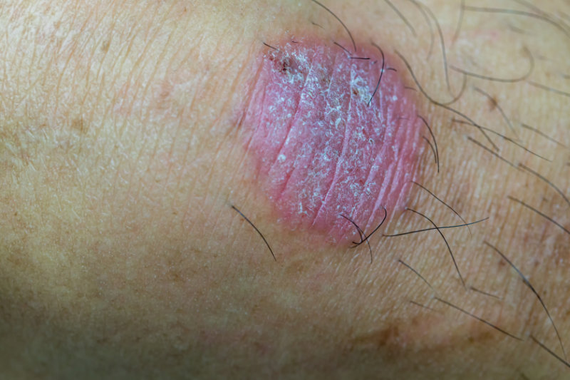
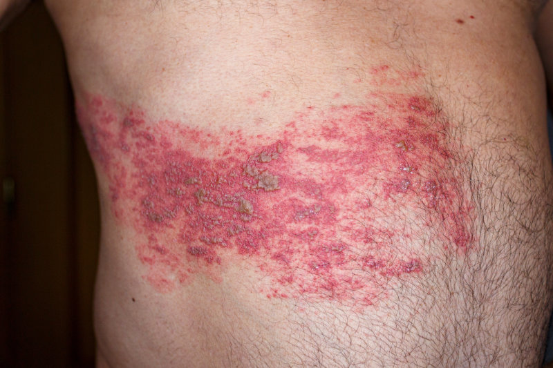
On physical examination, the evaluating nurse practitioner noted a unilateral eruption of grouped vesicles on an erythematous base, following the left T8 dermatome in a linear, dermatomal pattern. The lesions did not cross the midline, which is characteristic of herpes zoster. The vesicles were tense and clear, some beginning to crust at the margins. The surrounding skin showed signs of mild edema and tenderness to light palpation. No signs of systemic involvement, dissemination, or ocular complications were present. Lymphadenopathy was not appreciated, and the patient remained afebrile. Cranial nerves were intact, and the neurological exam was unremarkable beyond the localized hyperesthesia in the affected dermatome.

The nurse practitioner made a clinical diagnosis of herpes zoster based on the patient's age, symptomatology, and physical findings. Given the classic presentation and absence of red-flag features, no further laboratory or imaging studies were indicated. She discussed the natural course of shingles and emphasized the need for early antiviral therapy to reduce the duration and severity of symptoms and to mitigate the risk of postherpetic neuralgia, a common complication in older adults.

Nurse Practitioner Intervention and Management

The nurse practitioner prescribed valacyclovir 1,000 milligrams (mg) by mouth three times daily for seven days. This antiviral therapy was selected based on current clinical guidelines, which recommend initiating treatment early to reduce the severity and duration of symptoms. The goal was also to decrease the risk of complications such as postherpetic neuralgia, especially given the patient's age and history of type 2 diabetes. Acetaminophen was recommended for pain relief, and the patient was advised to take it regularly during the acute phase.

Patient education focused on safe practices to limit the spread of the virus and prevent secondary skin infections. Joseph was instructed to gently clean and cover the rash, wear loose clothing, and avoid touching or scratching the affected area. He was also advised to avoid contact with individuals at higher risk for severe varicella-zoster virus infection, including pregnant women, infants, and people with weakened immune systems. The nurse practitioner explained the warning signs of secondary infection, such as spreading redness, swelling, or discharge, and encouraged prompt follow-up if symptoms worsened or did not improve.

Preventive care was also addressed. The nurse practitioner provided information on the recombinant zoster vaccine (Shingrix), including its role in reducing the risk of future outbreaks and related complications. Although the vaccine cannot be given during active infection, the patient was encouraged to receive it after his skin lesions fully resolved. The discussion also included tips on supporting immune health, such as good blood sugar control, balanced nutrition, adequate sleep, and stress reduction.

Outcome

At his follow-up visit one week later, Joseph reported that the pain had decreased significantly. He described it as a dull ache, now rated 3 out of 10, compared to the initial sharp and burning discomfort. Most of the blisters had crusted, and there was no evidence of new lesions or signs of bacterial infection. His sleep had improved, and he expressed a greater sense of comfort and independence.

Joseph shared that he had followed the home care instructions carefully and appreciated the clear, supportive communication he received during his first visit. He stated that the education helped him understand the condition and feel more confident managing it. He expressed interest in getting the shingles vaccine once the rash healed and was provided with follow-up guidance and educational materials. The nurse practitioner reinforced the importance of continuing self-care and monitoring for any lingering pain that might indicate nerve involvement. Overall, the patient's recovery was progressing well, with no complications reported at this time.

Strengths of the Approach

  • Rapid recognition and diagnosis of a classic dermatomal rash.
  • Timely initiation of evidence-based antiviral therapy.
  • Proactive education on disease transmission, pain management, and preventive vaccination.
  • Patient-centered communication tailored to age and chronic condition management.

Opportunities for Growth

  • Consideration of adjunct neuropathic pain options (e.g., gabapentin) for potential postherpetic neuralgia.
  • More thorough assessment of psychosocial impact and sleep disturbances related to pain.

Reflection Questions

What is the role of the NP in reducing complications associated with shingles?

  • Nurse practitioners play a key role in early detection, prompt antiviral treatment, and preventive counseling to minimize risks such as postherpetic neuralgia.

Why is patient education essential in managing herpes zoster?

  • Understanding transmission, skin care, and vaccination empowers patients to reduce spread, improve outcomes, and make informed decisions about future prevention.

Conclusion

Skin conditions are a common reason patients of all ages seek care. As there are many different types of conditions affecting the skin and their presentations vary widely, it is important that healthcare professionals be aware of the different types and their symptoms. It is important to note that comorbid conditions can contribute to these diagnoses, requiring an individualized treatment approach. Effective management requires a deeper dive into the patient's history, lifestyle, and exposures, aiming to provide effective treatment and improve quality of life.

Select one of the following methods to complete this course.

Take TestPass an exam testing your knowledge of the course material.
OR
No TestComplete a course evaluation.

Implicit Bias Statement

CEUFast, Inc. is committed to furthering diversity, equity, and inclusion (DEI). While reflecting on this course content, CEUFast, Inc. would like you to consider your individual perspective and question your own biases. Remember, implicit bias is a form of bias that impacts our practice as healthcare professionals. Implicit bias occurs when we have automatic prejudices, judgments, and/or a general attitude towards a person or a group of people based on associated stereotypes we have formed over time. These automatic thoughts occur without our conscious knowledge and without our intentional desire to discriminate. The concern with implicit bias is that this can impact our actions and decisions with our workplace leadership, colleagues, and even our patients. While it is our universal goal to treat everyone equally, our implicit biases can influence our interactions, assessments, communication, prioritization, and decision-making concerning patients, which can ultimately adversely impact health outcomes. It is important to keep this in mind in order to intentionally work to self-identify our own risk areas where our implicit biases might influence our behaviors. Together, we can cease perpetuating stereotypes and remind each other to remain mindful to help avoid reacting according to biases that are contrary to our conscious beliefs and values.

References

  • Ahmad, B., Gurnani, B., & Patel, B. C. (2024). Herpes Simplex Keratitis. In StatPearls. StatPearls Publishing. Visit Source.
  • American Academy of Dermatology Association (AAD). (2025). Skin Conditions By The Numbers. American Academy of Dermatology Association. Visit Source.
  • Benitez Ojeda, A. B., & Mendez, M. D. (2023). Diaper Dermatitis. In StatPearls. StatPearls Publishing. Visit Source.
  • Centers for Disease Control and Prevention (CDC). (n.d.). Shingles (Herpes Zoster). Centers for Disease Control and Prevention. Visit Source.
  • Dinulos, J. (2020). Habif's Clinical Dermatology: A Color Guide to Diagnosis and Therapy. 7th edition. Elsevier.
  • Gupta, A. K., & Lipner, S. R. (2021). Skin Anatomy and Physiology. In Dermatology: Essentials for primary care (pp. 1–9). McGraw-Hill Education.
  • Johns Hopkins Medicine. (n.d.). Herpes: HSV-1 and HSV-2. Johns Hopkins Medicine. Visit Source.
  • Kottner, J., Everink, I., van Haastregt, J., Blume-Peytavi, U., & Schols, J. (2020). Prevalence of intertrigo and associated factors: A secondary data analysis of four annual multicentre prevalence studies in the Netherlands. International Journal of Nursing Studies, 104, 103437. Visit Source.
  • Leber, M. J. (2025). Balanitis. Medscape. Visit Source.
  • Loewen, K., Schreiber, Y., Kirlew, M., Bocking, N., & Kelly, L. (2017). Community-associated methicillin-resistant Staphylococcus aureus infection: Literature review and clinical update. Canadian family physician Medecin de famille canadien, 63(7), 512–520.
  • Oakley, A. (2017). Napkin dermatitis. DermNet. Visit Source.
  • Oakley, A. (2025). Herpes zoster. DermNet. Visit Source.
  • Patil, A., Goldust, M., & Wollina, U. (2022). Herpes zoster: A Review of Clinical Manifestations and Management. Viruses, 14(2), 192. Visit Source.
  • Powell, R. J., Leech, S. C., Till, S., Huber, P. A., Nasser, S. M., Clark, A. T., & British Society for Allergy and Clinical Immunology. (2015). BSACI guideline for the management of chronic urticaria and angioedema. Clinical and experimental allergy : journal of the British Society for Allergy and Clinical Immunology, 45(3), 547–565. Visit Source.
  • Schettini, N., Corazza, M., Schenetti, C., Pacetti, L., & Borghi, A. (2023). Urticaria: A Narrative Overview of Differential Diagnosis. Biomedicines, 11(4), 1096. Visit Source.
  • Shetty, B., Fernandes, R., Rodrigues, A. P., Chengoden, R., Bhattacharya, S., & Lakshmanna, K. (2022). Skin lesion classification of dermoscopic images using machine learning and convolutional neural network. Scientific reports, 12(1), 18134. Visit Source.
  • Sutaria, A. H., Masood, S., Saleh, H. M, & Schlessinger, J. (2023). Acne Vulgaris. In StatPearls. StatPearls Publishing. Visit Source.
  • World Health Organization (WHO). (n.d.). Scabies. World Health Organization. Visit Source.
  • Yee, G., Syed, H. A., & Al Aboud, A. M. (2025). Tinea Corporis. In StatPearls. StatPearls Publishing. Visit Source.
  • Zaenglein, A. L., Pathy, A. L., Schlosser, B. J., Alikhan, A., Baldwin, H. E., Berson, D. S., Bowe, W. P., Graber, E. M., Harper, J. C., Kang, S., Keri, J. E., Leyden, J. J., Reynolds, R. V., Silverberg, N. B., Stein Gold, L. F., Tollefson, M. M., Weiss, J. S., Dolan, N. C., Sagan, A. A., Stern, M., … Bhushan, R. (2016). Guidelines of care for the management of acne vulgaris. Journal of the American Academy of Dermatology, 74(5), 945–73.e33. Visit Source.