Exploring Secondary Skin Lesions: Unveiling Complications and Care
Written by Jennifer Huynh, BSN, RN, NCSN
Welcome back to our ongoing exploration of skin lesions. If you found our previous discussion on primary lesions enlightening, you're in for another informative treat. Our skin, our body's protective shield, faces constant challenges from everyday elements like the sun, gravity, and the natural aging process. But fear not; there are steps we can take to safeguard our skin's health, from wearing sun protection factor (SPF) to maintaining a healthy lifestyle. Yet, if skin injuries occur, proper care is crucial. Having covered primary lesions with their various origins, including pathology, physical factors, and infections, we're now poised to delve into the realm of secondary lesions. These lesions emerge as complications following primary conditions or injuries, revealing a deeper layer of skin health intricacies.
Our journey into secondary lesions presents a diverse spectrum of skin anomalies. Allow us to guide you through the intricacies of each type:
Scales are accumulations of flaky, irregularly shaped keratinized cells. Their thickness varies, influenced by factors like dryness or oiliness. Scales can arise from specific diseases, drug reactions, or even the neglect of regular dry skin care.
Lichenification involves the thickening and roughening of the epidermis due to persistent trauma or irritation. Chronic dermatitis is a common cause, and callouses can also exemplify this process.
Keloids are excessive collagen formation that follows wounds or lacerations, with keloids often emerging after surgical incisions. These irregular scars progressively expand beyond the incision boundaries, demanding attention.
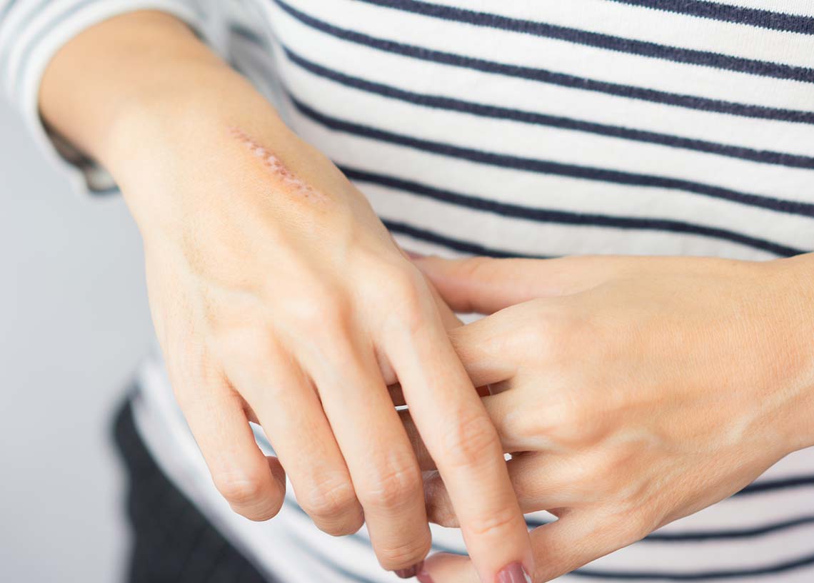
Scars commonly occur post-epidermal injury; scars may manifest as thin or thick fibrous skin, replacing normal skin post-healing.
Excoriation is characterized by the loss of the epidermis; excoriation presents as linear, hollowed-out areas with potential crusting along the edges. Abrasions or parasitic infections like scabies can cause such damage. Scratching can also cause excoriation. If it is deep enough within the dermis, it may cause scarring.
Fissures are linear cracks or epidermal breaks extending into the dermis, triggered by factors such as dryness or athlete's foot. Dermatitis can exacerbate fissures, possibly leading to scarring.
Erosion is the loss of the epidermis and creates depressions in the skin's surface, which may appear moist and glistening. This may occur after vesicle or bulla eruptions and chemical injuries.
Ulcers represent a more severe loss of skin; ulcers range from the epidermis to the deep dermal layers. Typically concave and varying in size, they include complications like stasis or pressure sores.
Atrophy is the thinning of the skin, leading to the loss of defined markings, and is a natural aspect of aging skin.
Special Secondary Skin Lesions
Having explored secondary lesions, we acknowledge that medical scenarios can be intricate, with exceptions aplenty. Our forthcoming discussion will cover particular lesions, presenting unique challenges and insights. Let’s review special lesions.
Comedones are a mixture of sebaceous and keratin material that blocks pores. Varying in size, comedones are different from acne. Acne are usually pustules filled with purlent fluid. At the same time, there are typically two types of comedones: open and closed. They are different in appearance as blackheads are open and whiteheads are closed. The color difference is due to the oxidizing process of exposed oils within the pores.
Burrows are narrow, irregular channels in the skin. These channels are raised due to the presence of parasites.
Petechia and purpuras are circumcised areas of blood that vary in size. Petechia are less than 0.5 cm, and purpuras are larger than 0.5 cm.
We understand that becoming an expert in defining every skin lesion is a tall order, but this knowledge can be invaluable when identifying lesions that may warrant further examination.
Many lesions share similarities, some stemming from similar primary causes like surgical care. However, understanding the distinction between them, such as keloids and scars, aids in determining appropriate treatment paths. Nurses play a pivotal role in recognizing the body's normal healing process and discerning when intervention is necessary to prevent excessive healing.
A crucial aspect I would like to emphasize is pressure ulcers. Regardless of your specialty, poor care makes every patient susceptible to developing these localized injuries. Pressure ulcers arise from unrelieved pressure, friction, shearing forces, and moisture. The ongoing pressure disrupts blood flow, leading to skin damage that progresses through different dermal layers. There are two types of pressure injury: decubitus and mucosal membrane injuries. We'll delve deeper into the nuances of pressure injuries in an upcoming post.
In wrapping up this comprehensive insight into secondary and special skin lesions, remember that while primary lesions may have diverse origins, secondary lesions can stem from poor care and complications. Some complications may be out of our control. Still, overall, our collective aim as professionals is to minimize the impact of secondary lesions, even as we continue to focus on preventing and addressing primary concerns. As mentioned before, dermatology has many components and nuances. As professionals, being diligent in our nursing assessments will provide the best outcome for patients with opportunities to intervene before damage is done.
About the Author:
Jennifer "Jenny" Huynh, BSN, RN, NCSN, graduated from the University of Massachusetts Lowell (Umass Lowell) and is certified as a school nurse. She has worked as an RN for six years, focusing on school nursing. Currently, Jenny is working on her Master's in Nursing Education and is an Adjunct Instructor at UMass Lowell.
Jenny is an independent contributor to CEUfast’s Nursing Blog Program. Please note that the views, thoughts, and opinions expressed in this blog post are solely of the independent contributor and do not necessarily represent those of CEUfast. This is not medical advice. Always consult with your personal healthcare provider for any health-related questions or concerns.
If you are interested in learning more about CEUfast’s Nursing Blog Program or would like to submit a blog post for consideration, please visit https://ceufast.com/blog/submissions.