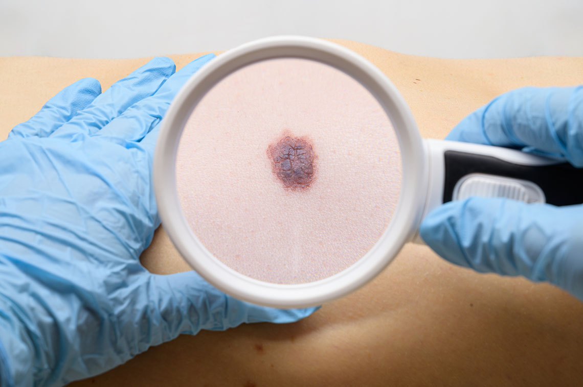
Decoding Primary Skin Lesions: A Comprehensive Guide
Written by Jennifer Huynh, BSN, RN, NCSN
Today, let's continue exploring the skin – the body's largest and most sensitive organ. With its remarkable complexity, the skin is a frontline defense against external factors and a canvas for age-associated changes. Our skin is an open book, reflecting genetics, environmental influences, and the natural passage of time. However, it's not invincible; the skin can succumb to ultraviolet (UV) radiation, inflammation, gravity, tobacco exposure, and the inevitable biological shifts that come with age. As we traverse the various stages of life, the skin can become thinner, lose elasticity, and develop wrinkles, making it susceptible to a range of diseases and infections.
When we embark on this journey, one initial sign we encounter is abnormalities like skin lesions. While these lesions might trigger concern, it's important to remember that not all indicate underlying severe issues. Some are intrinsic to the aging process or genetically determined. While a detailed exploration of diseases and their systemic effects will await future blog posts, let's delve into the fascinating world of skin lesions.
The landscape of skin lesions can be overwhelming due to its diversity, but it can be categorized into primary and secondary lesions. Primary lesions are the initial changes that manifest within the skin, while secondary lesions are the enduring effects that arise from primary lesions, often leading to further complications. Additionally, there are unique cases of skin lesions that deserve special attention. In our upcoming post, we'll delve into secondary and special lesions to provide a comprehensive understanding.
So, let's begin our journey by exploring the realm of primary skin lesions.
Unveiling the Diversity of Primary Skin Lesions
Macules: These are flat, small areas that alter the skin's color. Typically, macules are less than 1 cm in diameter. Examples range from freckles and nevi (flat moles) to petechiae, measles, and scarlet fever. While distinct from each other, understanding these variations is crucial for an accurate diagnosis.
Papules: Elevated and firm, papules encompass an area that’s less than 1 cm. Recognizable examples include elevated moles, warts, lichen planus, fibromas, and insect bites. As a side note, it's essential to recognize hyper-reactions in insect bites that might lead to anaphylaxis.
Patches: These are flat, irregularly shaped macules that are typically larger than 1 cm. Examples include vitiligo, port-wine stains, Mongolian spots, and cafe-au-lait spots. It's imperative for healthcare professionals to differentiate between normal birthmarks and bruises, such as the case with Mongolian spots, often found in the lumbosacral area.
Plaques: Elevations with a flat top surface; plaques are firm and rough lesions. Generally larger than 1 cm in diameter, they can include seborrheic and actinic keratoses. An important example is psoriasis, a chronic skin disease that increases susceptibility to secondary lesions.
Wheals: Irregular areas of cutaneous edema; wheals are solid and transient, varying in diameter. They are commonly linked to insect bites, hives (urticaria), and allergic reactions; close monitoring of the skin aids in diagnosing anaphylaxis.
Nodules: These elevated, firm, and circumscribed lesions reside deep in the dermis, typically measuring 1-2 cm in diameter. Examples include erythema nodosum and lipomas.
Tumors: Elevated, solid lesions that may be demarcated; tumors often lie deep within the dermis and are usually larger than 2 cm in diameter. Tumors can be benign or cancerous, necessitating proper diagnosis and follow-up care.
Vesicles: These elevated, superficial lesions are filled with serous fluid. Vesicles are smaller than 1 cm and do not penetrate the dermis. Common examples encompass chickenpox (varicella) and shingles (herpes zoster), both of which produce vesicles. Providers must be aware of the contagious nature of these conditions and provide guidance on returning to daily activities once the lesions dry and crust over.
Bullae: Larger than vesicles, bullae are vesicles greater than 1 cm in diameter. It is crucial to educate patients about blisters and the risk of infection from popping them.
Pustules: Elevated, superficial lesions filled with purulent fluid; pustules are commonly seen in conditions like acne and impetigo. Distinguishing between different pustular conditions is essential for accurate care.
Cysts: Elevated, encapsulated lesions within the dermis or subcutaneous layer; cysts can be filled with liquid or semisolid material. Distinguishing between different types of cysts, such as acne and cystic acne, is crucial for developing appropriate treatment plans.
Telangiectasia: These fine, irregular lines are a result of capillary dilation in the skin. Conditions like rosacea can lead to the production of telangiectasia lines.
Understanding the relationship between different types of lesions and associated diseases enhances our diagnostic accuracy. For instance, differentiating between chickenpox and measles, or understanding that scarlet fever may present with macules while chickenpox may manifest as vesicles, is vital. Similarly, recognizing the distinction between a cyst and a nodule is crucial, even though they both exist within the dermis, with varying consistencies.
Furthermore, it's imperative for healthcare providers to comprehend the tailored care required for different lesions. By staying vigilant and informed about various lesion types, we enhance our ability to provide effective assessment and care. Dermatology, like any specialized field, is intricate, but as nurses, it's our responsibility to be well-rounded professionals, ensuring our patients receive the comprehensive medical attention they deserve. We hope this discussion on primary skin lesions aids in your understanding and contributes to your holistic approach to patient care.
Stay tuned for more insightful explorations into the diverse world of dermatology.
About the Author:
Jennifer "Jenny" Huynh, BSN, RN, NCSN, graduated from the University of Massachusetts Lowell (Umass Lowell) and is certified as a school nurse. She has worked as an RN for six years, focusing on school nursing. Currently, Jenny is working on her Master's in Nursing Education and is an Adjunct Instructor at UMass Lowell.
Jenny is an independent contributor to CEUfast’s Nursing Blog Program. Please note that the views, thoughts, and opinions expressed in this blog post are solely of the independent contributor and do not necessarily represent those of CEUfast. This is not medical advice. Always consult with your personal healthcare provider for any health-related questions or concerns.
If you are interested in learning more about CEUfast’s Nursing Blog Program or would like to submit a blog post for consideration, please visit https://ceufast.com/blog/submissions.


