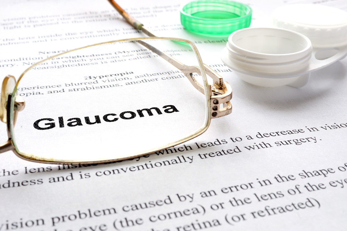
Glaucoma: The Silent Thief of Sight
Written by Mariya Rizwan, PharmD
Three million Americans have glaucoma, according to the Centers for Disease Control and Prevention. It was previously believed that glaucoma was caused by high intraocular pressure, but it is now recognized that optic nerve damage can occur at normal or even low pressures. Glaucoma is defined as a problem with the integrity of the optic nerve structure or function that causes changes in the optic nerve, leading to specific visual defects. By lowering the intraocular pressure, optic nerve damage can be slowed or stopped. However, some patients may have optic nerve damage with visual field defects even with normal intraocular pressure. Raised intraocular pressure leads to compromised optic nerve and retina blood flow. Due to low blood supply, tissue damage occurs and progresses. Increased intraocular pressure, if left untreated, can lead to blindness.
What Causes Glaucoma?
To understand glaucoma, it is important to understand facts about the aqueous humor. A balance exists between the aqueous humor's production, circulation, and outflow. The aqueous humor is produced in the posterior chamber ciliary processes and flows through the pupil into the anterior chamber. From the anterior chamber, it passes through Schlemm’s canal and the aqueous veins into the anterior ciliary veins.
Chronic open-angle glaucoma, also called primary open-angle glaucoma (POAG), is the most common form, accounting for 90% of all cases. It is caused by an overproduction of aqueous humor or obstruction to its flow through the trabecular meshwork or the canal of Schlemm. However, the chamber angle between the iris and cornea remains open. Intraocular pressure increases because the aqueous humor cannot leave the eye at the rate it is produced.
Acute glaucoma, closed-angle glaucoma, or narrow-angle glaucoma is less common. However, its onset is sudden and needs to be treated as an emergency. Anterior displacement of the iris occurs against the cornea, obstructing the outflow of aqueous humor and narrowing or obstructing the chamber angle.
Acute glaucoma attacks are caused because of injuries, pupil dilation, or stress. However, secondary glaucoma occurs with other diseases of the eye, such as uveitis, iritis, trauma, tumors, postsurgical procedures, or when the circulation of aqueous humor is disrupted with either a decreased angle or an increased intraocular volume.
Congenital glaucoma is caused by an autosomal recessive trait that results in the dysfunctional development of the trabecular meshwork through which aqueous humor flows.
Role of Nurses In The Management Of Glaucoma
When you suspect a patient has glaucoma, ask about recent eye trauma, infection, or eye surgery. Antihistamine use can also trigger glaucoma as it causes pupils to dilate, which may result in obstruction of fluid flow. Moreover, having a family history of glaucoma can also play a role in the development of open-angle glaucoma.
Since open-angle glaucoma develops gradually, the visual history should focus on foggy vision, diminished accommodation, frequent changes in eyeglass prescription, mild eye pain, headaches, visual field deficits, and halos around lights. Make sure to include these topics when reviewing the history.
Some patients with glaucoma do not have any symptoms. However, its common symptoms include eye redness or pain, headaches, and visual changes such as fogginess or multicolored halos.
When you gently palpate the covered eyeballs on physical examination, you will find a firmer globe because of raised intraocular pressure. Visual field examination helps in ruling out blind spots and peripheral field loss. Also, inspect the patient’s eyes for reddened sclera, turbid aqueous humor, and moderately dilated nonreactive pupils.
Other symptoms include extreme unilateral eye pain, blurred vision, and possibly nausea and vomiting. Moreover, symptoms of congenital glaucoma include photophobia, cloudy corneas, excessive tearing, muscle spasms around the orbital ridge, or blepharospasms.
Nurses should lend a hearing ear to the patient and understand their anxiety due to concerns of vision loss or already lost vision. Grieving for the potential of vision loss or already lost vision follows the stages of denial, anger, bargaining, depression, and acceptance.
Planning and Implementation
Medications are used for the management of glaucoma. Surgical intervention is required to lower intraocular pressure when drugs are not effective. Argon laser trabeculoplasty is often preferred because it has an 80% success rate in lowering intraocular pressure.
Surgical filtering treatment produces a permanent fistula from the anterior chamber and the subconjunctival space. Filtering procedures include trabeculectomy, cyclodialysis, peripheral iridectomy, deep sclerectomy, and ocular implantation devices such as the Molteno implant.
Post-operative care plans after surgical filtering include dilation and topical steroids to rest the pupil. After peripheral iridectomy, the postoperative care plan includes cycloplegic eye drops in only the affected eye to relax the ciliary muscle and decrease inflammation, which helps prevent adhesions.
When other surgical procedures fail, cyclocryotherapy may be helpful. The freezing effect of the probe destroys parts of the ciliary body, which reduces aqueous humor production.
The Bottom Line
Patients with glaucoma need lifelong medications. Vision loss due to glaucoma is permanent, but further deterioration can be prevented by keeping intraocular pressure under control.
To prevent self-care deficits, encourage independence with activities of daily living and assist them as necessary. Compel them to express anxiety, grieving, and concerns about glaucoma or blindness. Also, encourage compliance with the recommended treatment plan and follow-up care.
About the Author:
Mariya Rizwan is an experienced pharmacist who has been working as a medical writer for four years. Her passion lies in crafting articles on topics ranging from Pharmacology, General Medicine, Pathology to Pharmacognosy.
Mariya is an independent contributor to CEUfast’s Nursing Blog Program.
Please note that the views, thoughts, and opinions expressed in this blog post are solely of the independent contributor and do not necessarily represent those of CEUfast. This blog post is not medical advice. Always consult with your personal healthcare provider for any health-related questions or concerns.
If you want to learn more about CEUfast’s Nursing Blog Program or would like to submit a blog post for consideration, please visit https://ceufast.com/blog/submissions.