Sign Up
You are not currently logged in. Please log in to CEUfast to enable the course progress and auto resume features.

Course Library

Angina (FL Autonomous Practice INITIAL - Differential Diagnosis)

3 Contact Hours
Only FL APRNs will receive credit for this course.
Listen to Audio
CEUfast OwlGet one year unlimited nursing CEUs $39Sign up now
This course is only applicable for Florida nurse practitioners who need to meet the autonomous practice initial licensure requirement.
This peer reviewed course is applicable for the following professions:
Advanced Practice Registered Nurse (APRN)
This course will be updated or discontinued on or before Tuesday, April 15, 2025

Nationally Accredited

CEUFast, Inc. is accredited as a provider of nursing continuing professional development by the American Nurses Credentialing Center's Commission on Accreditation. ANCC Provider number #P0274.


Outcomes

≥92% of participants will recognize suffocating angina pain indicative of acutely life-threatening conditions from other sources of similar pain.

Objectives

Following the completion of this course, the participant will be able to accomplish the following objectives:

  1. Describe the prevalence of chest pain.
  2. Identify three potential causes of angina-type chest pain.
  3. Outline the classifications of Angina Pectoris.
  4. Identify methods to diagnose cardiac-related angina.
  5. Choose treatment options for patients with cardiac ischemia-related angina.
CEUFast Inc. and the course planners for this educational activity do not have any relevant financial relationship(s) to disclose with ineligible companies whose primary business is producing, marketing, selling, re-selling, or distributing healthcare products used by or on patients.

Last Updated:
  • $39 Unlimited Access for 1 Year
    (Includes all state required Nursing CEs)
  • No Tests Required
    (Accepted by most states & professions)
  • Instant Reporting to CE Broker
  • Instant Access to certificates of completion
Logo Audio
Now includes
Audio Courses!
Learn More
Restart
Restart
  • 0% complete
Hide Outline
Playback Speed

Narrator Preference

(Automatically scroll to related sections.)
Done
Angina (FL Autonomous Practice INITIAL - Differential Diagnosis)
0:00
0:15
 
To earn a certificate of completion you have one of two options:
  1. Take test and pass with a score of at least 80%
  2. Attest that you have read and learned all the course materials.
    (NOTE: Some approval agencies and organizations require you to take a test and "No Test" is NOT an option.)
Author:    David Tilton (RN, BSN)

Introduction

Angina is an attack of spasmodic pain of the anterior chest or throat that has a suffocating quality. Healthcare workers must recognize suffocating angina pain indicative of acutely life-threatening conditions from other sources of similar pain.

Angina is the term medical professionals have agreed to use for ischemic chest pain of cardiac origin. When someone says, "I am having angina" or "the client has angina," we automatically assume it is all about the heart. We further make the mental leap that an inadequate supply of oxygen-rich blood to tissues, e.g., ischemia, is at the heart of the pain (Aroesty, 2020).

Pain in the heart is not always the case. The term angina is not exclusive to any specific bodily organ, nor is a sensation of chest tightness or pain limited to being caused by the heart alone. Even those symptoms related to cardiac distress can be experienced in diverse ways other than the one stereotyped angina image. This means that descriptions that nicely fit into our concept of pain from cardiac origin may have nothing to do with the heart at all.

This course seeks to expand awareness of alternative causes of angina and review the types of pathologies that may cause this feeling. We will look at various means to distinguish between them and then discuss methods employed to treat this pain in the neck and chest when it relates to cardiac distress. So, deep breaths and hang on tight as we discover some of the hidden meanings that rest behind the term "angina."

Angina – pronounced an-jahy-nuh or an-juh-nuh

Noun

  1. A condition marked by severe pain in the chest, often also spreading to the shoulders, arms, and neck, caused by an inadequate blood supply to the heart.
  2. Any attack of painful spasms characterized by sensations of choking or suffocating.
  3. Any disease of the throat or the arched opening at the back of the mouth leading to the pharynx.
  4. Any of several disorders in which there is intense localized pain. E.g., "Ludwig's angina."
(Oxford Languages, 2021) (Dictionary.com, 2021)

Angina Constricts

We will refer to angina as the "anginal equivalents" seen in clients by health professionals much too frequently. The occurrence of distressing discomfort and constrictive pain characteristic of angina spasms may originate from many different conditions. Some causes require prompt medical attention (e.g., tearing of the aortic blood vessel, ischemic heart attack, or pulmonary embolism). At the same time, other sources of angina-type pain may allow more time for accurate diagnosis and establishment of a treatment regimen (e.g., gallbladder attacks, esophageal spasm, pericarditis, panic attacks, etc.).

Anginal Equivalent
Definition: a group of symptoms heralding myocardial ischemia that does not include chest pain (e.g., dyspnea, diaphoresis, profuse vomiting in a diabetic patient, or arm or jaw pain)
(Mahler, 2021)

The term angina does not mean heart. Angina is a descriptive word focused on the sensation being experienced, regardless of its source. The word angina can, and is, applied to a wide range of conditions that manifest in a painful intermittent strangling sensation reminiscent of suffocative constriction or anoxic spasms. Differentiating between causes of angina-type sensations can be complex and taxing. We will discuss some of the more common disease processes observed in conjunction with the feeling of angina and those individually unique variant symptoms referred to in recent literature as anginal equivalents.

In its most common medical use, the term angina is automatically associated with chest discomfort accompanying a decrease in the supply of oxygenated blood flow to the heart muscle in relation to oxygen demand. The reason for this association is, frankly, prevalence. According to the American Heart Association Heart Disease and Stroke Statistics 2021 Update, more than eleven million Americans experienced angina related to cardiovascular disease (Virani, 2021).

American Heart Association Heart Disease and Stroke Statistics - 2021 Update
  • The death rate of cardiovascular disease (CVD) and coronary heart disease (CHD) decreased by 28.1% from 2007 to 2017, with the actual number of deaths dropping by 10%.
  • An estimated 20.1 million Americans have CHD, or 7.2% of Americans older than 20 years (i.e., adult).
  • An estimated 11 million, or 4.1% of American adults, have angina related to CVD.
  • Around 655,000 Americans perish from heart disease every year, approximately 1 of every four men and 1 of every five women.
  • One American adult dies every 36 seconds from CVD.
(Virani, 2021) (CDC, 2020) (Health, 2020)

While cardiac ischemia-related chest pain is both life-threatening and prevalent, other causes of angina-type pain are frequently encountered by health professionals.

Causes of Chest Pain

Both cardiac and noncardiac disorders can cause angina. Therefore, it is crucial to have an awareness of possible alternative causes of anginal sensations and differentiate serious, imminently life-threatening conditions from sources of discomfort that, while still serious, are less emergent. It should also be emphasized that factors and conditions not directly related to the heart can increase myocardial oxygen demand and present credibly as angina pectoris.

Noncardiac Chest Pain (NCCP) accounts for an estimated 50% of all clients presenting at the emergency department and 80% of those seen in primary care with complaints of chest pain (Teragawa, 2020) (Newman, 2020).

These noncardiac imposters may produce believable anginal symptoms that can blur and confuse an initial diagnosis. NCCPs include disease processes such as GERD, anemia, fever, hypoxia, thyrotoxicosis (extreme hyperthyroidism), aortic stenosis, sympathomimetic drug effects, general anxiety disorder/ panic attack, or cocaine use. The following is a listing of some noncardiac causes of angina-type chest pain.

Possible Noncardiac Causes of Angina or Anginal Type Pain
Chest Wall Related Causes
  • Musculoskeletal pain
  • Isolated musculoskeletal chest pain syndromes
  • Costochondritis
    • Costovertebral joint dysfunction syndrome
  • Rheumatic diseases
    • Rheumatoid arthritis
    • Ankylosing spondylitis
    • Psoriatic arthritis
    • Fibromyalgia
  • Nonrheumatic systemic diseases
    • Stress fractures
    • Neoplasms, including pathologic fractures
    • Sickle cell anemia
    • Infections such as septic arthritis or osteomyelitis
  • Skin and sensory nerves
    • Herpes zoster (shingles)
    • Postherpetic or post-radiation neuralgia
Gastrointestinal Related Causes
  • Gastroesophageal reflux disease
  • Esophageal hyperalgesia
  • Esophageal spasm
  • Esophageal rupture
  • Medication-induced esophagitis
  • Radiating or referred visceral pain
    • Peptic ulcer disease
    • Cholecystitis
    • Pancreatitis
    • Kidney stones
    • Appendicitis
Pulmonary Related Causes
  • Acute pulmonary thromboembolism
  • Pulmonary hypertension, cor pulmonale
  • Pneumonia
  • Cancer
  • Sarcoidosis
  • Pneumothorax
  • Pleuritis
  • Pleural effusion
Psychogenic Related Causes
  • General anxiety disorder
  • Panic disorder
  • Depression
  • Hypochondriasis
  • Munchausen syndrome
  • Phobias
(Teragawa, 2020) (Medicine, 2020) (AMBOSS, 2021)

Cardiac or cardiac-associated chest pain can by itself have many different etiologies other than coronary artery disease. One example is aortic valve disease, where the aortic orifice is obstructed to some degree in its function. This occurrence can readily increase systolic pressure and result in a deficit in the level of myocardial oxygenation. Such an aortic valve impairment is frequently heart muscle hypertrophy with a chronic increase in myocardial oxygen demand and subsequent chest pain.

Another cardiac-related origin of chest pain can be seen in mitral valve prolapse, where the valvular function is compromised by an overabundance of mitral valve leaflet tissue. The result to that person is an increased tendency to experience chest pain related to an oxygen deficit.

Cardiac Associated Causes of Angina
  • Coronary heart disease  Coronary artery disease
    • Stable angina pectoris
    • Unstable angina
    • Non-ST elevation myocardial infarction
    • ST-elevation myocardial infarction
  • Aortic dissection
  • Valvular heart disease
    • Aortic stenosis
    • Mitral stenosis
    • Pulmonic stenosis
  • Pericarditis
  • Myocarditis
  • Stress-induced cardiomyopathy
  • Cardiac syndrome X
  • Coronary vasospasm
  • Idiopathic hypertrophic subaortic stenosis
(1MD, 2020) (Roland, 2019)

Idiopathic Hypertrophic Sub-aortic Stenosis (IHSS)

Idiopathic hypertrophic sub-aortic stenosis (IHSS) is another example of an unexpected angina producer. IHSS is a type of gross obstruction of the left ventricular outflow tract originating from a hypertrophied heart septum, not uncommon in a congestive heart. The increase in muscle mass that accompanies congestive heart failure produces an increased oxygen demand when a hyper-contractile state exists. This leads to a resultant overall decrease in blood volume flow and the experience of constrictive chest pain, angina (Maron, 2019).

When present, pulmonary hypertension can result in anginal symptoms. This can occur first by dilating the pulmonary arteries or the varying degree of right ventricular ischemia resulting from pulmonary hypertension.

Angina Pectoris Classifications

Angina pectoris is a clinical syndrome resulting from an imbalance between the oxygen demand present in heart tissue and an insufficient supply of oxygen-carrying blood due to changes in the coronary arteries supplying the heart itself. This imbalance, or oxygen starvation, results in myocardial ischemia. The supply of oxygenated blood can be impaired for a variety of reasons. These range from actual blood flow blockage to coronary atherosclerosis or coronary spasm to reduce available supply due to hypotension and anemia. Vascular processes such as preload, afterload, and heart rate affect the demand side of the equation. Preload is the volume of blood waiting to be pumped by the heart or from another angle. It is the resident pressure that distends the ventricle before ventricular contraction. Afterload is the resistance against which the heart's left ventricle must push the blood that is being pumped.

Angina Pectoris is the expected companion of ischemic heart disease. From the Latin "angere" meaning to choke, and "pectus" chest, or "the choking of the chest," which is true. Any oxygen deficit created by a reduced blood flow in the arteries feeding the myocardium typically results in what is often described as a vice-like squeezing, burning, an elephant sitting on the chest, crushing, a heaviness that is located behind the mid-sternum and may radiate into the arms, neck, or jaw.
Anginal Equivalents are alternate symptoms that may represent the same oxygen deficit condition experienced in the cardiac tissues. Some of the more common anginal equivalents conveyed to health professionals are nausea, unusual intense fatigue, back pain, upper epigastric discomfort, or shortness of breath.
Currently, the exact process that results in angina pectoris is not known. It is thought to be related to an increase in sympathetic and vagal efferent nerve fibers' excitement following bradykinin and adenosine release from the myocardium's ischemic tissues. A better understanding of the mechanism generating cardiac-specific angina will greatly enhance rapid differential diagnosis of angina.
(Haber, 2021) (McMahon, 2021)

Angina pectoris is itself broken into categories to better describe the qualities and conditions of the term.

Stable vs. Unstable Angina Pectoris

Stable Angina

Stable angina: chest pain occurs when the myocardial oxygen demand exceeds a coronary artery's ability to supply oxygenated blood to the heart muscle itself. As a person exercises, myocardial work increases and is accompanied by an increase in myocardial oxygen demand. Normal coronary arteries dilate in response to this demand, and coronary blood flow increases.

Stable angina is found frequently in an individual with a fixed obstruction in their coronary vessels, such as coronary artery disease. This fixed disease condition limits the blood flow consistently as it flows through the diseased artery regardless of tissue demand requirements. The result is a consistently reproducible oxygen deficit during exercise, increasing myocardial oxygen demand. Even though demand has increased, the oxygen supply remains fixed, resulting in chest pain secondary to mounting ischemia. With rest, myocardial oxygen demand again falls within the supply limit, and the pain is relieved as the deficit decreases. Therefore, these triggers initiating "stable angina" occur when the heart is working harder than usual, such as exercising, doing yard work, experiencing emotional stress, eating a large meal, or going out into cold weather. The pain associated with stable angina usually goes away after a few minutes of rest or angina medication. Often these attacks last from one to five minutes (Hanes, 2020).

Because stable angina occurs in a repetitive pattern, it is sometimes referred to as chronic stable angina or brief myocardial ischemia. A diagnostic history of this type of chest pain is characterized by the same precipitating factors, repetition in quality or duration, or a consistent response to the same treatment modes such as rest or nitroglycerin. As a general rule for diagnosis, the presentation's angina pattern should remain unchanged for at least one month.

Unstable Angina

Unstable angina, in contrast, implies an abrupt and critical reduction in coronary artery blood flow. Key factors related to the potential for diminution in coronary blood flow should be considered to diagnose unstable angina. Be aware, unstable angina can occur at any time, even when a person is at rest. It may ensue with or without physical exertion, and it should always be treated as an emergency (Hanes, 2020).

Coronary blood flow is reduced by the known presence of pre-existing arterial plaque, especially plaque rich in foam cells (lipid macrophages). Plaque composed of these cells may be easily fractured or dislodged. This easily dislodged plaque may result in a debris fragment that partially occludes a coronary artery. Such non-occlusive, partial blockages may be identified on angiography and coronary angioscopy. This type of occurrence is the presumed etiology for most of the individuals who present with unstable angina (Baruah, 2021).

Atherosclerotic plaque contains varying levels of high lipid macrophages known as foam cells. As arterial plaque accumulates, the production of macrophage proteases and neutrophil elastases may increase instability in foam cell-rich atherosclerotic plaque leading to fissuring or rupture and the creation of occlusive debris. Moderate to significant plaque disruptions commonly result in unstable angina or acute infarction.
(AMBOSS, 2020)

Prinzmetal's Variant Angina

Prinzmetal's variant angina, also known by the newer term vasospastic angina, is a focal spasm of a coronary artery that can occur at rest. Spasm of the arteries supplying oxygenated blood to the heart muscle can be caused by adrenergic input, hyperventilation, emotional stress, cold stimuli, or street drugs such as cocaine. Coronary artery vasospasm may also explain transient ischemia associated with microcirculatory angina, in which only the small intramural branches of coronary vessels constrict and lead to pain, while the larger epicardial supply arteries remain unaffected.

Prinzmetal angina is relatively rare, most often affecting people less than 50 years old, and has fewer risk factors for heart disease unless they are smokers. Low levels of the element magnesium are thought to play a role in coronary vasospasm, and more individuals of Japanese descent than Caucasians suffer from this form of angina (Wang, 2018).

Case Study:
Kami is a nurse and a returning veteran who has completed two tours of duty in the middle east. While there, she started smoking to relieve the stress of not knowing when a bomb would explode. She just turned 51 years old and, at night, started waking up with a crushing sensation in her chest radiating to her left arm. Kami does not have diabetes and considers herself physically fit. She has had this sensation for the past two weeks and finally decided to see her physician after the pain lasted about 30 minutes and radiated to her shoulder and jaw. She told the physician she ended up having a cigarette to relieve the tension. Kami is not pregnant and had not gone through menopause. She did notice when menstruating, she never had chest discomfort. She has no known allergies.
Her father grew up in Ohio and died of a heart attack at 71 years of age. He had diabetes. Her mother was from Osaka, Japan, and had no history of heart disease; however, her mother did have a stroke at the age of 70, is a diabetic, and is still alive.
Kami's vital signs were checked, and her oxygen saturation was 98%. Her heart rate was 82, and her blood pressure 128/70. The ER physician immediately ordered an EKG, a cardiac workup, chest X-ray, and labs. Kami's EKG was normal, chest X-ray, cardiac enzymes, and labs were normal. She was scheduled for a glucose tolerance test and stress test. Her physician suggested she stop smoking and recommended a class she could attend. He did not want to prescribe any anti-smoking medications at this time.
Kami's physician told her he thought she had Prinzmetal angina and prescribed the following:
  1. Nitroglycerin to stop the coronary spasm and pain.
  2. Long-acting nitrates and a calcium channel blocker help open the blood vessels and improve blood and oxygen flow to the heart muscle. He said she might have to change the calcium channel blocker or long-acting nitrate, depending on her response.
  3. If the above were not effective, he would order alpha-blockers to lower blood pressure and reduce blood vessel narrowing by blocking stress hormones.
Kami was told that medicines could help control coronary spasms, and drugs such as calcium antagonists and nitrates are used as the mainstays of treatment. If she continued to have discomfort and a blockage, an angioplasty or another surgical procedure might be needed. She was informed that coronary vasospasms tend to come in cycles appearing and disappearing. Her physician said that the medication would be reduced normally after 6 to 12 months of treatment, but this is considered a chronic condition.

Symptoms of Prinzmetal angina often share these characteristics: (Wang, 2018) (NIH, 2018).

  • Chest pain tends to be located under the breast bone
  • Pain described more as discomfort with tightness, pressure, squeezing, or crushing sensation with discomfort radiating to the neck, jaw, arm, or shoulder
  • Pain has a gradual onset, lasts from 5 to 15 minutes, and gradually fades
  • Nitroglycerin is often successful in relieving the pain
  • An episode may be accompanied by nausea, sweating, dyspnea, dizziness, or the feeling of palpitations

The causes of coronary spasms are not precisely known, and theories related to problems with the thin lining of the blood vessels' endothelium and the chemical produced by the vessel epithelial lining called nitric oxide is being researched. The presence of nitric oxide widens the blood vessel diameter allowing blood to flow easily. When there is a reduction in this chemical, a coronary spasm may occur. Levels of this artery widening chemical are higher when estrogen levels are also high at specific stages of the women's menstrual cycle. Interestingly, women have a lesser incidence of Prinzmetal angina. On the other hand, smokers tend to have a lower level of nitric oxide in their blood vessels than nonsmokers and a higher incidence of experiencing vasospastic angina.

Other research studies suggest the following (Wang, 2018):

  • Atherosclerosis or other conditions producing partial vasoconstriction create a reduced threshold for exceeding myocardial demand leading to reactive spasms in the coronary arteries.
  • Stimulation in the nervous system (brain and spinal cord) or muscle tissue from chemical triggers involved in making the arteries narrow and widen could contribute to coronary spasms. Some known spasm triggers in susceptible individuals include exposure to or elevation in chemicals associated with; tobacco or cocaine use, hyperventilation, acetylcholine, ergonovine, histamine, and serotonin.
  • The presence of inflammation or oxidative stress in coronary arteries resulting in hyperstimulation leading to coronary vasospasm.

Increased Thrombosis

A heightened platelet tendency to activate and adhere to coronary arteries can be affected, leading to progressive arterial narrowing until the resultant stenosis is sufficiently severe to produce pain, even at rest. This mechanism is sometimes observed following balloon angioplasty, stent placement, or other revascularization procedures when restenosis, the recurrence of narrowing, is observed. The gradual return to stenosis's stricture happens most often between 3 to 12 months after revascularization procedures such as stent placement. Typically, recurrent angina is a primary indication. However, an acute myocardial infarction may occur in around 10 percent of restenosis patients (Levin, 2020).

Increased thrombosis in coronary arteries can result from exposure to subendothelial components of the blood vessel wall coming in contact with blood components and triggering an increase in platelet adhesion and activation. Activated platelets aggregate in response to exposed vessel wall collagen or increased chemical aggregate triggers (e.g., thromboxane, adenosine diphosphate).
(Levin, 2020) (Narula, 2020)

Patients with a history of acute coronary syndrome often have a recurrent transient reduction in coronary blood supply due to vasoconstriction and thrombus formation at an atherosclerotic plaque rupture site. This is due to episodic platelet aggregation and complex interactions among the vascular wall, atherogenic lipoproteins, platelets, and leukocytes (Levin, 2020).

Coronary Inflammation

Evidence suggests that chronic low-grade inflammation plays an important role in atherogenesis and thrombogenesis. Experts are unsure exactly what role inflammation of the blood vessels takes in the atherosclerotic build-up. Evidence does show inflammation as the initiating event in many heart attacks and strokes. Inflammatory markers, such as C-reactive protein and amyloid A, have proven to be predictive of mortality and future cardiac events in unstable coronary syndromes (Narula, 2020).

Typical vs. Atypical Angina

Angina may also be classified as either typical or atypical. Typical angina has a gradual onset and is brought on by predictable precipitating factors such as exercise, shoveling snow, or chasing down a taxi.

Common Precipitating Factors in Typical Angina
Physical Factors:
  • Bathing, eating, dressing, sexual intercourse, straining during bowel movements, alcohol ingestion, cigarette smoking
Environmental Factors:
  • Extreme cold, extreme heat, high altitudes
Emotional Factors:
  • Stress, worry, anger, fear, excitement, any strong emotion
(Alaeddini, 2018)

Be aware that typical angina and stable angina are not the same things. Stable angina – a.k.a. angina pectoris brought about by decreased blood supply to the heart muscle has a gradual initiation, consistent symptoms, and tapered relief with rest. In typical angina, observation into the pattern of response to a causative event is the key. Physical exertion or intense emotional stress tends to be the consistent initiator of typical angina. Attacks of classic substernal chest discomfort accompany typical angina, which will last between three to five minutes and rarely exceed 30 minutes. Either rest or use of nitroglycerin works to end an episode. Suppose an angina attack persists for 30 minutes or longer, particularly if this is a deviation from this individual's typical presentation. In that case, it is wise to rule out myocardial infarction.

Typical angina, as a general rule, responds well to sublingual nitroglycerin. Most people with typical angina can predict fairly accurately when their angina will occur, responding to what provocation and how it will progress or resolve. This sort of forecast is beneficial during diagnostic workups, allowing the use of a 12-lead ECG to document changes.

The discomfort or pain associated with typical angina is usually located in the sub or retrosternal area of the anterior chest. Do not expect the client to be able to pinpoint the exact location. Vagueness in relation to describing the source of the discomfort is typical of this form of angina. Yet, you may often observe a clenching of the fist over the sternal region. This is known as Levine's Sign and is a useful diagnostic observation (Alaeddini, 2018).

In typical angina, the discomfort experienced is frequently transmitted to the left shoulder and radiates down one or both arms. It may also radiate to the throat, neck, and jaw. On questioning the client regarding the quality of their feelings, it is best to avoid using the actual word pain. Talking only about pain tends to lead people away from other descriptive terms. It may cause the person not to mention or describe any other feelings they are having, such as heaviness, crushing, burning, or squeezing sensations.

Atypical Angina

Atypical angina, or angina equivalents, are so named as they can manifest in unusual and sometimes bizarre fashions, often unique to that individual experiencing them. Equivalent sensations may be reported to the health professional as dyspnea, syncope with exertion, or epigastric sensations such as nausea and indigestion that occur even during rest periods while exercising.

As we have mentioned, variant angina known as Prinzmetal's angina is a form of atypical constrictive chest pain that falls into the category of unstable angina. It is most frequently caused by a normal coronary artery's focal spasm or a spasm superimposed on a fixed lesion. It tends to be cyclic and occurs at rest without relation to effort. Often, yet not always, it is associated with ST-segment elevation on the electrocardiogram (ECG)(NIH, 2018).

Equivalent angina encompasses the many forms of cardiac-induced chest pain that do not present as expected. They may manifest as dyspnea, belching, fatigue, or even faintness. This form of angina is often found in women who are experiencing an acute cardiac event. Do not neglect persistent symptoms just because they are not what might be expected in a middle-aged, overweight, beer-drinking, cigar-smoking man.

Women's Angina Similar Yet Not Identical to Male Chest Pain

Studies such as WISE, the National Institute of Health's Women's Ischemic Syndrome Evaluation, show that coronary artery disease is different from in men in many women.

Coronary Microvascular Syndrome is when plaque evenly coats exceedingly small arteries instead of forming more obvious obstructions in large ones. The resultant narrowing of small coronary arteries, challenging to see on angiogram studies, is often missed during diagnostic workups. In addition to narrowing in the smallest arteries, many women experience a change in the endothelium of coronary vessels, preventing the blood vessel from dilating in response to stress or other increased oxygen demands.
Narrowed small arteries mean less oxygen flow to the heart, resulting in chest pain or other equivalent anginal symptoms found in many women, such as back pain, fatigue, or indigestion.
(Kenkre, 2017) (AHA, 2021)

The classic description of chest pain from ischemia-related cardiac sensations is most commonly seen in the male, middle-aged population among those with risk factors for a coronary artery disease such as atherosclerosis. However, amongst the female population of all ages, clients with diabetes, and the elderly, many anginal events are described as different or atypical sensations.

Anginal equivalents are frequently not "noticed" (e.g., are "silent") or are not regarded as potentially serious indicators of cardiac risk by those experiencing them. Wearable ECG monitors and other equipment have revealed many ischemic episodes occur during regular daily activities and go ignored or unnoticed. Estimates are that about 45% of all MI's (myocardial infarctions) are silent or SMI (silent myocardial infarctions). This suggests that our preconceptions of what angina should look like must change to avoid misdiagnosing those whose presenting symptoms differ from our classically trained expectations (Harvard, 2020).

Classification Tools

Clinical tools are employed to help determine heart damage and make informed decisions regarding care. The following are two commonly used classification tools for describing angina. The chances are good that you will see one or both of these tools in active use in your local area of practice.

Canadian Cardiovascular Society Angina Scale
The Canadian Cardiovascular Society grading scale is commonly used for the classification of angina severity:
  • Class I – Angina only during strenuous or prolonged physical activity
  • Class II – Slight limitation, with angina only during vigorous physical activity (walking uphill, climbing stairs rapidly, etc.)
  • Class III – Symptoms with everyday living activities, i.e., moderate limitation (walking 1 to 2 blocks on a level surface, climbing 1 flight of stairs under normal conditions)
  • Class IV – Inability to perform any activity without angina or angina at rest, i.e., severe limitation
(FPNotebook, 2018)

In clients graded with low severity of the damage, or damage risk, lifestyle changes may focus on treatment. For higher grades of risk/ damage, immediate cardiac surgery in conjunction with medical intervention may be appropriate.

New York Heart Association Functional Classification System
The New York Heart Association classification system quantifies the quality of life issues such as the functional limitation imposed by patients' symptoms from difficulty breathing and angina. It is an especially useful basis for comparison between current and previous episodes.
  • Class I (Mild) – No limitation of physical activity. Ordinary physical activity does not cause undue fatigue, palpitation, or dyspnea (shortness of breath).
  • Class II (Mild) – Slight limitation of physical activity. Comfortable at rest, but ordinary physical activity results in fatigue, palpitation, or dyspnea.
  • Class III (Moderate) – Marked limitation of physical activity. Comfortable at rest, but less than ordinary activity causes fatigue, palpitation, or dyspnea.
  • Class IV (Severe) – Unable to carry out any physical activity without discomfort. Symptoms of cardiac insufficiency at rest. If any physical activity is undertaken, discomfort is increased.
(Conklin, 2021)

Diagnosing Angina Pectoris

Prompt diagnosis of cardiac ischemic angina combines the results of a person's history and physical presentation. The following are the most common signs and symptoms of an acute episode of cardiac ischemic angina.

Cardiac Ischemic Angina
  • Retrosternal chest discomfort rather than frank pain (i.e., pressure, heaviness, squeezing, burning, choking)
  • Positive Levine sign (presence of fist clenched over the sternum when describing the discomfort)
  • Diaphoresis with cold, clammy skin
  • Indigestion, nausea, or vomiting
  • Shortness of breath (dyspnea)
  • Lightheadedness
  • Anxiety with increased heart rate
  • Pain localized primarily in the epigastrium, back, neck, jaw, or shoulders
  • Report of recent exertion, eating, exposure to cold, or emotional stress
  • Discomfort duration approximately 1-5 minutes with relief following rest or nitroglycerin
(Steinbaum, 2019)

The first thing an emergency room visit for angina will bring is a search for myocardial infarction. It is well known that angina does not always signal myocardial infarction (MI), yet due to the immediate threat to life that such an emergent event causes, suspicion of a heart attack always takes precedence when the cause of angina symptoms is unknown.

Acute Management of Angina of Unknown Origins
Initial Management:
  • Monitoring of vital signs and oxygen saturation
  • Establishing intravenous access
  • Draw blood specimens: CBC, chemistry, coagulation studies, cardiac markers (CK-MB, myoglobin, troponin)
  • 12 lead electrocardiogram
  • Take a targeted history
Medical management may include:
  • Administering oxygen
  • Aspirin
  • Nitroglycerin or morphine to relieve chest pain
  • Assessing for thrombolytics
Diagnostic workup:
  • Cardiac stress testing
  • Cardiac catheterization
(Haber, 2021)

In many instances, cardiac origin's ischemic angina can be readily ruled out by history, physical examination findings, and basic laboratory panels. A complete blood count and electrolyte panels with BUN, creatinine, and glucose levels provide quick assistance for isolating chest pain's infectious or metabolic origins. Such lab results and chest x-ray findings are usually normal in those with true angina pectoris. Chest x-rays may also show cardiomegaly evidence in those who have had a previous MI, ischemic cardiomyopathy, pericardial effusion, or are experiencing acute pulmonary edema.

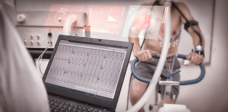
A cardiac stress test is a tool the physician may utilize to determine if the heart is getting the blood it needs under external stress, even if the patient has no signs of a problem when the heart is "at rest." When a patient has arthritis or another medical condition that prevents the patient from exercising during a stress test, the physician may give a medication that makes the heart work hard, as it would during exercise. This is called a pharmacological stress test.

On a less urgent event note, graded exercise stress testing is the most widely used diagnostic test for evaluating patients presenting chest pain. Those who have an established stable angina pectoris can also provide prognostic information about the disease's extent. Exercise stress testing can be performed alone or in conjunction with echocardiography and myocardial perfusion scintigraphy studies. For detecting cardiac ischemic processes, stress echocardiography has shown an overall sensitivity of 79 percent with a specificity of around 91 percent. While exercise stress testing alone generally has a somewhat lower diagnostic sensitivity, it is less expensive and therefore a reasonable choice in certain circumstances (Sicari, 2017).

Stress echocardiography testing can be beneficial in the evaluation of segmental wall motion during exercise. Visualization of the cardiac structure makes it possible to detect changes in the regional wall movements that tend to occur during myocardial ischemia. Experience has shown that normal myocardium becomes hyperdynamic during ischemia in response to stresses such as exercise, while affected ischemic segments become hypokinetic or akinetic. Stress echocardiography also benefits from allowing simultaneous evaluation of left ventricular function, cardiac dimensions, and visualization of any valvular disease effects. It is especially useful in patients with baseline ECG abnormalities and those with systolic murmurs suggestive of aortic stenosis or hypertrophic cardiomyopathy. Perhaps, most valuable of all is its help in the localization of ischemia and evaluation of its severity.

Thallium-201 (TI-201) and technetium-99m sestamibi (Tc-99m) are the most frequently used myocardial perfusion scintigraphy (imaging) tests. These tests are widely available and useful in individuals with unstable chest pain and baseline ECG abnormalities to localize the region of ischemic changes and prognostic indicators.

During the imaging scan, a camera takes pictures of the heart after a special test medicine (radioactive tracer) is given through an IV. The tracer travels through the blood and into the heart muscle. As the tracer moves through the heart muscle, areas that have good blood flow absorb the tracer. Areas that do not absorb tracer may not be getting enough blood or may have been damaged by a heart attack.

In essence, this type of testing is used to:

  • Assess the amount of blood reaching the heart muscle.
  • To identify areas of heart muscle lacking an adequate blood supply as a result of a heart attack.
  • To identify blocked coronary arteries and evaluate the effectiveness of coronary bypass grafts of angioplasty.

Contrary to many in the health professions, electrocardiography (EKG or ECG) is not definitive when attempting to determine the cause of acute angina. About 50 percent of patients experiencing angina specifically related to an ischemic cardiac process have normal findings on a resting ECG (Alaeddini, 2018).

Information obtained from an ECG is very helpful as it can provide evidence of prior MI, intraventricular conduction delay, various degrees of atrioventricular block, arrhythmias, or ST-T wave changes. The point is simply not to dismiss chest pain in the presence of a normal ECG, as evidenced by those patients with known coronary artery disease who have been documented as showing pseudonormalization of a resting ECG, even during active monitoring chest pain.

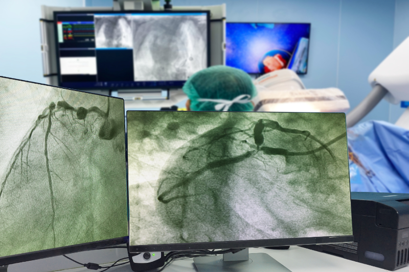
Cardiac Catheterization

Cardiac catheterization is an invasive procedure that involves aspects of both diagnosis and the potential for treating some types of ischemic cardiac disorders. Catheterization assesses angina by taking a close look at coronary hemodynamics. A cardiac cath (cardiac angiography) is usually performed in a special area under local anesthesia and sterile conditions. A flexible catheter is introduced into the heart via an artery or vein located in the groin or arm and advanced into the target area. The dye is injected through the catheter into the coronary arteries or heart's chambers, while an x-ray is taken and recorded on film. This procedure allows the physician to visualize the coronary arteries and their function and valves. Indications for cardiac catheterization include the following:

  • Confirm or exclude coronary atherosclerosis
  • Assess the feasibility of surgical correction of lesions
  • Detect valvular disease
  • Evaluate left ventricular hemodynamics
  • Assess for congenital cardiac anomalies
  • Confirm the presence of tumors, cardiomyopathies, or anomalous coronary arteries

Treating Angina Pectoris

Angina pectoris is a result of an imbalance between the oxygen supply to the heart and the demand. When the oxygen demand is greater than the supply, chest pain occurs. The pain is caused by an insufficient blood supply to the heart, causing a myocardium's temporary ischemia (inadequate oxygen for the myocardium to meet its metabolic needs). The coronary arteries are unable to provide enough oxygen-carrying blood to meet the heart muscle's oxygen requirements.

Dramatic lifestyle changes often accompany the onset of angina in a person's life. Treatment options for angina, particularly angina that is cardiac ischemic in origin, play an essential role in the dialog between clients and health professionals. We will discuss five methods for increasing the supply of needed oxygen to the heart's muscle itself.

The first, increasing the concentrations of oxygen present in the carrying medium, is mostly of aid on an acute basis. A small number of angina sufferers use oxygen routinely as part of their treatment regimen. Oxygen administration is undoubtedly a valid complementary treatment for the most challenging angina cases associated with ischemia, whether it is the cardiac or noncardiac origin.

The second method of increasing oxygen flow to the heart muscle is using medications to increase flow availability by the carrying medium. One way of speaking, the goal of pharmacologic intervention here is to convert unstable angina into stable, predictable angina. The following are medications commonly used in the treatment of angina.

Medications Used in Treatment of Angina Pectoris
Anti-anginal and Anti-ischemic drugs
  • Aspirin - Aspirin inhibits blood clotting, helping to maintain blood flow through narrowed heart arteries. Frequently given when presenting with angina, especially during a suspected heart attack, aspirin can decrease death rates by 25 percent, and its antiplatelet effect can last as long as seven days. Chewing the aspirin hastens its absorption.
  • Clopidogrel (Plavix®) - Selectively inhibits ADP binding to platelet receptor, thereby inhibiting platelet aggregation. Consider use in patients with contraindication to aspirin.
  • Nitroglycerine - This medication for treating angina temporarily opens narrowed blood vessels, improving blood flow to and from your heart. It comes in a small sublingual pill or sublingual spray form and a long-acting form (Isosorbide).
  • Beta-adrenergic blockers - Slow the heart rate and decrease overall oxygen demands.
    • Metoprolol
    • Atenolol
    • Propranolol
  • Angiotensin-converting enzyme (ACE) inhibitors - These drugs allow blood to flow from the heart more easily. For instance, a doctor may prescribe ACE inhibitors if a person has had a moderate to severe heart attack, reducing the heart's overall pumping capacity.
    • Ramipril
  • Calcium channel blockers - When treating coronary artery spasms, heart medications such as calcium channel blockers work to relax the coronary arteries and prevent the spasms. They can also aid in Prinzmetal angina (vasospastic angina).
    • Amlodipine
    • Diltiazem
    • Verapamil
  • Ranolazine (Ranexa®) - Cardio selective anti-ischemic agent (piperazine derivative) partially inhibits fatty acid oxidation. They are indicated for chronic angina unresponsive to other antianginal treatments. Unlike beta-blockers, calcium channel blockers, and nitrates, ranolazine does not reduce blood pressure or heart rate.
(Alaeddini, 2018)

The third choice for promoting increased oxygen flow to the heart tissue is that of revascularization procedures. These invasive procedures have the goal of opening partially occluded blood vessels or replacing completely blocked vessels. Coronary Artery Bypass Graft surgery (CABG) is a surgical procedure that involves the removal of a portion of a healthy blood vessel, such as a vein from the leg or the internal mammary artery (located in the chest), and using it to bypass the blockage(s) that are present in the coronary arteries.

Another revascularization procedure is Directional Coronary Atherectomy (DCA). This is the insertion of a specialized catheter into a compromised coronary artery, similar to an angioplasty, except that plaque is mechanically removed to decrease vessel blockage.

Another revascularization procedure is balloon angioplasty with or without stents placement to hold open a diseased cardiac artery.

An angioplasty is a procedure performed on patients with blocked or clogged coronary arteries. The technique is similar to a cardiac catheterization. A special catheter with a small balloon is inserted into the blockage and inflated to compress the plaque and open the artery. The uninflated balloon catheter is advanced to the area of blockage. The balloon is inflated and deflated several times until the blockage is compressed and the artery is widened. When using a stent, a fine wire mesh designed to expand and hold open the lumen of an artery is placed using a special catheter. These stents are springy, durable bundles that help provide renewed blood flow to ischemic tissue. Often angioplasty and stent placement occur during the same procedure, though either can certainly be done independently. These procedures have proven effective in the revascularization of ischemic cardiac tissues.

A fourth option is that of TMR. Transmyocardial laser revascularization is available for clients with persistent angina, considered inoperable or poor risks for the more traditional revascularization procedures. In this procedure, a surgeon exposes the heart's muscles. Using a special high-energy computerized CO2 laser creates between twenty and forty, one-millimeter wide channels (think of the width of a pinhead) in the muscle of the left heart ventricle. Body responses to these perforations close the outside of the channels. Yet, the cardiac muscle's unique characteristics allow the channels inside to remain open, providing new blood flow to ischemic tissue (Alaeddini, 2018).

The mechanism by which this works is not entirely understood even by those specialists who perform TMR. Evidence that has been gathered suggests that coronary blood flow is improved, however, and it may be that the procedure promotes angiogenesis, the growth of new, very small blood vessels that contribute to improved oxygenation of the heart.

A fifth and somewhat exciting option increasingly available for achieving improved oxygenation to the myocardium in cardiac ischemic angina sufferers is enhanced External Counter Pulsation (EECP). EECP is a noninvasive treatment using carefully timed, sequential inflations of pressure cuffs placed onto the client's calves, thighs, and buttocks. Inflation and deflation of these cuffs are timed to the patient's ECG, and the effectiveness is observed by noninvasive arterial pressure waveform monitoring.

The overall hemodynamic effect of EECP compressions is to:

  1. Provide diastolic augmentation and thus increase coronary perfusion pressure
  2. Unload systolic cardiac workload volumes and therefore decrease myocardial oxygen demand
  3. Increase venous return and, subsequently, cardiac output.

This pressure shift sequence works to displace the pressure of flowing blood backward into the coronary arteries during heart diastole when the cardiac tissue is in a state of relaxation. Resistance to backpressure in the coronary arteries is at its lowest point.

Remarkably, coronary collateral vessel development appears to be stimulated by this noninvasive increase in artery perfusion pressures. With time, a noticeable increase in perfusion capacities can be seen in both the new and pre-existing coronary arteries. Currently, this therapy is gaining favor as an exciting adjunct to the traditional treatment of cardiac ischemic-related angina. The investigation is being done concerning EECP's strong potential for treating other severe cardiac pathologies (Caceres, 2021).

The FDA approved EECP to treat angina in 1995 and later to treat congestive heart failure in 2002. Current treatment regimens vary, however for many clients who suffer from angina resistance to conventional approaches, a series of around 35 daily one-hour sessions of EECP appears to promote growth in the collateral coronary artery circulation with reduction of ischemic angina symptoms.
(Devabhaktuni, 2020)

Conclusion

Angina is the strangling, compressive discomfort common to a broad spectrum of disease processes. It may be the first visible sign of a disorder warranting emergency attention, including but not limited to anoxia and ischemia of the heart muscle. Angina must always be carefully investigated to identify the underlying cause. Awareness of both typical angina, and anginal equivalent symptoms, must be present to aid in the prompt recognition of cardiac ischemic angina and allow timely intervention to restore adequate oxygenation to tissues in distress. By our awareness of what angina is and the alert observations we make, we as health care workers can begin to get a firmer grip on this terrible pain in the neck and chest that angina is to us all.

Select one of the following methods to complete this course.

Take TestPass an exam testing your knowledge of the course material.
OR
No TestAttest that you have read and learned all the course materials.

Implicit Bias Statement

CEUFast, Inc. is committed to furthering diversity, equity, and inclusion (DEI). While reflecting on this course content, CEUFast, Inc. would like you to consider your individual perspective and question your own biases. Remember, implicit bias is a form of bias that impacts our practice as healthcare professionals. Implicit bias occurs when we have automatic prejudices, judgments, and/or a general attitude towards a person or a group of people based on associated stereotypes we have formed over time. These automatic thoughts occur without our conscious knowledge and without our intentional desire to discriminate. The concern with implicit bias is that this can impact our actions and decisions with our workplace leadership, colleagues, and even our patients. While it is our universal goal to treat everyone equally, our implicit biases can influence our interactions, assessments, communication, prioritization, and decision-making concerning patients, which can ultimately adversely impact health outcomes. It is important to keep this in mind in order to intentionally work to self-identify our own risk areas where our implicit biases might influence our behaviors. Together, we can cease perpetuating stereotypes and remind each other to remain mindful to help avoid reacting according to biases that are contrary to our conscious beliefs and values.

References

  • 1MD. (2020, July 7). Chest Pain: Symptoms, Causes, and When to See Your Doctor. Retrieved March 1, 2021, from 1MD.org. Visit Source.
  • AHA. (2021, February 10). Coronary Microvascular Disease (MVD). Retrieved March 2, 2021, from American Heart Association. Visit Source.
  • Alaeddini, J. (2018, July 19). Angina Pectoris. (E. Yang, Editor) Retrieved March 2, 2021, from Medscape. Visit Source.
  • AMBOSS. (2020, October 23). Atherosclerosis. Retrieved March 1, 2021, from AMBOSS. Visit Source.
  • AMBOSS. (2021, January 18). Chest Pain. Retrieved February 27, 2021, from AMBOSS.com. Visit Source.
  • Aroesty, J. (2020, July 1). Patient Education: Chest Pain (Beyond the Basics). UpToDate. Retrieved February 27, 2021. Visit Source.
  • Baruah, R. (2021, February 16). Unstable Angina. (A. Sidwell, Editor) Retrieved March 1, 2021, from BMJ Best Practice. Visit Source.
  • Caceres, J. (2021, January 7). Enhanced external counterpulsation: A unique treatment for the "No-Option" refractory angina patient. doi. Visit Source.
  • CDC. (2020, September 8). Heart Disease Facts. Retrieved February 27, 2021, from Centers for Disease Control and Prevention. Visit Source.
  • Conklin, L. M. (2021, February 10). These Are the 4 Stages of Heart Failure. Retrieved March 2, 2021, from The Healthy. Visit Source.
  • Devabhaktuni, S. R. (2020, December 24). Cardiac Syndrome X Treatment & Management. (R. A. Lange, Editor) Retrieved March 3, 2021, from Medscape. Visit Source.
  • Dictionary.com. (2021). Angina. Retrieved February 27, 2021, from Dictionary.com. Visit Source.
  • FPNotebook. (2018, July 24). Canadian Cardiovascular Society Angina Classification. Retrieved March 2, 2021, from Family Practice Notebook. Visit Source.
  • Haber, M. D. (2021, January 25). Angina Pectoris in Emergency Medicine. (B. E. Brenner, Editor) Retrieved March 1, 2021, from Medscape. Visit Source.
  • Hanes, E. (2020, December 7). Stable Angina vs. Unstable Angina: What's the Difference? Retrieved March 1, 2021, from HealthGrades. Visit Source.
  • Harvard. (2020, November 3). The Danger of "Silent" Heart Attacks. Retrieved March 2, 2021, from Harvard Health Publishing. Visit Source.
  • Health, G. (2020, October 1). 7 Impactful Heart Disease Statistics. Retrieved February 27, 2021, from GoMoHealth.com. Visit Source.
  • Kenkre, T. S. (2017, December 7). Ten-Year Mortality in the WISE Study (Women's Ischemia Syndrome Evaluation). Retrieved March 2, 2021, from Circulation. Visit Source.
  • Levin, T. (2020, November 12). Intracoronary Stent Restenosis. (S. Windecker, Editor) Retrieved March 2, 2021, from UpToDate. Visit Source.
  • Mahler, S. A. (2021, February 9). Angina Pectoris: Chest Pain Caused by Fixed Epicardial Coronary Artery Obstruction. Retrieved February 27, 2021, from UpToDate. Visit Source.
  • Maron, M. S. (2019, August 19). Hypertrophic cardiomyopathy: Medical therapy for heart failure. (W. J. McKenna, Editor,) Retrieved March 1, 2021, from UpToDate. Visit Source.
  • McMahon, M. (2021, February 18). What Is an Anginal Equivalent? Retrieved March 1, 2021, from WiseGeek. Visit Source.
  • Medicine, U. (2020, March 9). Chest Pain: It's Not Always a Matter of the Heart. Retrieved February 28, 2021, from University of Alabama at Birmingham. Visit Source.
  • Narula, N. (2020, July 16). Pathologic Disparities Between Peripheral Artery Disease and Coronary Artery Disease. doi. Visit Source.
  • Newman, D. P. (2020, October 6). Rib Mediated Non-Cardiac Chest Pain: A Case Report.doi. Visit Source.
  • NIH. (2018, January 18). Prinzmetal's Variant Angina. Retrieved March 1, 2021, from RareDiseaseInfo. Visit Source.
  • Oxford Languages. (2021). Angina. (Oxford University Press) Retrieved February 27, 2021, from Oxford Dictionary. Visit Source.
  • Roland, J. (2019, July 29). 30 Causes for Chest Pain and When to Seek help. Retrieved March 1, 2021, from Healthline. Visit Source.
  • Sicari, R. (2017, March 21). The clinical use of stress echocardiography in ischemic heart disease. doi. Visit Source.
  • Steinbaum, S. R. (2019, July 4). What Is Ischemia? Retrieved March 2, 2021, from WebMD. Visit Source.
  • Teragawa, H. (2020, June 15). Is Noncardiac Chest Pain Truly Noncardiac? doi:Teragawa, H., Oshita, C., & Orita, Y. (2020). Is Noncardiac Chest Pain Truly Noncardiac? Visit Source.
  • Virani, S. S. (2021, January 27). Heart Disease and Stroke Statistics—2021 Update. Retrieved February 27, 2021, from American Heart Association. Visit Source.
  • Wang, S. S. (2018, November 14). Coronary Artery Vasospasm. (E. H. Yang, Editor) Retrieved March 1, 2021, from Medscape. Visit Source.