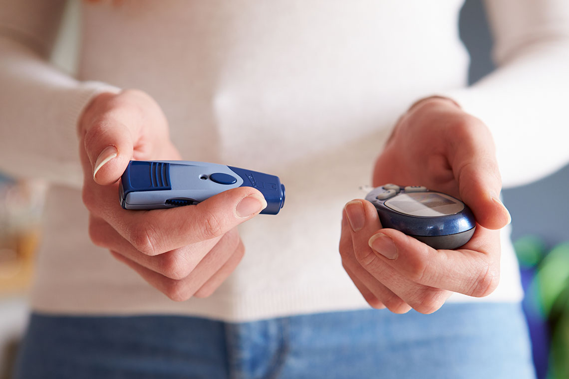
In the United States (U.S.), nearly thirty-nine million people live with diabetes. This dangerous metabolic derailment, if left unchecked, can cause multi-organ failure and or death. Diabetic ketoacidosis (DKA) leads to over two hundred thousand hospitalizations each year. Yet, with timely recognition and disciplined intervention, it is preventable and manageable.
Every nurse, provider, and allied professional, whether stationed in a rural clinic, a bustling emergency department, or an intensive care unit, must grasp DKAs underlying causes and the rationale behind each treatment step. Solid understanding empowers safer decisions at the bedside and fosters better outcomes, one patient at a time.
Understanding DKA Pathophysiology: The Foundation for Clinical Practice
DKA does not strike without warning. It takes shape through a series of metabolic missteps, which are clear in theory but relentless in real life once insulin falls short. Knowing this chain reaction matters. Lets break down how this crisis escalates in the body, step by step.
The Root Causes: Insulin Deficiency Patterns
Absolute Insulin Deficiency (Type 1 Diabetes)
In type 1 diabetes, the immune system destroys pancreatic beta cells. No beta cells, no insulinsimple as that. When insulin disappears, glucose stays stuck in the bloodstream instead of feeding the bodys cells. With glucose locked out, the body raids its fat and muscle stores for energy.
This breakdowncalled catabolismunleashes fatty acids into the bloodstream. The liver turns these fatty acids into ketones: beta-hydroxybutyrate and acetoacetate. Ketones then pile up faster than the bodys buffers can handle. Acid levels rise, while pushing the blood pH low.
Relative Insulin Deficiency (Type 2 Diabetes)
Type 2 diabetes follows a different script. The pancreas still makes insulin, just not enough when stress or infection pushes up demand on the body. Meanwhile, the bodys cells resist what little insulin remains. So, glucose production goes unchecked, cells starve, fat stores break down, and the same ketone flood appears.
Whether insulin deficiency stems from autoimmune destruction or overwhelming metabolic demand, the body's response follows a predictable, devastating cascade. The metabolic derailment unfolds through five interconnected stages, each amplifying the next.
The Metabolic Cascade: How DKA Unfolds Step by Step Through Five Interconnected Stages
- Hyperglycemia: Inadequate insulin lets the liver churn out excess glucose through gluconeogenesis and glycogen breakdown. Blood sugar shoots up.
- Osmotic Diuresis: High glucose draws water out of cells into the bloodstream. The kidneys work overtime to flush this sugar, taking electrolytes and fluids along. Dehydration sets in fast.
- Accelerated Lipolysis and Ketogenesis: Starved cells demand energy. The body doubles down on fat breakdown. The liver converts the flood of fatty acids into ketones, outpacing the bodys ability to neutralize the acid.
- Respiratory Compensation: The body tries to fix the acid problem by breathing faster and deeperwhat we call Kussmaul respirations. Every rapid breath blows off carbon dioxide, easing the acid burden just enough to buy time.
- Multi-Organ Consequences: If nothing stops this cycle, severe dehydration and unchecked acid buildup short-circuit other organs. The brain clouds overconfusion, comaand the heart strains to pump through a low-volume, acid-loaded bloodstream. Shock looms next.
Understanding this progression explains why precise, fast diagnosis is critical. Heres how the American Diabetes Association (ADA) helps clinicians confirm DKA without delay.
2024 American Diabetes Association Diagnostic Criteria: The Triangle Approach
Diagnosing DKA demands speed and accuracy. One overlooked clue can cost precious time. To help clinicians pin down DKA early and avoid missteps, the ADA, working alongside the Endocrine Society, updated the core criteria for U.S. practice in 2024.
Think of this method as a triangle: three sides, all considered critical. Miss one side, and the diagnosis wobbles.
The Three Sides of the DKA Triangle
1. Diabetes or Hyperglycemia
- Blood glucose at or above 200 milligrams per deciliter (mg/dL) (11.1 millimoles per liter [mmol/L]).
- Or, a known diabetes diagnosis, no matter the current glucose level.
A striking point: in the U.S., twenty-four percent of children with new-onset type 1 diabetes show up in DKA at first diagnosis.
2. Ketonemia
- Beta-hydroxybutyrate level is at or above 3.0 mmol/L, which is the gold standard according to the ADA.
- Blood measurement beats urine strips for catching euglycemic cases, which hide behind normal or only elevated glucose.
3.Metabolic Acidosis
- Arterial pH under 7.30, or venous pH under 7.25.
- Serum bicarbonate under 18 milliequivalents per liter (mEq/L).
- Anion gap greater than 12 mEq/L if electrolytes are available.
A patient must meet all three parts of this triangle for a confident DKA diagnosis. If something doesnt add up, double-check labs, medications, and the patients recent history. But not every case fits the classic mold. Some patients develop dangerous ketosis with normal glucose a trap every provider should watch for.
Euglycemic DKA: The Hidden Threat
Not every DKA case comes with sky-high glucose. Some patients slip into ketosis and acid buildup while their blood sugar stays under 200 (mg/dL). This so-called euglycemic DKA is increasing in frequency.
The primary culprit? Sodium-glucose transporter type 2 (SGLT2) inhibitors. Over three million people worldwide use these drugs to control blood sugar yet they raise the risk for hidden DKA, especially under certain conditions.
What Are SGLT2 Inhibitors?
These drugs force the kidneys to dump extra glucose into urine instead of letting the body reabsorb it. Helpful for blood sugar control, but they can mask rising ketone levels.
Common SGLT2 inhibitors:
- Canagliflozin (Invokana)
- Dapagliflozin (Farxiga)
- Empagliflozin (Jardiance)
- Ertugliflozin (Steglatro)
Key Triggers for Euglycemic DKA
1. Surgery
Surgical stress spikes hormones like cortisol and glucagon. Patients often fast before and after. The FDA advises stopping SGLT2 inhibitors at least three days before surgery to lower DKA risk.
2. Acute Illness with Poor Food Intake
Infections or stomach bugs curb appetite and fluid intake. Less food plus ongoing glucose loss in urine pushes the body to burn fat faster. More fat breakdown equals more ketones.
3. Low-Carb or Ketogenic Diets
These diets push the body to rely on fat for energy. Combined with SGLT2 effects, ketone levels can climb too fast for the body to buffer.
4. Dehydration from Heat or Excessive Sweating
Heat stress and diuretic effects from SGLT2 inhibitors double fluid loss. Dehydration concentrates ketones and limits kidney clearance, pushing acid levels higher.
Key Point: When treating anyone on SGLT2 inhibitors who shows vague nausea, fatigue, or mild breathing changes check for ketones early. Normal glucose does not rule out DKA anymore. After diagnosing DKA, whether classic or hidden, determine its severity to create the safest care plan.
DKA Severity Assessment and Hospital Care Standards
After confirming DKA, the next step is to classify its severity, which determines all subsequent steps. The wrong level of care wastes resources or worse, delays life-saving treatment. Clear classification aligns the patient with the right unit, meets Medicare length-of-stay expectations, and satisfies Joint Commission quality targets.
Use this straightforward guide to grade DKA into mild, moderate, or severe. Each level comes with its own lab thresholds, mental status clues, preferred unit, and Medicare Diagnosis-Related Group (DRG) code.
Mild DKA
- pH: 7.25 to 7.30
- Serum bicarbonate: 1518 mEq/L
- Mental status: Alert and oriented
- Recommended setting: Medical/surgical ward
- Expected length of stay: One to two days
Moderate DKA
- pH: 7.00 to < 7.25
- Serum bicarbonate: 1015 mEq/L
- Mental status: Drowsy but responsive
- Recommended setting: Intermediate care or step-down unit
- Expected length of stay: Two to three days
Severe DKA
- pH: Below 7.00
- Serum bicarbonate: Under 10 mEq/L
- Mental status: Stupor or coma
- Recommended setting: Intensive care unit
- Expected length of stay: Three to five days
Practical Points for Clinicians
Reassess severity with each new lab draw and mental status check. Classification alone isnt enough. Catching DKA early before it escalates remains the frontline defense.
Early Recognition Across Care Settings
Knowing the severity helps, but catching DKA early saves lives. Every provider must pick up the early signs, whether at a primary care office, an urgent care triage, or inside the emergency department.
Three Stages of Recognizable Progression
Stage 1: Early Warning - Home or Primary Care
- Polyuria: Over three liters of urine in twenty-four hours.
- Polydipsia: Intense thirst driving constant fluid intake.
- Polyphagia: Persistent hunger despite eating.
- Unexplained weight loss: More than five pounds in seven days.
- Fatigue: Tiredness that does not improve with sleep.
- Slow healing or frequent infections.
Patients often chalk these up to stress or overwork. Providers must dig deeper.
Stage 2: Intermediate - Urgent Care or Emergency Department (ED) Triage
- Kussmaul respirations: Deep, labored breathing.
- Acetone breath: Sweet, fruity odor noticeable near the patient.
- Dehydration signs: Dry tongue, poor skin turgor.
- Gastrointestinal upset: Nausea, vomiting, abdominal pain.
- Headache, muscle weakness.
- These signs demand immediate labs and treatment orders.
Stage 3: Critical - ED Priority or Intensive Care Unit (ICU) Admission
- Confusion, disorientation, or lethargy.
- Severe dehydration with orthostatic hypotension: Drop in systolic blood pressure by at least twenty millimeters of mercury (mmHg) or diastolic by ten mmHg when standing.
- Repeated vomiting with no oral intake tolerance.
- Tachycardia plus low blood pressure shock may follow without prompt fluids and insulin.
Once severity and early signs are clear, treatment must move fast and follow proven steps to reverse the crisis safely.
Evidence-Based DKA Treatment: American Hospital Standards
Treating DKA requires more than just insulin and fluids. American hospitals follow a six-step protocol that aligns with Joint Commission safety goals, Centers for Medicare & Medicaid Services (CMS) quality benchmarks, and up-to-date ADA standards. Every step calls for rapid action, clear documentation, and tight teamwork.
Step 1: Immediate Assessment and Stabilization (First 30 Minutes)
- Assess airway, breathing, and circulation right away.
- Insert two large-bore IVs (16 or 18 gauge); use central access if needed.
- Attach continuous cardiac monitoring to catch electrolyte-induced arrhythmias.
- Order stat labs: Point-of-care glucose, comprehensive metabolic panel (CMP), arterial blood gas, beta-hydroxybutyrate, phosphorus, and magnesium.
- Start a time-stamped flow sheet to meet CMS core measure requirements.
Step 2: Fluid Resuscitation (Top Priority)
Begin with 0.9 percent normal saline or balanced crystalloids like Lactated Ringers.
- Infuse at 15 to 20 mL per kilogram (kg) per hour roughly 1 to 1.5 liters hourly for an average adult.
- Adjust rate for patients older than sixty-five, or those with heart failure or renal impairment.
- American studies show balanced crystalloids shorten time to resolution by about two and a half hours and reduce hospital stays.
- Target mean arterial pressure above sixty-five mmHg and urine output over 0.5 mL/kg/hour.
Step 3: Insulin Therapy (ADA Protocol)
Administer continuous IV regular insulin at 0.1 units per kg per hour.
- For mild cases, subcutaneous rapid-acting insulin analogs (aspart, lispro) every one to two hours work well.
- Aim to lower glucose by fifty to seventy-five mg/dL each hour never exceed a drop of one hundred mg/dL/hour to prevent cerebral edema.
- Once glucose drops to 200250 mg/dL, cut the insulin infusion to 0.050.1 units/kg/hour.
- Add D5W or D10W to prevent hypoglycemia at this stage.
If a hospital faces insulin shortages, use established substitution protocols and document alternatives.
Step 4: Electrolyte Management (Patient Safety First)
- Replace potassium if levels fall below 5.2 mEq/L.
- Keep potassium between 4.0 and 5.0 mEq/L during treatment.
- Typical replacement: Add twenty to forty mEq/L to IV fluids.
Never push insulin if potassium sits below 3.3 mEq/L replenish potassium first.
- Replace phosphate only if levels drop below 1.0 mg/dL and the patient shows muscle weakness or respiratory distress.
- Check magnesium if potassium does not respond to replacement.
- Follow hospital electrolyte order sets to prevent errors.
Step 5: Continuous Monitoring (Joint Commission Standards)
- Nurses check vital signs and neurological status every hour.
- Labs: Electrolytes every two to four hours; acid-base status every four to six hours.
- Keep a clear medication administration record and update flow sheets for CMS compliance.
- Assess fall risk, skin integrity, and IV sites as per the hospitals policy.
- Track time to insulin start, glucose normalization, and ketone clearance for quality audits.
Step 6: Resolution and Safe Transition
Confirm complete DKA resolution before stepping down care:
- Blood glucose below 200 mg/dL.
- Serum bicarbonate at or above 15 mEq/L.
- Venous pH at or above 7.30.
- Beta-hydroxybutyrate under 0.6 mmol/L.
- Closed anion gap (below 12 mEq/L).
- Patient is eating and drinking without nausea.
- Plan follow-up: Switch to subcutaneous insulin, educate the patient and family, and schedule prompt endocrinology review to prevent relapse.
Your Commitment to Safer DKA Care
DKA challenges even the most experienced teams. Yet, each crisis brings an opportunity to stabilize a patient in danger, to teach better prevention, and to strengthen trust at the bedside.
This guide has walked through the chain reaction behind DKA, the clear-cut diagnostic triangle, the practical severity tiers, and the evidence-based steps that American hospitals rely on. Follow them step by step check labs early, classify severity accurately, move fast on fluids and insulin, replace electrolytes safely, and monitor without pause.
Most importantly, do not treat DKA as just a complication to fix once. Teach each patient and family how to spot early warning signs, adjust medications during illness, and get help before a simple sick day turns into a life-threatening spiral.
Every correctly managed DKA case means fewer ICU admissions, shorter hospital stays, and more patients walking out the door healthier than they arrived. Stay sharp, stay prepared, and use this knowledge to protect the next patient who trusts you with their life.
About the Author:
Glenel Loring is a dedicated nurse transitioning into freelance health writing. With 20 years of bedside nursing experience, Glenel deeply understands healthcare. She is now looking to apply her medical expertise to craft engaging, informative content for diverse audiences. Glenel's writing highlights her ability to make complex health topics accessible and relatable. Her interest in herbal supplements as alternative health solutions showcases her dedication to lifelong learning. As a mother of two and avid gardener, Glenel brings a personal touch to her work. With her strong nursing background and emerging writing skills, Glenel seeks opportunities to help brands and publications educate and empower readers to take control of their well-being.
Glenel is an independent contributor to CEUfast's Nursing Blog Program. Please note that the views, thoughts, and opinions expressed in this blog post are solely those of the independent contributor and do not necessarily represent those of CEUfast. This blog post is not medical advice. Always consult with your personal healthcare provider for any health-related questions or concerns.
If you want to learn more about CEUfast's Nursing Blog Program or would like to submit a blog post for consideration, please visit https://ceufast.com/blog/submissions.